J. Braz. Chem. Soc., Vol. 18, No. 5, 1077-1080, 2007. Printed in Brazil - ©2007 Sociedade Brasileira de Química 0103 - 5053 $6.00+0.00
Short Report
*e-mail: [email protected]; [email protected]
Coumarins and Xanthones from the Seeds of
Mammea siamensis
Surat Laphookhieo,*,a Phunrawie Promnart,a John Keith Syers,a Akkharawit Kanjana-Opas,b
Chanita Ponglimanontc and Chatchanok Karalaic
a
School of Science, Mae Fah Luang University, Tasud, Muang, Chiang Rai 57100, Thailand
b
Department of Industrial Biotechnology, Faculty of Agro-Industry, Prince of Songkla University, Hat-Yai, Songkhla 90112, Thailand
c
Department of Chemistry, Faculty of Science, Prince of Songkla University, Hat-Yai, Songkhla 90112, Thailand
Uma cumarina inédita, mammea E/BB ciclo D (1), juntamente com cinco compostos conhecidos, mammea E/BA ciclo D (2), suragina C (3), terapina B (4), 1,7-dihidroxixantona (5) e 1-hidróxi-5-metoxyxantona (6), foram isolados de sementes de Mammea siamensis. Suas estruturas foram caracterizadas usando dados de RMN 1D e 2D. Suragina C e terapina B mostraram atividade citotóxica contra adenocarcinoma de mama (MCF-7), câncer cervical humano (HeLa), câncer de colon (HT-29) e câncer oral humano (KB).
A new coumarin, mammea E/BB cyclo D (1), together with five known compounds, mammea E/BA cyclo D (2), suragin C (3), therapin B (4), 1,7-dihydroxyxanthone (5) and 1-hydroxy-5-methoxyxanthone (6), were isolated from the seeds of Mammea siamensis. Their structures were characterized using 1D and 2D NMR spectral data. Suragin C and therapin B showed cytotoxic activity against breast adenocarcinoma (MCF-7), human cervical cancer (HeLa), colon cancer (HT-29) and human oral cancer (KB).
Keywords: mammea E/BB cyclo D, cytotoxic activity, Mammea siamensis, guttiferrae
Introduction
Mammea siamensis (Miq) T. Anders. (Guttiferae), known in Thai as “Sarapi”, is a small evergreen tree distributed in Thailand, Laos, Cambodia, Vietnam and Myanmar. The flowers of this plant have been used in traditional Thai medicine as a heart tonic. Investigations of different parts of the plant have revealed the presence of several coumarins
and xanthones.1-4 We have previously reported the isolation
and structure determination of phenolic compounds from
the seeds of this species.5 In a continuation of our study on
this plant, we now report herein the isolation and structure elucidation of a novel compound, mammea E/BB cyclo D
(1), together with three known coumarins, mammea E/BC
cyclo D (2),3 suragin C (3),6 therapin B (4)7 and two known
xanthones, 1,7-dihydroxyxanthone (5)8 and
1-hydroxy-5-methoxyxanthone (6)9 from the CH
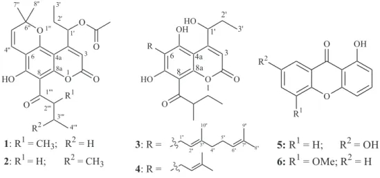
2Cl2 extract (Figure 1).
The cytotoxic activity of all isolates is also reported.
Experimental
General procedures
Melting points were determined using a Fisher-John
melting point apparatus. The optical rotation [α]D values
were determined with a JASCO P-1020 polarimeter. UV spectra were measured with a UV-160A spectrophotometer (Shimadzu). The IR spectra were measured with a
Perkin-Elmer FTS FT-IR spectrophotometer. The 1H and 13C NMR
spectra were recorded using 500 MHz Varian UNITY
INOVA and 300 MHz Bruker FTNMR Ultra ShieldTM
spectrometers. Chemical shifts were recorded in parts per
million (δ) in CDCl3 with tetramethylsilane (TMS) as an
internal reference. The EIMS was obtained from a MAT 95 XL mass spectrometer. Quick column chromatography (QCC) and column chromatography (CC) were carried out
on silica gel 60 F254 (Merck, 230-400 Mesh ASTM) and
silica gel 100 (Merck, 70-230 Mesh ASTM), respectively.
Precoated plates of silica gel 60 F254 and reversed-phase
1078 Coumarins and Xanthones from the Seeds of Mammea siamensis J. Braz. Chem. Soc.
Plant material
The seeds of M. siamensis were collected from Mae
Fah Luang University, Tasud, Muang, Chiang Rai Province, northern Thailand in August 2005. The identification was made by Professor Puangpen Sirirugsa, Department of Biology, Faculty of Science, Prince of Songkla University and a voucher specimen (No. SC09) was deposited at Prince of Songkla University Herbarium.
Extraction and Isolation
The seeds (224.5 g) of M. siamensis were extracted
successively with CH2Cl2 (500 mL) at room temperature
for 5 days. The filtered samples were combined and the solvents were evaporated under reduced pressure to
provide the CH2Cl2 extracts (44.4 g).
The CH2Cl2 extract (44.4 g) was chromatographed by
QCC and eluted with hexane-EtOAc mixtures to give seven fractions (F1-F7). Fraction F2 (1.92 g) was purified by
RP-18 CC with acetone: H2O (3:1) and followed by RP-18
preparative TLC with acetone: H2O (3:1) to yield 1 (3.1
mg) and 2 (4.3 mg). Fraction F4 (3.35 g) was separated by
CC with EtOAc: hexane (3:17) and followed by RP-18
preparative TLC with MeOH:H2O (4:1) to provide five
subfractions (F4a-F4e). Subfraction F4b (12.8 mg) was purified by preparative TLC with EtOAc: hexane (1:3, v/
v) to give 5 (2.1 mg). Subfraction F4d (1.02 g) was purified
by RP-18 CC with MeOH:H2O (4:1) and followed by CC
with EtOAc: hexane (1:3) to afford 4 (16.8 mg) and 3 (32.6
mg). Fraction F6 (167.0 mg) was separated by CC with
EtOAc: hexane (2:3, v/v) to give 6 (6.3 mg).
Mammea E/BB cyclo D(1)
Yellowish viscous oil; 1H NMR (δ, CDCl
3, 300 MHz):
14.44 (7-OH), 6.74 (1H, d, J 10.0 Hz, H-4′′), 6.60 (1H,
dd, J 6.8, 2.8 Hz, H-1′), 6.30 (1H, s, H-3), 5.60 (1H, d, J
10.0 Hz, H-5′′), 4.02 (1H, sextet, J 6.3 Hz, H-2′′′), 2.17
(3H, s, H-1′-COCH
3), 1.97 (1H, m, H-2′a), 1.80 (1H, m,
H-3′′′a), 1.78 (1H, m, H-2′b), 1.58 (3H, s, H-7′′), 1.56
(3H, s, H-8′), 1.45 (1H, m, H-3′′′b), 1.26 (3H, d, J 6.3 Hz,
H-5′′′), 1.07 (3H, t, J 7.2 Hz, H-3′) and 1.06 (3H, t, J 7.2
Hz, H-4′′′); 13C NMR data (CDCl
3, 75 MHz): 210.8
(C-1′′′), 170.3 (CH3CO), 163.5 7), 159.3 2), 157.5
(C-4), 156.7 (C-8a), 155.7 (C-5), 126.8 (C-5′′), 115.8
(C-4′′), 106.5 (C-6), 106.4 (C-3), 103.7 (C-8), 100.9 (C-4a),
80.2 (C-6′′), 73.0 (C-1′), 46.9 (C-2′′′), 29.6 (C-3′′′), 28.6
(C-2′), 28.4 (C-7′′), 27.8 (C-8′′), 21.0(CH3CO), 16.9
(C-5′′′), 10.6 (C-4′′′), 10.0 (C-3′); EIMS m/z (rel. int.): 428
[M]+ (39), 413 (100), 371 (45), 353 (13), 311 (29), 283
(5); HREIMS m/z [M]+ 428.1813 (calc. for C
24H28O7,
428.1835); UV(MeOH) λmax/nm: 225, 280, 285, 300, 373;
IR(CHCl3) νmax/cm-1: 3454, 1738, 1655, 1605; [α]
D
27-15.0°
(c 0.10, MeOH).
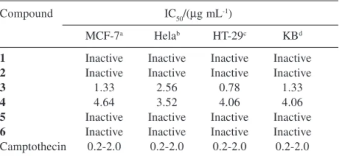
Cytotoxicity assay
The procedure for cytotoxic assay was performed by sulphorhodamine B (SRB) assay as described by Skehan et al.10 In this study, four cancer cell lines, MCF-7 (breast
adenocarcinoma), HeLa (human cervical cancer), HT-29 (colon cancer) and KB (human oral cancer) were used. Camptothecin, the reference substance, exhibited activity
toward MCF-7, HeLa, HT-29 and KB cell lines, with IC50
range of 0.2-2.0 µg mL-1 (Table 1).
Results and Discussion
Mammea E/BB cyclo D (1) was isolated as a yellowish
viscous oil, with a molecular formula C24H28O7, established
Figure 1. Structures of compounds 1-6.
O O O HO O O O R1 R2 1 3 4a 6 8 8a 1' 2' 3' 4'' 1'' 6'' 7'' 8'' 1''' 2''' 3'''
1: R1= CH3; R2= H
2: R1= H; R2= CH3
4'''
O
O OH
R2
R1
5:R1= H; R2= OH
6:R1= OMe; R2= H
O O OH HO R HO O 1 3 4a 6 8 8a
3: R =
4: R =
1079 Laphookhieo et al.
Vol. 18, No. 5, 2007
by HREIMS analysis of its molecular ion [M]+ at m/z
428.1813 (Calc. for C24H28O7 m/z 428.1835). The UV
spectrum of 1 showed absorption bands at 225, 280, 285,
300 and 373 nm suggesting the presence of conjugation in the molecule. The IR spectrum exhibited the
characteristic of carbonyl (1738 and 1655 cm-1) and
hydroxyl (3454 cm-1) functionalities. The 13C NMR and
DEPT spectra revealed 24 carbons, including six methyls
(δ 10.0, 10.6, 16.9, 21.1, 27.8 and 28.4), two methylenes
(δ 28.6 and 29.6), five methines (δ 46.9, 73.0, 106.4, 115.8
and 126.8) and eleven non-hydrogenated carbons (δ 80.2,
100.9, 103.7, 106.5, 155.7, 156.7, 157.5, 159.3, 163.5,
170.3 and 210.8). The 1H NMR spectral data showed a
chelated hydroxyl proton at δ 14.44 assignable to 7-OH
on the basis of HMBC correlations (Figure 2). The 1H
NMR spectrum also displayed a singlet signal at δ 6.30,
which is a typical chemical shift for H-3 of
4-alkylcoumarin skeleton.3,11 In addition, the 1H NMR
spectrum also showed the signals of chromene ring,
2-methyl-1-oxobutyl and 1-acetoxypropyl moieties. The 1H
NMR signals of chromene ring were appeared atδ6.74
(1H, d, J 10.0 Hz, H-4′′), 5.60 (1H, d, J 10.0 Hz, H-5′′),
1.58 (3H, s, H-7′′) and 1.56 (3H, s, H-8′′), while the
2-methyl-1-oxobutyl group showed signals at δ 4.02 (1H,
sextet, J 6.3 Hz, H-2′′′), 1.80 (1H, m, H-3′′′a), 1.45 (1H,
m, H-3′′b), 1.26 (3H, d, J 6.3 Hz, H-5′′′) and 1.06 (3H, t,
J 7.2 Hz, H-4′′′). Finally, the 1-acetonylpropyl moiety
showed the 1H NMR signals atδ6.60 (1H, dd, J 6.8, 2.8
Hz, H-1′), 2.17 (3H, s, H-1′-COCH3), 1.97 (1H, m,
H-2′a), 1.78 (1H, m, H-2′b), and 1.07 (3H, t, J 7.2 Hz, H-3′).
The locations of the three moieties were established based on the observed key HMBC correlations (Figure 2). The 1-acetoxypropyl unit was placed at C-4 due to the
oxymethine proton H-1′ (δ 6.60) showed 2J and 3J
correlation with C-4a (δ 100.9), C-4 (δ157.5) and C-3 (δ
106.4) in the HMBC spectrum. In addition, the olefinic
proton H-3 (δ 6.30) also showed 2J and 3J correlations
with C-1′ (δ 73.0), C-2 (δ 159.3) and C-4a (δ100.9). The
chromene ring was located at C-5/C-6 because the olefinic
proton H-4′′ (δ 6.74) displayed HMBC correlations to
C-5 (δ 155.7), C-6 (δ 106.5) and C-7 (δ 163.5). Finally, the
hydroxyl group was located at C-4 because the chelated
hydroxyl proton showed HMBC correlations to C-6 (δ
106.5), C-7 (δ 163.5) and C-8 (δ103.7) and the
2-methyl-1-oxobutyl moiety had to be placed at C-8 by process of elimination. Therefore, the structure of mammea E/BB
cyclo D was characterized as 1.
The reported compounds were tested for their cytotoxicity against MCF-7 (breast adenocarcinoma), HeLa (human cervical cancer), HT-29 (colon cancer) and KB human oral cancer) cell lines. The results are
summarized in Table 1. Only two coumarins, 3 and 4,
were found to be active in this study. Suragin C (3)showed
cytotoxic activities against all four cancer cell lines better
than therapin B (4)(Table 1). It should be noted that the
structural difference between suragin C (3) and therapin
B (4) is only at C-6 (3 possesses a geranyl group while 4
contains a prenyl group). The presence of a geranyl moiety seems to be important for enhancing the cytotoxic activity. The anticancer drug used as a standard in our cytotoxic
assay is camptothecin, which has an IC50 in the range of
0.2-2.0 µg mL-1.
It is worth noting that the genus Mammea of the family
Guttiferae has been known to be rich in coumarins and
xanthones,1-5,9,12-17 with more than 30 compounds having
been isolated from this genus. In this study, we have observed an additional new coumarin from the seeds of M. siamensis.
Acknowledgments
We are grateful to Mae Fah Luang University for financial support (Grant No. 49101020005). We also thank Professor Puangpen Sirirugsa, Department of Biology, Faculty of Science, Prince of Songkla University for plant identification.
Table 1. Cytotoxic activity of compounds 1-6
Compound IC50/(µg mL-1)
MCF-7a Helab HT-29c KBd
1 Inactive Inactive Inactive Inactive 2 Inactive Inactive Inactive Inactive
3 1.33 2.56 0.78 1.33
4 4.64 3.52 4.06 4.06
5 Inactive Inactive Inactive Inactive 6 Inactive Inactive Inactive Inactive Camptothecin 0.2-2.0 0.2-2.0 0.2-2.0 0.2-2.0
aMCF-7 (breast adenocarcinoma), bHeLa (human cervical cancer), c HT-29 (colon cancer) and d KB (human oral cancer).
Figure 2. Selective HMBC correlations of compound 1.
O O
O
O
HO
O
1080 Coumarins and Xanthones from the Seeds of Mammea siamensis J. Braz. Chem. Soc.
References
1. Thebtaranonth, C.; Imraporn, S.; Padungkul, N.; Phytochemistry 1981, 20, 2305.
2. Poobrasert, O.; Constant, H. L.; Beecher, C. W. W.; Farnsworth, N. R.; Kinghorn, A. D.; Pezzuto, J. M.; Cordell, G. A; Santisuk, T.; Reutrakul, V.; Phytochemistry 1998, 47, 1661.
3. Mahidol, C.; Kaweetripob, W.; Prawat, H.; Ruchirawat, S.; J. Nat. Prod. 2002, 65, 757.
4. Prachyawarakorn, V.; Mahidol, C.; Ruchirawat, S.; Phytochemistry 2006, 67, 924.
5. Laphookhieo, S.; Maneerat, W.; Kiattansakul, R.; Can. J. Chem. 2006, 84, 1546.
6. Mahandru, M. M.; Ravindran, V. K.; Phytochemistry 1986, 25, 555.
7. Lee, K. H.; Chai, H. B.; Tamez, P. A.; Pezzuto, J. M.; Cordell, G. A.; Win, K. K.; Tin-Wa, M.; Phytochemistry 2003, 64, 535. 8. Nagam T. J.; de Oliveira, F. F.; J. Braz. Chem. Soc. 1997, 8,
505.
9. Gunasekera, S. P.; Ramachandran, S.; Selliah, S.; Sultanbawa, M. U. S.; J. Chem. Soc., Perkin Trans. 1 1975, 2447.
10. Skehan, P.; Storeng, R.; Scudiero, D.; Monks, A.; Mcmahon, J.; Vistica, D.; Warren, J. T.; Bokesch, H.; Kenney, S.; Boyd, M. R.; J. Natl. Cancer Inst. 1990, 82, 1107.
11. Cruz, F. G.; da Silva-Neto, J. T.; Guedes, M. L. S.; J. Braz. Chem. Soc. 2001, 12, 117.
12. Reutrakul, V.; Leewanich, P.; Tuchinda, P.; Pohmakotr, M.; Jaipetch, T.; Sophasan, S.; Santisuk, T.; Planta Med. 2003, 69, 1048.
13. Kaweetripob, W.; Mahidol, C.; Prawat, H.; Ruchirawat, S.; Pharm. Biol. 2000, 38, 55.
14. Prachyawarakorn, V.; Mahidol, C.; Ruchirawat, S.; Pharm. Biol. 2000, 38, 58.
15. Combie, L.; Jones, R. C. F.; Palmer, C. J.; J. Chem. Soc. Perkin Trans. 1, 1987, 345.
16. Tosa, H.; Iinuma, M.; Murakami, K-I.; Ito, T.; Tanaka, T.; Chelladurai, V.; Riswan, S.; Phytochemistry 1997, 45, 133. 17. Iinuma, M.; Tosa, H.; Tanaka, T.; Riswan, S.; Phytochemistry
1996, 42, 245.
Received: January 9, 2007