Chro nic hype rte nsio n alte rs
the e xpre ssio n o f Cx43 in
cardio vascular muscle ce lls
1Department of Internal Medicine B, University Hospital, Lausanne, Switzerland 2Department of Morphology, University of Geneva, Geneva, Switzerland
J.-A. Haefliger1
and P. Meda2
Abstract
Connexin43 (Cx43), the predominant gap junction protein of muscle cells in vessels and heart, is involved in the control of cell-to-cell communication and is thought to modulate the contractility of the vascular wall and the electrical coupling of cardiac myocytes. We have investigated the effects of arterial hypertension on the expression of Cx43 in aorta and heart in three different models of experimental hypertension. Rats were made hypertensive either by clipping one renal artery (two kidney, one-clip renal (2K,1C) model) by administra-tion of deoxycorticosterone and salt (DOCA-salt model) or by inhib-iting nitric oxide synthase with NG-nitro-L-arginine methyl ester (L-NAME model). After 4 weeks, rats of the three models showed a similar increase in intra-arterial mean blood pressure and in the thickness of the walls of both aorta and heart. Analysis of heart mRNA demonstrated no change in Cx43 expression in the three models compared to their respective controls. The same 2K,1C and DOCA-salt hypertensive animals expressed twice more Cx43 in aorta, and the 2K,1C rats showed an increase in arterial distensibility. In contrast, the aortae of L-NAME hypertensive rats were characterized by a 50% decrease in Cx43 and the carotid arteries did not show increased distensibility. Western blot analysis indicated that Cx43 was more phosphorylated in the aortae of 2K,1C rats than in those of L-NAME or control rats, indicating a differential regulation of aortic Cx43 in different models of hypertension. The data suggest that localized mechanical forces induced by hypertension affect Cx43 expression and that the cell-to-cell communication mediated by Cx43 channels may contribute to regulating the elasticity of the vascular wall. Co rre spo nde nce
J.-A. Haefliger
Département de Medecine Interne B Laboratoire de Biologie
Moléculaire 19-135S Centre Hospitalier Universitaire Vaudois
CHUV-1011 Lausanne
Switzerland Fax: + 41-21-314-0968 E-mail: jhaeflig@ chuv.hospvd.ch
Presented at the Meeting “Gap Junctions in the Nervous and
Cardiovascular Systems: Clinical Implications”, Rio de Janeiro, RJ, Brazil, June 6-11, 1998.
J.-A. Haefliger is supported by a career award from the Max Cloëtta Foundation. Research supported by
grants from the Swiss National Science Foundation (No. 31-46770.96 to J.-A. Haefliger and No. 31-53720.98 to P. Meda), from the European Union (Q LRT-1999-00516) and from the Foundation de Reuter (No. 265) to P. Meda.
Received July 30, 1999 Accepted September 15, 1999
Ke y words
·Cell-cell communication
·Gap junctions
·Connexins
·Vasculature
·Heart
·Hypertension
·Blood pressure
·Renin
·Deoxycorticosterone
·Nitric oxide
·Distensibility
Intro ductio n
Gap junctions are seen at sites where the plasma membranes of two adjacent cells be-come closely apposed. In these regions, the two interacting membranes feature special-ized microdomains characterspecial-ized by the con-centration of large protein assemblies named connexons. These structures provide the wall of specialized intercellular channels that
cardiovas-cular system (4). As yet, little is known about their physiological role and their possible changes in cardiovascular diseases.
Gap junctions ensure the electrical and mechanical coupling of different types of muscle cells (5,6). Such a role is critical in the heart, since proper propulsion of blood in the circulation obrigatorily depends on the coordinated contraction of both atrial and ventricular cardiomyocytes (7,8). This con-traction in turn is mediated by the rapid propagation of action potentials to multiple cells that should depolarize in coordination. These events are dependent on gap junc-tional communications, whereby adjacent cardiomyocytes exchange current-carrying ions. By diffusing from one cell to the next, these ions synchronize the electrical and mechanical activity of neighboring cells (7). Coordination of smooth muscle cells of the vascular wall is also critical to the local modulation of vasomotor tone, thus contrib-uting to the proper function of large vessels. The aorta, which is a sparsely innervated and electrically quiescent vascular tissue, is likely to be particularly dependent on gap junc-tional communications for coordinating the responses of smooth muscle cells to diverse neural and endothelial signals (9,10).
Characterization of the expression of the proteins that form gap junctions may pro-vide insights into the possible involvement of junctional channels during cardiac and arterial hypertrophy associated with hyper-tension (11-13). Thus, conditions perturbing the function of the aortic wall, as observed during chronic hypertension, are expected to be associated with alterations of connexins, gap junctions or coupling.
To test this hypothesis, we have studied the expression of Cx43, the physiologically predominant connexin of myocardial and aortic smooth muscle cells (14), during chronic hypertension. To this end, we have investigated three rat models characterized by a similar degree of hypertension and by hypertrophy of both aortic and heart walls,
but differing markedly by the mechanism causing these changes. In the mineralocorti-coid salt-induced model (deoxycorticoster-one, DOCA-salt), hypertension resulted from increased retention of sodium in the pres-ence of suppressed renin secretion (15,16). In the two kidney, one-clip model (2K,1C), hypertension was produced by clipping one renal artery, leading to stimulation of renin secretion and to an angiotensin II (AngII)-dependent elevation of blood pressure. Un-der the conditions provided by these two models, the isobaric distensibility of the ca-rotid has been shown to increase due to a reduced elastic modulus, corresponding to a reduction of wall material stiffness (17,18). These biomechanical changes are not ob-served in a third rat model, in which a degree of hypertension similar to that observed in the two kidney, one-clip and the DOCA-salt models can be induced by inhibiting nitric oxide synthase (NOS) with NG
-nitro-L-argi-nine-methyl ester (L-NAME) (19,20). In this model, hypertension is associated with lim-ited cardiovascular hypertrophy (20,21), and with no increase in the isobaric distensibility of the carotid (22). Comparison of the three models is therefore of interest to determine whether Cx43 changes are consistently asso-ciated with hypertension and whether they vary, qualitatively and/or quantitatively, as a function of the mechanisms that lead to in-creased blood pressure and to cellular changes in the aortic wall (23).
Re sults
Effe cts of tre atme nts on blood pre ssure
Blood pressure levels were 1.4-1.6-fold higher in the 2K,1C, DOCA-salt and L-NAME animals compared to control (Figure 1).
Effe cts of hype rte nsion on the he art
DOCA-salt models showed a 30% increase in heart index (i.e., weight of myocardium per unit animal weight) compared to their normotensive counterparts (Figure 2). In agreement with this change, Northern blot analysis showed a two-fold increase in the expression of a-skeletal actin mRNA in the two groups of hypertensive rats and histol-ogy revealed a thickening of the left ventric-ular wall (11). Hearts of L-NAME hyperten-sive rats were also hypertrophied compared to those of normotensive controls, as indi-cated by a 17% increase in cardiac weight index (Figure 1). In agreement with this change, Northern blot analysis of heart tis-sue showed a three-fold increase in the ex-pression of atrial natriuretic peptide (ANP) mRNA and a two-fold increase in the ex-pression of skeletal a-actin mRNA in the hypertensive rats (23).
Quantitative assessment by Northern blot analysis showed that Cx43 expression was similar in the hypertrophied hearts of hyper-tensive rats of the three models studied, and in those of normotensive controls (Figure 2). This finding was correlated with the absence of significant differences in the levels and distribution of Cx43 which was
immunola-beled in the intercalated discs of heart muscle in the three models studied (11,23).
Effe cts of hype rte nsion on the aorta
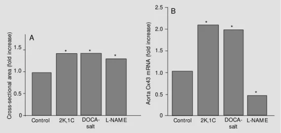
The thickness of the intima plus media layers of aorta was significantly larger in the 2K,1C and DOCA-salt hypertensive rats than in normotensive animals, resulting, in spite of a constant lumen radius, in a 40% increase of the vessel cross-sectional area (CSA), which was given by the formula: CSA = p [(lumen radius + media-intima thickness)2
-(lumen radius)2
] (Figure 3). These changes were due to an enlargement of smooth muscle
B
lo
o
d
p
re
s
s
u
re
(
fo
ld
i
n
c
re
a
s
e
) 2
*
* *
Control 2K,1C
DOCA-salt
L-NAM E
Figure 1 - Increase in blood pres-sure in the 2K,1C-, DOCA-salt-and L-NAM E-treated rats. In the t hree m odels st udied, blood pressure w as found to be in-creased 1.4-1.6-fold over control values (* P<0.05) four w eeks af-ter the beginning of treatment (Bonferroni-Dunn test). 1
0
Figure 2 - Thickening of the cardiac w all and expression of Cx43 in hearts of hypertensive rats. A, As compared to normotensive animals, the 2K,1C and DOCA-salt hypertensive rats show ed a similar 30% increase in heart index. In L-NAM E rats, this increase w as only 17% , on average. Values are reported as means of about 10 measurements (one measurement per rat) compared to the control value w hich w as set at 1. * P<0.01 compared to control (Fisher’s protected least significant difference test). B, Analysis of heart RNA revealed that the levels of the Cx43 transcript, w hich w as mostly provided by cardiomyocytes, w ere not altered in the three types of hypertensive rats investigated. Values represent ratios of densitometric measurements of Cx43 and glyceraldehyde-3-phosphate dehydrogenase (GAPDH) mRNAs, and are reported as means of about 10 measurements, relative to the control value, w hich w as given a value of 1.
H
e
a
rt
i
n
d
e
x
(
fo
ld
i
n
c
re
a
s
e
)
1.5
1.0
0.5
0
Control 2K,1C
DOCA-salt
L-NAM E
H
e
a
rt
C
x
4
3
m
R
N
A
(
fo
ld
i
n
c
re
a
s
e
)
1.5
1.0
0.5
0
Control 2K,1C
DOCA-salt
L-NAM E
* *
*
cells, whose numerical density was slightly reduced, and were paralleled by a significant increase in the expression of a-skeletal ac-tin. In comparison to controls, the L-NAME hypertensive animals also showed a 25% thickening of the aortic wall (intima plus media) resulting in an increased cross-sec-tional area of the aorta (Figure 3).
Quantitative assessment of Cx43 gene expression by Northern blotting of total aorta RNA, which was mostly contributed to by smooth muscle cells, showed significantly higher values in the aorta of 2K,1C and DOCA-salt hypertensive rats than in that of normotensive rats (Figure 3). In contrast, when compared to controls, the transcript of Cx43 was reduced by about 50% in the aorta of L-NAME-treated animals (Figure 3).
Cx43 was immunolocated on smooth muscle cells of the aortic media in both hypertensive and normotensive rats. The number of immunofluorescence spots re-flecting the abundance of Cx43 was larger in
the aorta of control rats than in that of L-NAME-treated rats. In contrast, the smooth muscle cells of 2K,1C and DOCA-salt rats showed a modest but sizeable increase in the amount of Cx43, as monitored by immuno-labeling of aorta cryosections (11).
Western blot analysis of total proteins extracted from the aorta showed that Cx43 was significantly decreased in L-NAME hy-pertensive rats. Extracts from the aorta of these animals contained one major immu-noreactive band (of about 44 kDa) whose average intensity was less than 50% that of controls. In contrast, extracts from aortas of 2K,1C hypertensive rats contained three im-munoreactive bands indicating that Cx43 was consistently more phosphorylated in the aorta of these animals than in that of both controls and L-NAME animals. Also the levels of Cx43 in 2K,1C were significantly higher than in controls (23).
A trend towards a reduced distensibility-pressure curve for the carotid artery was
Figure 3 - Thickening of the aortic w all in hypertensive rats and expression of Cx43 in aortae of hypertensive rats. A, After four w eeks of hypertension, the aorta of tw o kidney, one-clip renal (2K,1C) and DOCA-salt hypertensive animals show ed a thickened w all, resulting in a 40% increase in the cross-sectional area (control value w as set at 1), despite a constant internal diameter. The aorta of L-NAM E hypertensive rats w as thickened to a lesser extent, resulting in a 25% increase in cross-sectional area. Values are reported as means of 4-8 measurements. * P<0.01 compared to control (Scheffé test). B, Analysis of aorta RNA revealed that the transcript for Cx43, w hich w as mostly provided by smooth muscle cells, w as increased about tw o-fold in the hypertensive rats of the 2K,1C and DOCA-salt models. In contrast, the hypertensive rats treated w ith L-NAM E show ed a 50% reduction of the Cx43 transcript compared to the control value (w hich w as set at 1 in all groups). Values represent ratios of densitometric measurements of Cx43 and glyceraldehyde-3-phosphate dehydrogenase (GAPDH) mRNAs, and are reported as means of 6-10 experiments. * P<0.01 compared to control (Scheffé test).
C
ro
s
s
-s
e
c
ti
o
n
a
l
a
re
a
(
fo
ld
i
n
c
re
a
s
e
)
1.5
1.0
0.5
Control 2K,1C
DOCA-salt
L-NAM E
A
o
rt
a
C
x
4
3
m
R
N
A
(
fo
ld
i
n
c
re
a
s
e
)
1.5
1.0
0.5
0
Control 2K,1C
DOCA-salt
L-NAM E A
B
2.0 2.5
* *
*
*
*
*
seen in the L-NAME-treated rats, in the range of blood pressures (120-140 mmHg) that could be compared. In contrast, carotid dis-tensibility has been shown to be markedly increased in the 2K,1C model (20,22).
D iscussio n
We have examined the effects of chronic hypertension on the expression of Cx43, the major native connexin of the cardiovascular system, in three different experimental rat models.
After one month, the increase in blood pressure achieved in the three models was comparable. We found that all hypertensive animals exhibited cardiac hypertrophy (11) in the absence of differences in the levels of Cx43 connecting myocardial cells. This find-ing suggests that Cx43 is not involved in the myocardial adaptation that accompanies a hypertension-induced increase in heart load. This conclusion does not rule out that other connexins such as the Cx45, Cx40, and Cx37 isoforms (24) may participate in the heart changes induced by hypertension. Indeed, the inactivation of the Cx43 gene in trans-genic mice suggests that, at least under cer-tain conditions, Cx43 may be functionally replaced by other connexins (25).
Under the conditions used in the present study, all hypertensive animals also exhib-ited a thickening of the aortic wall, which was mostly accounted for by the hypertro-phy of smooth muscle cells and the accumu-lation of extracellular materials. The 2K,1C and DOCA-salt rats also exhibited a compa-rable increase in the level of Cx43 that was expressed by the smooth muscle cells of the aortic media. In contrast, decreased levels of Cx43 were found in the same cells of L-NAME-treated rats.
In the 2K,1C model, the development of hypertension results from the constriction of one renal artery and the ensuing activation of the renin-angiotensin system, as reflected by enhanced renin mRNA levels in the
hypo-perfused kidney and by elevated plasma re-nin activity (26). The further proteolytic cleavage of angiotensinogen by renin and the processing of angiotensin I by angio-tensin-converting enzyme leads to the gen-eration of the biologically active AngII. AngII is a most potent vasoconstrictor peptide which also plays a role in the development of vas-cular and cardiac hypertrophy (27,28). There-fore, the cellular and connexin changes ob-served in the 2K,1C rats could be due to both the increased blood pressure and the in-creased levels of AngII. To discriminate be-tween these possibilities, we have studied the DOCA-salt model, which is character-ized by the functional suppression of the renin-angiotensin system. In this model, hy-pertension is induced by administration of a salt-retaining mineralocorticoid in associa-tion with a high sodium intake (15). The finding in this model of vascular and con-nexin changes similar to those observed in the 2K,1C model indicates that these changes could not be related to the circulating levels of AngII, which differed considerably in the 2K,1C and the DOCA-salt hypertensive rats and, hence, are likely to be associated with the elevation of blood pressure. The molec-ular mechanism leading to the pressure-in-duced increase in the expression of the Cx43 gene remains to be elucidated. The presence of multiple promoters in the 5' untranslated region of this gene (29,30) raises the possi-bility that the tissue-specific regulation of this increase observed here is controlled by distinct transcription factors (31). Of par-ticular interest in this context is the recent finding that transcription of the Cx43 gene may be promoted by an increase in the ex-pression of c-fos (32), since the mRNA cod-ing for this transcription factor accumulates in smooth muscle cells of rat aortas follow-ing exposure to angiotensin II (33,34), which contributes to hypertension in the 2K,1C model.
and structural changes characterized by an outward hypertrophic remodeling with pre-served isobaric luminal diameter (18,35). This remodeling, which results from hyper-trophy of smooth muscle cells and alter-ations of extracellular matrix, may be re-garded as an adaptation to normalize wall stress. However, this adaptation is also likely to modify the mechanical properties of ar-teries, which could be detrimental in the long term (18). Previous studies have shown that the distensibility and compliance of vari-ous arteries are increased under isobaric con-ditions in the 2K,1C hypertensive animals (18,20), suggesting that changes in tissue composition and architecture permit arteries to maintain adequate elastic properties in spite of increased blood pressure. In con-trast, the hypertension caused by inhibition of nitric oxide is not associated with an increase in the isobaric distensibility of the carotid artery, despite a thickening of the arterial wall which is similar to that observed in other experimental models of hyperten-sion. The different viscoelastic properties of arteries in the 2K,1C and L-NAME-treated rats implies a differential structural and/or functional organization of tissues making up the wall of resistance arteries. Thus, the L-NAME-induced hypertension was associ-ated with a decrease in the expression of Cx43 in the smooth muscle cells of the aorta, contrasting with the findings for both the 2K,1C and the DOCA-salt models (11,12). The significance of the decrease in Cx43 in the L-NAME-treated rats remains to be elu-cidated. Several electrophysiological stud-ies have suggested that gap junction proteins may be important to coordinate the mechan-ical contractions of smooth muscle cells, possibly to insure a proper modulation of the vasomotor tone of the aortic wall (9). Cer-tainly, Cx43 can provide an intercellular pathway for the syncytial functioning of dis-tant smooth muscle cells that could be re-cruited for synchronous contraction through propagation of gap junction-permeant
sec-ond messengers (36).
Eventually, a different post-translational regulation of Cx43 was observed in the aor-tic smooth muscle cells of the different hy-pertension models. Thus, whereas the de-gree of Cx43 phosphorylation was found to be increased in the 2K,1C animals, it was decreased in the L-NAME-treated rats, which essentially expressed a non-phosphorylated form of Cx43. Since connexin phosphoryla-tion can affect the extent of juncphosphoryla-tional com-munication, this difference could result in a selective cell-to-cell exchange of the mol-ecules involved in both hypertrophy (2K,1C model) and polyploidy (L-NAME model) of vascular smooth muscle cells. Blockade of the nitric oxide production by endothelial cells after treatment with L-NAME is ex-pected to decrease the apoptosis and to pro-mote the proliferation of smooth muscle cells (37,38), thus accounting for their accumula-tion and polyploidy on the aortic wall. The reduced expression of Cx43 in the aorta of L-NAME-treated rats may also contribute, as in atherosclerotic lesions (39), to upregu-lating the adhesion of monocytes/macrophag-es to the aorta. After inhibition of NO pro-duction by L-NAME treatment, this adhe-sion increases (40).
Re fe re nce s
1. Beyer EC, Goodenough DA & Paul DL (1988). The connexins, a family of related gap junction proteins. In: Herzberg EL & Johnson RG (Editors), Gap Junction. Alan R. Liss, New York, 167-175.
2. Loew enstein WR (1981). Junctional inter-cellular communication. The cell-to-cell m em brane channel. Physiological Re-view s, 61: 829-913.
3. Spray DC (1998). Gap junction proteins. Where they live and how they die. Circu-lation Research, 83: 679-681.
4. Haefliger J-A, Bruzzone R, Jenkins NA, Gilbert DJ, Copeland NG & Paul DL (1992). Four novel members of the connexin fam-ily of gap junction proteins: molecular cloning, expression and chrom osom e mapping. Journal of Biological Chemistry, 267: 2057-2064.
5. Spray DC & Burt JM (1990). Structure-activity relations of cardiac gap-junction channel. American Journal of Physiology, 258: C195-C205.
6. Christ GJ (1995). M odulation of a 1-adre-nergic contractility in isolated vascular tis-sue by heptanol: A functional demonstra-tion of the potential importance of inter-cellular communication to vascular re-sponse generation. Life Sciences, 56: 709-721.
7. Severs NJ (1994). Pathophysiology of gap junctions in heart disease. Journal of Car-diovascular Electrophysiology, 5: 462-475. 8. Peters NS (1997). Gap junctions and clini-cal cardiology: from molecular biology to molecular medicine. European Heart Jour-nal, 18: 1697-1702.
9. Segal SS (1994). Cell-to-cell communica-tion coordinates blood flow control. Hy-pertension, 23: 1113-1120.
10. Larson DM , Haudenschild CC & Beyer EC (1990). Gap junction messenger RNA ex-pression by vascular w all cells. Circulation Research, 66: 1074-1080.
11. Haef liger J-A, Cast illo E, W aeber G, Bergonzelli GE, Aubert J-F, Sutter E, Nicod P, Waeber B & M eda P (1997). Hypertension increases connexin43 in a tissue-specific manner. Circulation, 95: 1007-1014.
12. Haef liger J-A, Cast illo E, W aeber G, Aubert J-F, Nicod P, Waeber B & M eda P (1997). Hypertension differentially affects the expression of the gap junctional pro-tein connexin43 in cardiac myocytes and aortic smooth muscle cells. Advances in Experimental M edicine and Biology, 432: 71-82.
13. Watts SW & Webb RC (1996). Vascular
gap junct ional com m unicat ion is in-creased in mineralocorticoid-salt hyper-tension. Hypertension, 28: 888-893. 14. Bruzzone R, Haefliger J-A, Gimlich RL &
Paul DL (1993). Connexin40, a compo-nent of gap junctions in vascular endothe-lium, is restricted in its ability to interact w ith other connexins. M olecular Biology of the Cell, 4: 7-20.
15. Gavras H, Brunner HR, Larah JH, Vaughn ED, Koss M , Cote LJ & Gavras I (1975). M alignant hypertension resulting from deoxycorticosterone acetate and salt ex-cess. Circulation Research, 36: 300-309. 16. Liu DT, Birchall I, Hew itson T,
Kincaid-Smith P & Whitw orth JA (1994). Effect of dietary calcium on the development of hypertension and hypertensive vascular lesions in DOCA-salt and tw o-kidney, one clip hypertensive rats. Journal of Hyper-tension, 12: 145-153.
17. M ulvany M J (1992). A reduced elastic modulus of vascular w all components in hypertension. Hypertension, 20: 7-9. 18. Zanchi A, Wiesel P, Aubert J-F, Brunner
HR & Hayoz D (1997). Tim e course changes of the mechanical properties of the carotid artery in renal hypertensive rats. Hypertension, 29: 1199-1203. 19. Arnal J-F, El Amrani AI, Chatellier G,
M énard J & M ichel JB (1993). Cardiac w eight in hypertension induced by nitric oxide synthase blockade. Hypertension, 22: 380-387.
20. Delacrétaz E, Zanchi A, Nussberger J, Hayoz D, Aubert J-F, Brunner HR & Waeber B (1995). Chronic nitric oxide syn-thase inhibition and carotid artery disten-sibility in renal hypertensive rats. Hyper-tension, 26: 332-336.
21. Devlin AM , Brosnan M J, Graham D, M orton JJ, M cPhaden AR, M cIntyre M , Hamilton CA, Reid JL & Dominiczak AF (1998). Vascular smooth muscle cell poly-ploidy and cardiomyocyte hypertrophy due to chronic NOS inhibition in vivo. American Journal of Physiology, 274: H52-H59.
22. Delacrétaz E, Hayoz D, Osterheld M C, Genton CY, Brunner HR & Waeber B (1994). Long-term nitric oxide synthase inhibition and distensibility of carotid ar-tery in intact rats. Hypertension, 23 (Part 2): 967-970.
23. Haefliger J-A, M eda P, Formenton A, Wiesel P, Zanchi A, Brunner HR, Nicod P & Hayoz D (1999). Aortic connexin43 is decreased during hypertension induced by inhibition of nitric oxide synthase.
Arte-riosclerosis, Thrombosis, and Vascular Bi-ology, 19: 1615-1622.
24. Bastide B, Neyses L, Ganten D, Paul M , Willecke K & Traub O (1993). Gap junction protein connexin40 is preferentially ex-pressed in vascular endothelium and con-ductive bundles of rat and is increased under hypertensive conditions. Circulation Research, 73: 1138-1149.
25. Gros DB & Jongsma HJ (1996). Connex-ins in mammalian heart function. Bioes-says, 18: 719-730.
26. Haefliger J-A, Bergonzelli G, Waeber G, Aubert J-F, Nussberger J, Gavras H, Nicod P & Waeber B (1995). Renin and angio-tensin II receptor gene expression in kid-neys of renal hypertensive rats. Hyperten-sion, 26: 733-737.
27. Levy BI, M ichel J-B, Salzmann J-L, Azizi M , Poitevin P, Safar M & Camilleri J-P (1988). Effects of chronic inhibition of con-verting enzyme on mechanical and struc-tural properties of arteries in rat renovas-cular hypertension. Circulation Research, 63: 227-239.
28. M orishita R, Higaki J, M iyazaki M & Ogihara T (1992). Possible role of the vas-cular renin-angiotensin system in hyper-tension and vascular hypertrophy. Hyper-tension, 19: II-62-II-67.
29. Yu W, Dahl G & Werner R (1994). The connexin43 gene is responsive to oestro-gen. Proceedings of the Royal Society of London. B, Biological Sciences, 255: 125-132.
30. Chen Z-Q, Lefebvre DL, Bai X-H, Reaume A, Rossant J & Lye SJ (1995). Identifica-tion of tw o regulatory elements w ithin the promoter region of the mouse Cx-43 gene. Journal of Biological Chemistry, 270: 3863-3868.
31. Piersanti M & Lye SJ (1995). Increase in messenger ribonucleic acid encoding the myometrial gap junction protein, con-nexin-43, requires protein synthesis and is associated w ith increased expression of the activator protein-1, c-fos. Endocri-nology, 136: 3571-3578.
32. Lefebvre DL, Piersanti M , Bai X-H, Chen Z-Q & Lye SJ (1996). M yometrial tran-scriptional regulation of the gap junction gene, connexin-43. Reproduction, Fertil-ity, and Development, 7: 603-611. 33. Taubman M B, Berk BC, Izumo S, Tsuda T,
Alexander RW & Nadal-Ginard B (1989). Angiotensin II induces c-fos mRNA in aor-tic smooth muscle. Journal of Biological Chemistry, 264: 526-530.
& Dzau VJ (1989). Angiotensin II induces c-fos expression in smooth muscle via transcriptional control. Hypertension, 13: 706-711.
35. Weber R, Stergiolpulos N, Brunner HR & Hayoz D (1996). Contributions of vascular tone and structure to elastic properties of a medium size artery. Hypertension, 27: 816-822.
36. Christ GJ, Spray DC, El-Sabban M , M oore LK & Brink PR (1996). Gap junction in vascular tissues. Evaluating the role of
the intercellular communication in the modulation of vasomotor tone. Circula-tion Research, 79: 631-646.
37. Dubrey R & Lüscher T (1995). Nitric oxide inhibits angiotensin II-induced migration of rat aortic smooth muscle cell. Journal of Clinical Investigation, 96: 141-149. 38. Pollman M J, Yamada T, Horiuchi M &
Gib-bons GH (1996). Vasoactive substances regulate vascular smooth muscle cell ap-optosis. Countervailing influences of ni-tric oxide and angiotensin II. Circulation
Research, 79: 748-756.
39. Blackburn JP, Peters NS, Yeh H-I, Rothery S, Green CR & Severs NJ (1995). Upregu-lation of Cx43 gap junctions during early stages of human coronary atherosclero-sis. Arteriosclerosis, Thrombosis, and Vas-cular Biology, 15: 1219-1228.