Muscle Cells in a Rat Model of Cavernous Neurectomy
Fan Yang1., Jian F. Zhao1., Qi Y. Shou2
, Xiao J. Huang1, Gang Chen1, Ke B. Yang1, Shi G. Zhang1, Bo D. Lv1,4*, Hui Y. Fu3,4*
1Department of Urology, The Second Affiliated Hospital, Zhejiang Chinese Medical University, Hangzhou, China,2Laboratory Animal Research Center, Zhejiang Chinese Medical University, Hangzhou, China,3Central Laboratory, The Second Clinical Medical College, Zhejiang Chinese Medical University, Hangzhou, China,4Andrology Laboratory on Integration of Chinese and Western Medicine, Zhejiang provincial Key Laboratory of Traditional Chinese Medicine, Hangzhou, China
Abstract
Background:Patients undergoing radical prostatectomy (RP) are at high risk for erectile dysfunction (ED) due to potential cavernous nerve (CN) damage during surgery. Penile hypoxia after RP is thought to significantly contribute to ED pathogenesis.
Aim:We previously showed that corpora cavernosum smooth muscle cells (CCSMCs) undergo phenotypic modulation under hypoxic conditionsin vitro. Here, we studied such changes in anin vivopost-RP ED model by investigating CCSMCs in
bilateral cavernous neurectomy (BCN) rats.
Methods:Sprague-Dawley rats underwent sham (n = 12) or BCN (n = 12) surgery. After 12 weeks, they were injected with apomorphine to determine erectile function. The penile tissues were harvested and assessed for fibrosis using Masson trichrome staining and for molecular markers of phenotypic modulation using immunohistochemistry and western blotting. CCSMC morphological structure was evaluated by hematoxylin-eosin (H&E) staining and transmission electron microscopy (TEM).
Results:Erectile function was significantly lower in BCN rats than in sham rats. BCN increased hypoxia-inducible factor-1a
and collagen protein expression in corpora cavernous tissue. H&E staining and TEM showed that CCSMCs in BCN rats underwent hypertrophy and showed rough endoplasmic reticulum formation. The expression of CCSMC phenotypic markers, such as smooth musclea-actin, smooth muscle myosin heavy chain, and desmin, was markedly lower, whereas vimentin protein expression was significantly higher in BCN rats than in control rats.
Conclusions: CCSMCs undergo phenotype modulation in rats with cavernous neurectomy. The results have unveiled physiological transformations that occur at the cellular and molecular levels and have helped characterize CN injury– induced ED.
Citation:Yang F, Zhao JF, Shou QY, Huang XJ, Chen G, et al. (2014) Phenotypic Modulation of Corpus Cavernosum Smooth Muscle Cells in a Rat Model of Cavernous Neurectomy. PLoS ONE 9(8): e105186. doi:10.1371/journal.pone.0105186
Editor:Aamir Ahmed, University College London, United Kingdom
ReceivedApril 1, 2014;AcceptedJuly 17, 2014;PublishedAugust 15, 2014
Copyright:ß2014 Yang et al. This is an open-access article distributed under the terms of the Creative Commons Attribution License, which permits unrestricted use, distribution, and reproduction in any medium, provided the original author and source are credited.
Data Availability:The authors confirm that all data underlying the findings are fully available without restriction. All relevant data are within the paper and its Supporting Information files.
Funding:This work was supported by the following: WKJ2010-2-020 (http://www.zjwst.gov.cn/), 2009ZA009 (http://zgj.zjtcm.net/), and Y2111167 (http://www. zjnsf.gov.cn/). The funders had no role in study design, data collection and analysis, decision to publish, or preparation of the manuscript.
Competing Interests:The authors have declared that no competing interests exist.
* Email: [email protected] (BDL); [email protected] (HYF)
.These authors contributed equally to this work.
Introduction
Radical prostatectomy (RP) is considered to be the most effective therapy for patients with early-stage prostate cancer, with approximately 25% of them undergoing RP [1]. Due to the potential risk of damage to the cavernous nerves (CNs), erectile dysfunction (ED) is a highly prevalent complication of RP surgery [2] and significantly impairs the man’s self-esteem and quality of life. Hypoxia is thought to be an etiological factor of RP-induced ED [3-5]. Provoked or spontaneous nocturnal erections play a critical role in the maintenance of male sexual health through reoxygenation of the corpus cavernosa [6]. Furthermore, at 12–15
weeks after CN injury, the collagen to smooth muscle ratio increases and this is accompanied by overexpression of the hypoxia inducible factor-1a(HIF-1a), an important transcription factor that responds to changes in oxygen pressure in the cellular environment [7,8]. The exact mechanism underlying these changes is not well understood yet.
plasticity in cellular phenotype and can change from a contractile (differentiated) state to a synthetic (dedifferentiated) state in response to extracellular cues [9]. The synthetic state is characterized by a high level of proliferation, migration, extracel-lular matrix production, and vimentin overexpression and low-level expression of contractile cytoskeletal proteins such as smooth muscle (SM) a-actin (a-SMA), SM myosin heavy chain (SMMHC), and desmin [10]. Recently emerging evidence has shown that the phenotypic modulation of SM cells (SMCs) plays an important role in the pathogenesis of various diseases of the cardiovascular and respiratory systems, such as atherosclerosis, hypertension, and asthma among others [11-13]. However, to our knowledge, changes in CCSMCs in post-neurectomy rats have not yet been reported.
The present study was designed to evaluate the phenotypic alterations in CCSMCs of penile tissue in anin vivorat model of post-RP ED.
Materials and Methods
Animals
Adult male Sprague-Dawley rats (SLRC Laboratory Animals, Shanghai, China) weighing 275–325 g were used in the experi-ments. The rats were raised using a 12:12 light cycle at 2461uC. The rats had free access to food and drinking water. They were separated into two groups: sham-operated (sham, n = 12) group and bilateral cavernous neurectomy (BCN, n = 12) group. All animals were handled in strict accordance with the recommenda-tions in the ARRIVE guidelines [14]. The protocol was approved by the Committee on the Ethics of Animal Experiments of the University of Zhejiang Chinese medical university. All surgery was performed under sodium pentobarbital anesthesia, and all efforts were made to minimize suffering. Animals were sacrificed by an anaesthetic overdose intraperitoneal administration of sodium pentobarbital and then cervical dislocation was applied to rats for euthanasia.
Establishment of a BCN Rat Model
The BCN procedures were performed according to methods described in a previous report [15]. All the rats were anaesthetized with an intraperitoneal injection of 3% sodium pentobarbital (0.1 ml/100 g); supplementary doses of sodium pentobarbital were used, when needed, to maintain anesthesia. A hypogastrium midline incision was made from the symphysis pubis to the mid-abdomen region. The dorsal lobes of the prostate were exposed, and then the CNs were identified and dissected and 5-mm segments were removed on both sides. For the sham-operated group, the CNs were dissected but not cut. Antibiotics were fed orally to all rats for 3 days after the operation.
Measurement of Erectile Responses
A penile erection experiment was performed using the methods described in a previous report [16]. All lights (except some indirect light for observation in the quiet room) were turned off, after which the rats were kept in a transparent box for approximately 10 min so that they could adapt to the new surroundings. Then, they were subcutaneously injected with apomorphine (100mg/kg; Sigma Chemical Company, St. Louis, Mo, USA), dissolved in normal saline solution, in the loose skin at the back of the neck. The status of penile erection was observed and recorded by two individuals for 30 min after the injection. Penis erections were identified with glans engorgement and appearance of the penile shaft. Erection rate was measured as the ratio of the number of rats that showed erections to the total number of rats.
Staining and Immunohistochemistry
Penises were excised and then placed in 4% neutral buffered formalin overnight at 4uC and subsequently processed, embedded in paraffin, and sectioned at 4mm. Hematoxylin-eosin and Masson trichrome staining was performed. The slides were photographed using a Nikon Eclipse 80i microscope (Nikon, Tokyo, Japan). Five non-overlapping images of Masson trichrome staining were captured from each slide at 4006magnification and semi-quantitative image analysis of Masson trichrome staining was performed as described previously using the Image-Pro Plus 6.0 software [17].
Sections were deparaffinized and rehydrated, and then retrieved with heat-induced epitope retrieval. Endogenous peroxidase was inhibited with 3% hydrogen peroxide (H2O2) and nonspecific antigen was blocked with 5% bovine serum albumin (BSA; Amresco, Solon, OH, USA). The slides were then incubated with the primary antibody (a-SMA [1:100], HIF-1a [1:100], and vimentin [1:250], all from Abcam, Cambridge, UK) overnight at 4uC, rinsed 3 times in phosphate-buffered saline (PBS) for 5 min at room temperature, and incubated with a biotinylated secondary antibody (diluted 1:100); this was followed by incubation with the streptavidin–biotin peroxidase complex (diluted 1:100). Immuno-histochemical detection was performed with 3,39 -diaminobenzi-dine tetrahydrochloride (DAB) following the manufacturer’s instructions. Tissue sections were viewed with a Nikon Eclipse 80i microscope (Nikon, Tokyo, Japan) equipped with a camera. Images were captured using the NIS-Element S.F. 2.30 software at 406, 1006, and 4006 magnification. Five non-overlapping images were captured from each slide at 4006magnification. Semiquantitative image analysis was performed with the Image-Pro Plus 6.0 software, as described in a previous study [18].
Transmission Electron Microscopy
The penile tissues were prepared as approximately 2 mm-thick-sections and fixed in 2.5% glutaraldehyde, and were further fixed in 1% osmium tetroxide and dehydrated in graded concentrations of ethanol; subsequently, they were infiltrated in graded resins and finally embedded in Epon epoxy resin. Ultrathin sections were cut with a diamond knife and stained with 5% uranyl acetate and lead citrate. Images were acquired by a fully trained expert by using a Tecnai 10 transmission electron microscope (Philips, Amsterdam, The Netherlands).
Western blot analysis
Immunoreactive proteins were visualized with the Odyssey Infrared Imaging System (Li-Cor).
Statistical Analysis
All experiments were performed in triplicates. Data were analyzed using one-way ANOVA followed by Student’s t-test using the SPSS 15.0 software. Data have been expressed as mean 6SEM.P,0.05 was considered to indicate statistical significance.
Results
Assessment of erectile response
The erectile response in the BCN group was significantly lower than that in the sham group (Table 1). After being injected with apomorphine, the rats in the BCN group exhibited a lower erection rate than the sham-operation group (P,0.05); no rats in the BCN group showed any erection within 30 min, while 11 rats showed a clear erection and 1 rat showed a slight erection in the sham group.
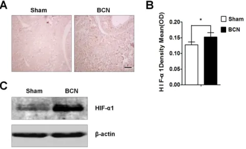
Assessment of HIF-1aexpression
The HIF-1aprotein expression in penile tissue from the sham and BCN groups was evaluated by immunohistochemistry. The
results showed that, at 12 weeks after surgery, very few areas in the tissues from the sham group were positive for HIF-1a protein expression, whereas tissues from the BCN group exhibited strong expression in many areas (Figure 1A). Total HIF-1a levels were markedly higher in penile tissues of the BCN group than in those of the sham group (P,0.05; Figure 1B). Western blot results also
showed increased expression of HIF-1ain BCN rats (Figure 1C). Assessment of collagen fiber expression
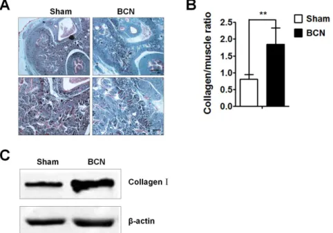
The extent of corpus cavernosum fibrosis was evaluated by Masson trichrome staining of the penile tissue from the sham and BCN groups. The degree of penile fibrosis in BCN rats was more severe than that in the sham group; photomicrographs from a typical experiment are showed in Figure 2A. Computer-assisted analysis indicated that the collagen/muscle ratio was 0.8060.14 in sham group rats. BCN significantly increased this ratio (1.8460.49,P,0.01, Figure 2B). Western blot results also showed a remarkable increase in the expression of collagen-I in BCN rats (Figure 2C).
Assessment of CCSMC morphological features
Prolonged hypoxia for 12 weeks resulted in significant cellular hypertrophy. H&E staining images were acquired under high
Table 1.Penile erectile response to injection of apomorphine (100mg/kg).
Group Reaction Non-reaction Total Erectile rate (%)
Sham 11 1 12 91.67
BCN 0 12 12 0*
Total 11 13 24 55
Data were analyzed by chi-square test (xc2= 12.93). *P,0.05. BCN group, n = 12; sham-operation group, n = 12. BCN = bilateral cavernous neurectomy; Sham = sham operation.
doi:10.1371/journal.pone.0105186.t001
Figure 1. Expression of HIF-1ain the corpus cavernosum tissues of BCN rats.(A) Rats were killed at 12 weeks after BCN, and penis samples were prepared for detection of HIF-1aexpression using immunohistochemical staining (1006, Scale bars = 200mm). (B) Semi-quantitative image analysis of HIF-1aexpression in corpus cavernosum tissues was performed using the Image-Pro Plus 6.0 software. *P,0.05 compared with the sham
rats (n = 5/group). (C) Rat corpus cavernosum tissues were harvested and its lysates were used for western blotting with primary antibodies againsta -SMA, Vim, SMMHC, desmin, andb-actin (Sham group: n = 3, BCN group: n = 4). All results are representative of three independent experiments. BCN = bilateral cavernous neurectomy; Sham = sham operation.
magnification (10006), using an oil immersion technique to aid visualization. Compared with the CCSMCs from the sham group, those from the BCN group showed an increased cell diameter, suggesting hypertrophy (Figure 3A). We also observed ultrastruc-tural changes along with changes in cellular morphological features that included high levels of myofilament loss and rough endoplasmic reticulum (RER) formation (Figure 3B).
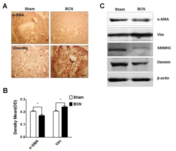
Assessment of proteins associated with the CCSMC phenotype. The expression of phenotypic protein markers was evaluated by immunohistochemical staining of penile tissue from the sham and BCN groups. Photomicrographs from a typical experiment are shown in Figure 4A. Computer-assisted analysis of these images indicated that a-SMA expression had significantly decreased and vimentin expression had dramatically increased in the BCN rats as compared to that in the sham group (P,0.05, Figure 4B). Using western blot analyses, we found that the expression ofa-SMA, SMMHC, and desmin was downregulated, whereas the expression of vimentin was upregulated in the BCN rats (Figure 4C).
Discussion
To our knowledge, our study is the first to demonstrate that CCSMCs undergo phenotypic modulation in BCN rats. BCN was also shown to cause a significant decrease in erectile function in these rats. HIF-1aand collagen proteins were highly expressed in the penile corpora cavernous tissues of rats following BCN exposure. Additionally, SMC contractile proteins of the corpora cavernosum, such as a-SMA, SMMHC, and desmin, were downregulated in BCN rats, whereas synthesis of the SMC phenotypic marker protein vimentin was significantly upregulated. Increase in collagen synthesis and in hypoxia inducible factor-1a (HIF-1a) and transforming growth factor-b1 (TGF-b1) expression has been observed after cavernous neurectomy. [7] Our results confirm the direct relationship that has been hypothesized to exist between hypoxia and corpus cavernosum fibrosis in penile tissue after neurectomy. It is widely believed that the lack of sleep-related erectile episodes, induced by BCN, is one of the most important reasons for hypoxia in the corpus cavernosum [19] suggesting that improvements in cavernous Figure 3. Observation of the morphology and structure of
corpus cavernosum smooth muscle cells in BCN rats. Represen-tative H&E photomicrograph (10006, Scale bars = 10mm) and trans-mission electron microscopy photomicrographs (40,0006, Scale
bars = 0.2mm) of CCSMCs. The protocol used is described in the methods section. M: mitochondria; RER: rough endoplasmic reticulum; G: Golgi apparatus. BCN = bilateral cavernous neurectomy; Sham = sham operation.
doi:10.1371/journal.pone.0105186.g003
Figure 2. Analysis of corpus cavernosum tissue fibrosis in BCN rats. (A) Penis samples were prepared for the detection of corpus cavernosum tissue fibrosis using Masson trichrome staining (Upper-line: 406, Scale bars = 500mm, down-line: 1006, Scale bars = 200mm). (B) Semi-quantitative image analysis of collagen/muscle ratio in corpus cavernosum tissues was performed using the Image-Pro Plus 6.0 software. **P,0.01
compared with the sham rats (n = 5/group). (C) Western blot analyses measuring collagen-I protein expression levels in penile tissues of both groups (Sham group: n = 3, BCN group: n = 4). All results are representative of three independent experiments. BCN = bilateral cavernous neurectomy; Sham = sham operation.
doi:10.1371/journal.pone.0105186.g002
blood flow and reoxygenation of the penis provide effective therapies for preserving the erectile function after RP.
CCSMCs are known to be the most important cells in the male erection process [20]. It has been observed that the abnormalities in CCSMCs that are attributed to CN injury reduce the ability of the tissue to establish sufficient intracavernous pressure (ICP) for blocking the veins that traverse under the tunica albuginea and egress from the corporal bodies.[21] SMCs exist in contractile and synthetic phenotypic states, even in mature organs. SMCs maintain high plasticity and undergo phenotypic modulation in response to local cellular stimuli, such as hypoxic conditions [22]. Cellular morphological features are an important indicator of SMC behavior [23]. In culture, a single cell in the contractile state exhibits a spindle or rhomboid shape and contains a large number of rich myofilaments [24]. SMCs with a synthetic phenotype appear broader with a bigger diameter and show ECM deposition and formation of RER [25]. In this study, the CCSMCs in BCN rat penile tissues displayed similar changes in their morphology (increased cell diameter) and structure (increased loss of myofil-aments and increased RER formation).
A variety of SMC-specific target gene and gene products have been identified as useful markers of the phenotypic state of the SMCs. These include a large number of contractile proteins, including a-SMA, SMMHC, and desmin. a-SMA was the first protein found to be expressed in the contractile state of the SMC in mature organs and serves as a reliable marker [11]. SMMHC is
highly restricted to SMCs and therefore serves as an ideal SMC marker. Both desmin and vimentin are cytoskeletal proteins of the intermediate filament. Desmin is believed to be another contractile SMC marker [26], while high expression of vimentin in impaired vascular tissue has been used as a marker for synthetic SMC [27]. These four proteins (a-SMA, SMMHC, desmin, and vimentin) have been used in this study to characterize SMC phenotypes. Our results showed that CCSMCs subject to BCN-induced hypoxic conditions displayed changes in protein expression, that is, decreased expression of a-SMA, SMMHC, and desmin and increased expression of vimentin, that were consistent with other published data on phenotype modulation in SMCs.
Conclusions
Our results demonstrated that CCSMCs undergo a shift in phenotypes from a contractile state to a synthetic state in a rat model of BCN. This phenotypic modulation could play a key role in the pathogenesis of post-RP ED. The exact molecular mechanism underlying this effect remains to be further clarified.
Author Contributions
Conceived and designed the experiments: HYF BDL XJH. Performed the experiments: FY JFZ QYS. Analyzed the data: FY. Contributed reagents/ materials/analysis tools: GC SGZ KBY. Contributed to the writing of the manuscript: HYF FY.
Figure 4. Measurement of the phenotypic modulation of CCSMCs in BCN rats. (A) Representative immunohistochemical staining photomicrographs for smooth musclea-actin (a-SMA) and vimentin proteins (1006, Scale bars = 200mm). (B) Semi-quantitative image analysis ofa -SMA and vimentin expression in both groups. *P,0.05 compared to the sham rats (n = 5/group). (C) Western blot analysis of the levels ofa-SMA, vimentin, desmin, SM myosin heavy chain (MHC), andb-actin proteins in penile tissue of both groups (Sham group: n = 3, BCN group: n = 4). All results are representative of three independent experiments. BCN = bilateral cavernous neurectomy; Sham = sham operation.
References
1. Meng MV, Elkin EP, Harlan SR, Mehta SS, Lubeck DP, et al. (2003) Predictors of treatment after initial surveillance in men with prostate cancer: results from CaPSURE. J Urol 170: 2279–2283.
2. Meuleman EJ, Mulders PF (2003) Erectile function after radical prostatectomy: a review. Eur Urol 43: 95–102.
3. Welliver RC Jr, Mechlin C, Goodwin B, Alukal JP, McCullough AR (2014) A Pilot Study to Determine Penile Oxygen Saturation Before and After Vacuum Therapy in Patients with Erectile Dysfunction After Radical Prostatectomy. J Sex Med 11: 1071–1077.
4. Liu K, Liu XS, Xiao L, Shang J, Li MC, et al. (2012) NADPH oxidase activation: a mechanism of erectile dysfunction in a rat model of sleep apnea. J Androl 33: 1186–1198.
5. Magheli A, Burnett AL (2009) Erectile dysfunction following prostatectomy: prevention and treatment. Nat Rev Urol 6: 415–427.
6. Montorsi F, Guazzoni G, Strambi LF, Da Pozzo LF, Nava L, et al. (1997) Recovery of spontaneous erectile function after nerve-sparing radical retropubic prostatectomy with and without early intracavernous injections of alprostadil: results of a prospective, randomized trial. J Urol 158: 1408–1410.
7. Leungwattanakij S, Bivalacqua TJ, Usta MF, Yang DY, Hyun JS, et al. (2003) Cavernous neurotomy causes hypoxia and fibrosis in rat corpus cavernosum. J Androl 24: 239–245.
8. Hu WL, Hu LQ, Song J, Li SW, Zheng XM, et al. (2004) Fibrosis of corpus cavernosum in animals following cavernous nerve ablation. Asian J Androl 6: 111–116.
9. Owens GK (1995) Regulation of differentiation of vascular smooth muscle cells. Physiol Rev 75: 487–517.
10. Orr AW, Lee MY, Lemmon JA, Yurdagul A Jr, Gomez MF, et al. (2009) Molecular mechanisms of collagen isotype-specific modulation of smooth muscle cell phenotype. Arterioscler Thromb Vasc Biol 29: 225–231.
11. Owens GK, Kumar MS, Wamhoff BR (2004) Molecular regulation of vascular smooth muscle cell differentiation in development and disease. Physiol Rev 84: 767–801.
12. Prakash YS (2013) Airway smooth muscle in airway reactivity and remodeling: what have we learned? Am J Physiol Lung Cell Mol Physiol 305: L912–933. 13. Hansson GK, Hermansson A (2011) The immune system in atherosclerosis. Nat
Immunol 12: 204–212.
14. Kilkenny C, Browne WJ, Cuthill IC, Emerson M, Altman DG (2010) Improving bioscience research reporting: the ARRIVE guidelines for reporting animal research. PLoS Biol 8: e1000412.
15. Zhang X, Hu L, Yin J, Mo Z, Chen J (2002) Rat model of erectile dysfunction caused by cavernous nerve ablation. Chin Med J (Engl) 115: 1179–1182. 16. Wei AY, He SH, Zhao JF, Liu Y, Hu YW, et al. (2012) Characterization of
corpus cavernosum smooth muscle cell phenotype in diabetic rats with erectile dysfunction. Int J Impot Res 24: 196–201.
17. Vignozzi L, Filippi S, Morelli A, Ambrosini S, Luconi M, et al. (2006) Effect of chronic tadalafil administration on penile hypoxia induced by cavernous neurotomy in the rat. J Sex Med 3: 419–431.
18. Li FF, Shen J, Shen HJ, Zhang X, Cao R, et al. (2012) Shp2 plays an important role in acute cigarette smoke-mediated lung inflammation. J Immunol 189: 3159–3167.
19. Moreland RB (1998) Is there a role of hypoxemia in penile fibrosis: a viewpoint presented to the Society for the Study of Impotence. Int J Impot Res 10: 113– 120.
20. Nehra A, Goldstein I, Pabby A, Nugent M, Huang YH, et al. (1996) Mechanisms of venous leakage: a prospective clinicopathological correlation of corporeal function and structure. J Urol 156: 1320–1329.
21. Ferrini MG, Kovanecz I, Sanchez S, Umeh C, Rajfer J, et al. (2009) Fibrosis and loss of smooth muscle in the corpora cavernosa precede corporal veno-occlusive dysfunction (CVOD) induced by experimental cavernosal nerve damage in the rat. J Sex Med 6: 415–428.
22. Halayko AJ, Solway J (2001) Molecular mechanisms of phenotypic plasticity in smooth muscle cells. J Appl Physiol (1985) 90: 358–368.
23. Thakar RG, Cheng Q, Patel S, Chu J, Nasir M, et al. (2009) Cell-shape regulation of smooth muscle cell proliferation. Biophys J 96: 3423–3432. 24. Chamley-Campbell J, Campbell GR, Ross R (1979) The smooth muscle cell in
culture. Physiol Rev 59: 1–61.
25. Hedin U, Thyberg J (1987) Plasma fibronectin promotes modulation of arterial smooth-muscle cells from contractile to synthetic phenotype. Differentiation 33: 239–246.
26. Mericskay M, Parlakian A, Porteu A, Dandre F, Bonnet J, et al. (2000) An overlapping CArG/octamer element is required for regulation of desmin gene transcription in arterial smooth muscle cells. Dev Biol 226: 192–208. 27. Asada H, Paszkowiak J, Teso D, Alvi K, Thorisson A, et al. (2005) Sustained
orbital shear stress stimulates smooth muscle cell proliferation via the extracellular signal-regulated protein kinase 1/2 pathway. J Vasc Surg 42: 772–780.