Pregnant Women in France: A Cross-Sectional Analysis
Jean-Noel Vergnes1,2*, Monique Kaminski1,3, Nathalie Lelong1,3, Anne-Marie Musset4, Michel Sixou2, Cathy Nabet1,2,5,6, for the EPIPAP group"
1INSERM UMRS 953, Epidemiological Research Unit on Perinatal Health and Women’s and Children’s Health, Villejuif, France,2Department of Epidemiology, Faculty of Dentistry, Paul Sabatier University, Toulouse, France,3UMPC UnivP06, UMRS 953, Paris, France,4Faculty of Dentistry, Louis Pasteur University, Strasbourg, France, 5Faculty of Dentistry, Paris Descartes University, Paris, France,6Charles Foix Hospital, Ivry/Seine, France
Abstract
Introduction:Little is known on the prevalence of tooth decay among pregnant women. Better knowledge of tooth decay risk indicators during pregnancy could help to develop follow-up protocols for women at risk, along with better prevention strategies. The aim of this study was to assess the frequency of tooth decay and the number of decayed teeth per woman in a large sample of pregnant women in France, and to study associated risk indicators.
Methods:A secondary cross-sectional analysis of data from a French multicentre case-control study was performed. The sample was composed of 1094 at-term women of six maternity units. A dental examination was carried out within 2 to 4 days post-partum. Socio-demographic and behavioural characteristics were obtained through a standardised interview with the women. Medical characteristics were obtained from the women’s medical records. Risk indicators associated with tooth decay were identified using a negative binomial hurdle model.
Results:51.6% of the women had tooth decay. The mean number of decayed teeth among women having at least one was 3.1 (s.d. = 2.8). Having tooth decay was statistically associated with lower age (aOR = 1.58, 95%CI [1.03,2.45]), lower educational level (aOR = 1.53, 95%CI [1.06,2.23]) and dental plaque (aOR = 1.75, 95%CI [1.27,2.41]). The number of decayed teeth was associated with the same risk indicators and with non-French nationality and inadequate prenatal care.
Discussion:The frequency of tooth decay and the number of decayed teeth among pregnant women were high. Oral health promotion programmes must continue to inform women and care providers about the importance of dental care before, during and after pregnancy. Future research should also assess the effectiveness of public policies related to oral health in target populations of pregnant women facing challenging social or economic situations.
Citation:Vergnes J-N, Kaminski M, Lelong N, Musset A-M, Sixou M, et al. (2012) Frequency and Risk Indicators of Tooth Decay among Pregnant Women in France: A Cross-Sectional Analysis. PLoS ONE 7(5): e33296. doi:10.1371/journal.pone.0033296
Editor:Nick Harvey, University of Southampton, United Kingdom
ReceivedOctober 8, 2011;AcceptedFebruary 10, 2012;PublishedMay 7, 2012
Copyright:ß2012 Vergnes et al. This is an open-access article distributed under the terms of the Creative Commons Attribution License, which permits unrestricted use, distribution, and reproduction in any medium, provided the original author and source are credited.
Funding:This study was supported by the National Programme for Hospital Clinical Research (national PHRC2004, AOM04047) and National Institute of Health and Medical Research (INSERM). The funders had no role in study design, data collection and analysis, decision to publish, or preparation of the manuscript. Competing Interests:The authors have declared that no competing interests exist.
* E-mail: jn.vergnes@gmail.com
"Membership of the EPIPAP Group is provided in the Acknowledgments.
Introduction
Tooth decay is a widespread, infectious disease classically related to the interplay of biological, behavioural and socio-economic influences. It affects about 40–50% of adults in industrialised countries [1,2]. It has been hypothesised that pregnancy could increase the risk of caries initiation or progres-sion, by changes in saliva composition [3], repeated gastric reflux or less effective oral health care [4]. However, given the relatively short time frame of pregnancy and the kinetics of dental caries progression [5,6], it is unlikely that tooth decay will develop from initial carious lesion to major tooth damage within this period. Indeed, pregnancy in itself has never been clearly associated with an increased incidence of dental caries. Nevertheless, tooth decay is worth studying during pregnancy because the disease has potentially more critical consequences during this particular
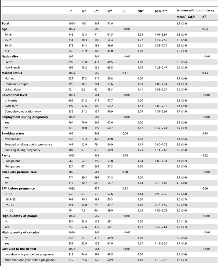
Table 1.Frequency of tooth decay and number of decayed teeth, according to women’s characteristics.
na %a nb %b pc ORd 95% CIe Women with tooth decay
Meanf(s.d.g) ph
Total 1094 100 565 51.6 3.1 (2.8)
Age 1094 565 ,0.01 0.24
18–24 148 13.5 91 61.5 2.54 1.61–3.99 3.8 (2.9)
25–29 331 30.3 180 54.4 1.77 1.22–2.55 3.0 (2.8)
30–34 375 34.3 186 49.6 1.21 0.85–1.74 2.8 (2.5)
$35 240 21.9 108 45.0 1.00 3.5 (3.2)
Nationality 1091 564 ,0.01 ,0.01
French 892 81.8 433 48.5 1.00 2.8 (2.6)
Non-French 199 18.2 131 65.8 1.74 1.23–2.47 4.2 (3.3)
Marital status 1094 565 0.41 0.14
Married 627 57.3 319 50.8 1.00 3.1 (2.6)
Unmarried couple 395 36.1 204 51.6 1.09 0.83–1.44 3.1 (3.1)
Living alone 72 6.6 42 58.3 1.41 0.83–2.39 4.0 (3.4)
Educational level 1093 564 ,0.01 ,0.01
University 669 61.2 319 47.7 1.00 2.8 (2.4)
Sixth form 192 17.6 106 55.2 1.53 1.08–2.17 3.4 (2.9)
Compulsory education only 232 21.2 139 59.9 2.09 1.51–2.91 3.7 (3.5)
Employment during pregnancy 1092 563 ,0.01 ,0.01
Yes 764 70.0 364 47.6 1.00 2.9 (2.6)
No 328 30.0 199 60.7 1.82 1.37–2.41 3.7 (3.2)
Smoking status 1091 562 0.04 0.74
Non-smoker 843 77.3 420 49.8 1.00 3.1 (2.6)
Stopped smoking during pregnancy 141 12.9 79 56.0 1.19 0.80–1.75 3.5 (3.4)
Smoking during pregnancy 107 9.8 63 58.9 1.73 1.11–2.67 2.9 (2.4)
Parity 1093 564 0.78 0.52
Primiparous 569 52.1 295 51.8 1.04 0.80–1.34 3.1 (2.7)
Multiparous 524 47.1 269 51.3 1.00 3.2 (3.0)
Adequate prenatal care 1091 563 0.60 ,0.01
Yes 974 89.3 499 51.2 1.00 3.1 (2.6)
No 117 10.7 64 54.7 1.14 0.70–1.85 4.0 (4.0)
BMI before pregnancy 1082 557 0.13 0.66
,18.5 9.2 8.5 53 57.6 1.44 0.90–2.30 3.7 (3.4)
[18.5–25[ 761 70.3 383 50.3 1.00 3.0 (2.7)
[25–30[ 151 14.0 75 49.7 1.10 0.76–1.59 3.2 (2.9)
.= 30 78 7.2 46 59.0 1.65 1.00–2.72 3.6 (3.0)
High quantity of plaque 1094 565 ,0.01 ,0.01
No 354 32.4 135 38.1 1.00 2.0 (1.2)
Yes 740 67.6 430 58.1 1.92 1.42–2.61 3.5 (3.1)
High quantity of calculus 1094 565 ,0.01 ,0.01
No 843 77.1 412 48.9 1.00 2.9 (2.6)
Yes 251 22.9 153 61.0 1.67 1.18–2.36 3.7 (3.2)
Last visit to the dentist 1092 564 ,0.01 ,0.01
Less than one year before pregnancy 813 74.4 394 48.5 1.00 2.9 (2.6)
More than one year before pregnancy 279 25.6 170 60.9 1.60 1.18–2.16 3.6 (3.1)
aNumber and percentage of women in each class of the variables. b
Number and percentage of women with tooth decay in each class of the variables. cWald
x2test adjusted for examiner. dOdds Ratio adjusted for examiner. e95% Confidence Interval.
The few studies assessing the frequency of tooth decay during pregnancy report values between 47% and 69% [7,10,11]. These recent studies were conducted on relatively small populations in Pakistan, Brazil and Hungary. To our knowledge, there is no previous study reporting data on the frequency of tooth decay among pregnant women in France. Neither have we found any international study investigating the risk indicators specifically related to decayed teeth in pregnant women. Better knowledge of the prevalence of tooth decay and associated risk indicators during pregnancy would help to develop follow-up protocols for women at risk, along with better prevention strategies.
The objectives of this study were to assess the frequency of tooth decay and the number of decayed teeth in a large sample of pregnant women in France, and to study associated risk indicators.
Methods
Ethics Statement
The study was approved by the French data protection authority, and all the women included gave their written informed consent.
Study Sample
The study sample was made up of 1094 women who had given birth to a singleton live-born infant at term ($37 weeks), randomly selected between 2003 and 2006 in 6 maternity units of 3 French regions (Ile-de-France, Midi-Pyre´ne´es and Alsace). This sample formed the control group of the EPIPAP study, a multicentre case-control study which primarily aimed to analyse the association between periodontitis and preterm birth, according to the main causes of preterm birth [12]. Oral health comparisons between cases (women with delivery term at ,37 weeks’ gestation) and controls (women with delivery term at$37 weeks’ gestation) have been published elsewhere [12,13]. Only the control group of the
EPIPAP case-control study was used in this cross-sectional analysis. Non-inclusion criteria were: age under 18, not under-standing the French language, HIV infection, unbalanced diabetes or any medical condition that required antibiotic prophylaxis for dental examination and periodontal probing, fewer than 6 teeth, and infant born with a severe congenital malformation.
Data
Examinations were performed within 2 to 4 days post-partum, in the post-delivery wards of the maternity units. It was considered that tooth decay observed within 4 days post-partum was already present during pregnancy. Women were examined in a sitting position. The eleven dentists in charge of the oral examinations performed intra-oral screening to ascertain the amount of plaque, calculus and gingival inflammation, clinical attachment level, periodontal pocket depth, bleeding on probing, and presence of tooth decay and fillings. Examiners were given instructions to assess carious lesions according to the World Health Organisation (WHO) diagnosis criteria [14]. The presence of carious lesions was recorded at the surface level of the teeth using sterile dental mirrors and explorers. A carious lesion was defined as a cavity that appeared as a darkened hole with irregular breakdown of the enamel surface. Stain and pigmentation alone were not considered as carious lesions neither were white spot lesions, nor apparent tooth wear or erosion. Four surfaces were examined and coded for incisors and canines, and five surfaces for premolars and molars. Third molars were excluded from the assessment and radiographs were not taken. A decayed surface was recorded when at least one carious lesion could be observed on a surface, including carious lesion contiguous with the margin of a filling. Analyses were performed at tooth level. A decayed tooth was a tooth with at least one decayed surface. A woman was considered as having tooth decay if at least one of her teeth was decayed.
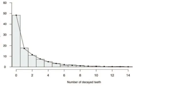
Figure 1. Distribution of number of decayed teeth per woman.The bars represent the values observed in the sample of 1094 women. The curve represents the values predicted by the Hurdle model.
doi:10.1371/journal.pone.0033296.g001
gStandard deviation.
hGeneral linear models (F-test) adjusted for examiner. doi:10.1371/journal.pone.0033296.t001
Amounts of plaque and calculus were measured at four sites per tooth, using the Silness-Lo¨e plaque index [15] and the Greene and Vermillion calculus index [16]. A woman was classified as having a high quantity of plaque if the examiner reported at least one site with visible plaque on at least one tooth. Similarly, a woman was classified as having a high quantity of calculus if the examiner reported at least one site with calculus covering more than one third of the exposed tooth surface of at least one tooth. Adequacy of dental attendance was assessed through the variable ‘time since last visit to dentist’ (consistent with clinical guidelines for patients aged 18 years and older: less than one year before pregnancy; or
not consistent with clinical guidelines: more than one year before pregnancy or never) [17].
Socio-demographic and behavioural characteristics were obtained through a standardised interview of the women after the dental examination. Medical characteristics were obtained from the women’s medical records. All examiners were blinded to the medical and socio-demographic data. Socio-demographic characteristics included age (18–24,25–29,30–34 and $35 years), nationality (French or not), marital status (married, unmarried couple, living alone), educational level (primary and secondary compulsory education, sixth form, university), and
Table 2.Risk indicators for tooth decay: results from the multivariate analysisa.
Variables Logistic portionb Negative binomial portionc
aORd 95% CIe p-value
Adjusted
exp(b)f 95% CI p-value
Age (years)
18–24 1.58 [1.03,2.45] 0.03* 1.37 [1.06,1.78] 0.01*
25–29 1.59 [1.16,2.17] ,0.01* 1.05 [0.85,1.29] 0.62
$30 1.00 1.00
Nationality
French 1.00 1.00
Non-French 1.48 [1.00,2.19] 0.05 1.30 [1.04,1.62] 0.02*
Educational level
University 1.00 1.00
Sixth form 1.15 [0.78,1.67] 0.45 1.19 [0.92,1.54] 0.17
Compulsory education only 1.53 [1.06,2.23] 0.02* 1.40 [1.09,1.79] ,0.01*
Employment during pregnancy
Yes 1.00 1.00
No 1.35 [0.97,1.86] 0.07 1.15 [0.93,1.41] 0.18
Smoking
Non-smoker 1.00 1.00
Stopped smoking during pregnancy 1.11 [0.73,1.68] 0.60 1.19 [0.87,1.47] 0.18
Smoker during pregnancy 1.49 [0.93,2.39] 0.09 0.83 [0.51,1.14] 0.26
Adequate prenatal care
Yes 1.00 1.00
No 0.80 [0.47,1.37] 0.41 1.46 [1.08,1.99] 0.01*
High quantity of plaque
Yes 1.75 [1.27,2.41] ,0.01* 1.82 [1.38,2.42] ,0.01*
No 1.00 1.00
High quantity of calculus
Yes 1.41 [0.97,2.05] 0.06 1.23 [1.00,1.53] 0.05
No 1.00 1.00
Last visit to the dentist
Less than one year before pregnancy 1.00 1.00
More than one year before pregnancy 1.31 [0.94,1.80] 0.09 1.18 [0.97,1.44] 0.08
aHurdle model, adjusted for all the variables in the table plus examiner to account for inter-examiner variability. bModels the probability of the women having tooth decay.
cModels the number of decayed teeth among the women having at least one. dAdjusted odds ratio.
e95% Confidence Interval. fexp(
b) can be interpreted as follows: while holding all other variables constant in the model, among women having tooth decay, a woman in a given class has on average exp(b) more decayed teeth than a woman in the reference class. For example, among women having tooth decay, a woman aged 18–24 years has on average 1.37 more decayed teeth (or 37% more decayed teeth) than a woman aged 30 or more (reference class for age).
*Significant ata= 5%.
employment during pregnancy (yes/no). Behavioural character-istics were smoking status during pregnancy (non smoker before pregnancy, stopped smoking during pregnancy, smoker during pregnancy), and adequacy of prenatal care, assessed by the number of prenatal visits according to gestational age at delivery with reference to French regulations. Medical characteristics were the Body Mass Index (BMI) and the parity of the mother (primiparous vs multiparous). BMI was calculated by dividing weight (in kilograms) by the square of the height (in metres), and assessed using self-reported values of height and weight before pregnancy. BMI values were classified into four categories: less than 18.5, 18.5 to 24.9, 25 to 29.9 and more or equal to 30.
Statistical Analysis
Descriptive analysis of the sample was performed using relative percentages for each class of categorical variables. The proportion of women with tooth decay was also presented according to the women’s characteristics. Bivariate analyses were conducted to identify women’s characteristics associated with tooth decay and number of decayed teeth. Risk indicators associated with tooth decay were identified using the Wald Chi-2 test adjusted for examiner, and odds ratios (ORs) and their 95% confidence intervals (95% CI) were calculated. In order to avoid losing quantitative information, the number of decayed teeth per woman was also calculated, hypothesising that the risk of caries-related problems during pregnancy increased with the number of decayed teeth. Among women having at least one decayed tooth, risk indicators associated with the number of decayed teeth were identified using general linear models (F-test) adjusted for examiner.
Risk indicators associated with tooth decay or the number of decayed teeth were analysed together using a hurdle model, a two-component regression model for count outcomes [18]. Hurdle models are appropriate for modelling count data with excess zeros [19], which is the case for the number of decayed teeth per person in adult populations of contemporary industrialised countries [20]. This model first uses logistic regression to predict the probability of the woman having any decayed teeth, then it calculates the conditional expectation of the number of decayed teeth for the subsample of only the women who have at least one. The count part of the model is a truncated negative binomial regression (with log link) [18]. All women’s risk indicators significantly associated with tooth decay and/or the number of decayed teeth in the bivariate analysis were included in the model. Ordinal variables with more than two classes were dummy coded for the regression procedures. The multivariate analysis was also adjusted for examiner to take the inter-examiner variability into account. The adequacy of the model was assessed using the Wald Chi-2 test, and predicted values of number of decayed teeth were calculated.
The significance level was set at p,0.05. Statistical analyses were performed using SAS software version 9, and R software version 2.7.1, with the additional pscl package version 1.02 (hurdle() function).
Results
18.2% of the women were not French, 6.6% were living alone, 21.2% had a low educational level, and 30.0% were not employed during pregnancy (Table 1). On the whole, 51.6% of the women had tooth decay. Among women who had tooth decay, the mean number of decayed teeth was 3.1 (sd = 2.8). Both the frequency of tooth decay and the mean number of decayed teeth were
significantly associated with non-French nationality, lower educa-tional level, unemployment during pregnancy, high quantity of plaque, high quantity of calculus and time since last visit to the dentist. The frequency of women with tooth decay was higher among lower age groups and among smokers during pregnancy (Table 1). The mean number of decayed teeth was also higher when prenatal care was inadequate (Table 1). In contrast, neither presence of tooth decay nor number of decayed teeth were associated with marital status, parity, or BMI before pregnancy.
Figure 1 represents the observed distribution of the number of decayed teeth per woman. The distribution is skewed to the right, with 48.4% of ‘caries-free’ women. The curve shows the predicted distribution of the number of decayed teeth per woman obtained from the multivariate analysis and indicates a good fit of the model to the data (Figure 1). The hurdle model was significant (Wald Chi-2 test, p,0.0001), meaning that at least one of the regression coefficients was not equal to zero.
Table 2 shows the results of the multivariate analysis (Hurdle model) between women’s characteristics and both existence of tooth decay and number of decayed teeth. In the logistic portion of the hurdle model, lower age groups, low educational level and high quantity of plaque were independently associated with a higher risk of tooth decay. Non-French nationality was borderline significant. In the negative binomial portion, the number of decayed teeth was associated with the same risk factors and with non-French nationality and inadequate prenatal care. Compara-tively to women aged 30 years and more, women in the 18–24 age group presented 1.37 more decayed teeth on average, i.e. 37% more decayed teeth. Non-French women had on average 30% more decayed teeth than French women. Women with an educational level of primary or compulsory secondary school had on average 40% more decayed teeth than more highly educated women, and women with inadequate prenatal care during pregnancy presented on average 46% more decayed teeth than women with adequate prenatal care.
Discussion
We showed that more than 50% of the pregnant women had tooth decay. Having tooth decay was associated with lower age and lower educational level. The number of decayed teeth was associated with the same risk indicators, and with non-French nationality and inadequate prenatal care.
based is weaker than the results of longitudinal studies. Another limitation of this study is that we did not explore some variables that could be considered as important risk indicators for tooth decay, such as dietary habits or dental hygiene habits. The primary EPIPAP study was designed to analyse the association between maternal periodontitis and preterm birth according to causes of preterm birth, so dietary habits were not collected. Given the putative overestimation of self-reported oral hygiene practices, we considered the presence of plaque and calculus as more direct risk indicators for tooth decay.
The frequency of dental caries in our sample was similar to the prevalence observed in the general adult population of the same age [2,20,24], although dental studies among the general adult population remain rare. Our results (51% of women with tooth decay) are in agreement with the frequency of dental caries among pregnant or post-partum women reported in previous studies from other countries. In a Pakistani cohort study of 1152 pregnant women (mean age 26.5 years), nearly 47% of the women had at least one decayed tooth [10]. The prevalence of tooth decay was 61% in a sample of 504 low-income Brazilian pregnant women (mean age 24 years) [7]. A Hungarian study found that 69% of postpartum mothers (mean age 27.5 years) required one or more restorations [11]. In all these studies, as well as ours, the conditions of dental examination might have led to an underestimation of both frequency of tooth decay and number of decayed teeth.
Lower age, non-French nationality and low educational level were related to both frequency of tooth decay and number of decayed teeth. We found that 18–24 year-old women were at higher risk for tooth decay than the older ones, independently of the amount of dental plaque and adequacy of dental attendance. Lower age as a risk indicator for tooth decay has already been described in France [24]. In 2004, a study involving about 600 000 adults showed that the highest proportion of subjects with at least one untreated carious lesion was among the 20–24 age group [24]. Although not explored in this study, younger women (aged 18–24 years) could be at higher risk of dental caries because snacking has been shown to be common in this age group [25]. Another explanation from the literature could be that 18–24 year-old adults are less likely to regularly visit a dental professional than other age groups [26]. Even if we adjusted for adequacy of dental attendance, it is likely that this binary variable would not fully reflect the preventive behaviour of the included women.
Non-French nationality was found to be associated with higher risk of having tooth decay. In France, the current nationality of the mother is a variable widely used in epidemiological studies [27,28,29] as ethnic category does not cover the notion of migration. It was important to take the woman’s nationality into account in the multivariate analysis because it has been shown that a lack of regular medical care stems from social obstacles, especially in foreign women [30]. For example, it has been shown that immigrant women are at risk of poor pregnancy outcomes [31], and that immigrant status is a significant caries predictor in children living in a deprived area [32]. Poor availability of
translations and of culturally competent services may constitute an obstacle to a contributive medical visit [31]. Further studies are needed to elucidate the obstacles to optimal management of these women in the French model of healthcare organisation, which is based on the principle of universal access to care [31].
A lower educational level was also found to be a significant risk factor for the frequency of tooth decay and the number of decayed teeth, which is consistent with previous studies showing that low educational level can be considered as a major risk factor for dental caries [33]. In the present study, women in the lower educational levels were more likely to declare insufficient dental attendance (data not shown). These data corroborate a French national study showing that subjects of lower educational levels were less likely to visit a dentist annually [34].
In conclusion, the frequency of tooth decay and the number of decayed teeth among pregnant women were high. Oral health promotion programmes need to inform pregnant women, prenatal care providers and oral health professionals about the particular importance of dental care before, during and after pregnancy. Future research should also assess the effectiveness of public policies related to oral health among some target groups of pregnant women facing challenging social or economic situations.
Acknowledgments
The authors would like to thank Laetitia Marchand and Nicolas Drewniak for stimulating discussions and helpful assistance. This article was prepared as part of J.N. Vergnes’ PhD, mentored by C. Nabet and M. Sixou. All the co-authors are members of the EPIPAP study group, for which the full list of members is given below.
The EPIPAP (EPIdemiological Study on the Relation between Periodontitis and Adverse Pregnancy Outcomes) Study Group
Members of steering committee
C. Nabet (Project leader, INSERM UMRS 953, Paris; Faculty of Dentistry, Paris Descartes University; Charles Foix Hospital, Ivry/Seine), M-L. Colombier (Faculty of Dentistry, Paris Descartes University, Paris), F. Goffinet (INSERM UMRS 953; Port Royal Hospital, Paris), M. Kaminski (INSERM UMRS 953, Paris), N. Lelong (INSERM UMRS 953, Paris).
Members of the EPIPAP group
A. Berrebi (Paule de Viguier Hospital, Toulouse), B. Carbonne (Saint-Antoine Hospital, Paris), P. Kassab (INSERM UMRS 953, Paris), G. Kayem (INSERM UMRS 953, Paris; Intercommunal Hospital, Cre´teil), B. Langer (Hautepierre Hospital, Strasbourg), A-M. Musset (Faculty of Dentistry, Louis Pasteur University, Strasbourg), I. Nisand (Hautepierre Hospital, Strasbourg), O. Parant (Paule de Viguier Hospital, Toulouse), M. Sixou (Faculty of Dentistry, Paul Sabatier University, Toulouse), N. Tordjeman (Victor Dupouy Hospital, Argenteuil), C. Vayssie`re (Haute-pierre Hospital, Strasbourg), J-N. Vergnes (INSERM UMRS 953, Paris; Faculty of Dentistry, Paul Sabatier University, Toulouse).
Author Contributions
Conceived and designed the experiments: MK CN. Performed the experiments: JNV. Analyzed the data: JNV NL. Contributed reagents/ materials/analysis tools: MS AMM MK CN. Wrote the paper: JNV MK CN.
References
1. Brown LJ, Wall TP, Lazar V (2002) Trends in caries among adults 18 to 45 years old. J Am Dent Assoc 133: 827–834.
2. Hescot P, Bourgeois D, Doury J (1997) Oral health in 35–44 year old adults in France. Int Dent J 47: 94–99.
3. Laine MA (2002) Effect of pregnancy on periodontal and dental health. Acta Odontol Scand 60: 257–264.
4. Silk H, Douglass AB, Douglass JM, Silk L (2008) Oral health during pregnancy. Am Fam Physician 77: 1139–1144.
5. Shwartz M, Gro¨ndahl HG, Pliskin JS, Boffa J (1984) A longitudinal analysis from bite-wing radiographs of the rate of progression of approximal carious lesions through human dental enamel. Arch Oral Biol 29: 529–536.
6. Berkey CS, Douglass CW, Valachovic RW, Chauncey HH (1988) Longitudinal radiographic analysis of carious lesion progression. Community Dent Oral Epidemiol 16: 83–90.
7. de Oliveira BH, Nadanovsky P (2006) The impact of oral pain on quality of life during pregnancy in low-income Brazilian women. J Orofac Pain 20: 297–305. 8. McKenna L, McIntyre M (2006) What over-the-counter preparations are
pregnant women taking? A literature review. J Adv Nurs 56: 636–645. 9. Kumar J, Samelson R (2009) Oral health care during pregnancy
10. Mobeen N, Jehan I, Banday N, Moore J, McClure EM, et al. (2008) Periodontal disease and adverse birth outcomes: a study from Pakistan. Am J Obstet Gynecol 198: e511–518.
11. Radnai M, Gorzo I, Nagy E, Urban E, Eller J, et al. (2007) The oral health status of postpartum mothers in South-East Hungary. Community Dent Health 24: 111–116.
12. Nabet C, Lelong N, Colombier ML, Sixou M, Musset AM, et al. (2010) Maternal periodontitis and the causes of preterm birth: the case-control Epipap study. J Clin Periodontol 37: 37–45.
13. Vergnes JN, Kaminski M, Lelong N, Musset AM, Sixou M, et al. (2011) Maternal dental caries and pre-term birth: results from the EPIPAP study. Acta Odontol Scand 69: 248–256.
14. WHO (1997) Oral Health Surveys - Basic methods, 4th edn. Geneva: World Health Organization.
15. Silness J, Loe H (1964) Periodontal Disease In Pregnancy. Ii. Correlation Between Oral Hygiene And Periodontal Condition. Acta Odontol Scand 22: 121–135.
16. Greene JC, Vermillion JR (1960) Oral hygiene index: a method for classifying oral hygiene status. J Am Dent Assoc 61: 172–177.
17. NICE: Dental recall: recall interval between routine dental examination Available: www.nice.org.uk/CG019NICEguideline via the internet. Accessed 2012 Apr 13.
18. Mullahy J (1986) Specification and testing of some modified count data models. J Econom 33: 341–365.
19. Khan A, Ullah S, Nitz J (2011) Statistical modelling of falls count data with excess zeros. Inj Prev 17: 266–270.
20. Broadbent JM, Thomson WM, Poulton R (2006) Progression of dental caries and tooth loss between the third and fourth decades of life: a birth cohort study. Caries Res 40: 459–465.
21. Blondel B, Supernant K, Du Mazaubrun C, Breart G (2006) Trends in perinatal health in metropolitan France between 1995 and 2003: results from the National Perinatal Surveys. J Gynecol Obstet Biol Reprod 35: 373–387.
22. Reilly M, Torrang A, Klint A (2005) Re-use of case-control data for analysis of new outcome variables. Stat Med 24: 4009–4019.
23. Burt BA (2001) Definitions of risk. J Dent Educ 65: 1007–1008.
24. Dauphinot V, Dupre´ C, Gueguen R, Naudin F (2006) Ge´ographie de la sante´ dans les centres d’examens de sante´: donne´es re´gionalise´es. CETAF 2006: 1–87. pp 1–87.
25. Akarslan ZZ, Sadik B, Sadik E, Erten H (2008) Dietary habits and oral health related behaviors in relation to DMFT indexes of a group of young adult patients attending a dental school. Med Oral Patol Oral Cir Bucal 13: E800–807.
26. Slack-Smith LM, Mills CR, Bulsara MK, O’Grady MJ (2007) Demographic, health and lifestyle factors associated with dental service attendance by young adults. Aust Dent J 52: 205–209.
27. Blondel B, Norton J, du Mazaubrun C, Bre´art G (2001) Development of the main indicators of perinatal health in metropolitan France between 1995 and 1998. Results of the national perinatal survey. J Gynecol Obstet Biol Reprod 30: 552–564.
28. Le Vu S, Le Strat Y, Barin F, Pillonel J, Cazein F, et al. (2010) Population-based HIV-1 incidence in France, 2003–08: a modelling analysis. Lancet Infect Dis 10: 682–687.
29. Bonet M, Blondel B, Khoshnood B (2010) Evaluating regional differences in breast-feeding in French maternity units: a multi-level approach. Public Health Nutr 13: 1946–1954.
30. Blondel B, Marshall B (1996) Women with little or no prenatal care during pregnancy. Results of a study of twenty departments. J Gynecol Obstet Biol Reprod 25: 729–736.
31. Philibert M, Deneux-Tharaux C, Bouvier-Colle MH (2008) Can excess maternal mortality among women of foreign nationality be explained by suboptimal obstetric care? BJOG 115: 1411–1418.
32. Tubert-Jeannin S, Riordan PJ, Manevy R, Lecuyer MM, Pegon-Machat E (2009) Caries prevalence and fluoride use in low SES children in Clermont-Ferrand (France). Community Dent Health 26: 23–28.
33. Paulander J, Axelsson P, Lindhe J (2003) Association between level of education and oral health status in 35-, 50-, 65- and 75-year-olds. J Clin Periodontol 30: 697–704.