EXPERIMENTS IN NATURE AND LABORATORY OBSERVATIONS
WITH
NAUSITHOE AUREA (SCYPHOZOA: CORONATAE) SUPPORT
THE CONCEPT OF PERENNATION BY TISSUE SAVING AND
CON-FIRM DORMANCY
1 Departamento de Zoologia, Instituto de Biociências, Universidade de São Paulo, Caixa Postal 11461, 05422-970, São
Paulo, SP, Brasil.
Tel.:+55-11-30917619 Fax: +55-11-30917513
2 Zoologisches Institut und Zoologisches Museum, Universität Hamburg, Martin-Luther-King Platz 3, 20146 Hamburg,
Germany,
Tel.: +49-40-428382086 Fax: +49-40-428382086
e-mails: [email protected], [email protected], [email protected]
Abstract
Stephanocyphistomae of Nausithoe aurea from São Paulo State, Brazil (in subtropical western South Atlantic wa-ters), were relocated with their substrata in nature to study their survivorship under control and and experimental series — i.e. the polyps in the original orientation and inverted, and in each series exposed and buried polyps. We found that N. aurea survives over 13 months in nature, between 1/3 – 1/4 of 268 stephanoscyphistomae as normal feeding polyps, by segmentation produces planuloids and rejuvenates the polyps — an additional explanation for clustering of the solitary stephanocyphistomae. Dormant living tissues within the periderm of the tube were considered resting stages. The results support the concept that coronates in general have the capacity to save all living tissue and transform it to the energy saving sessile stage — the perennial polyp.
Key-words: Cnidaria, Scyphozoa, Nausithoe aurea, perennation, resting stages, Brazil.
Resumo
Estefanocifístomas de Nausithoe aurea no Estado de São Paulo, Brasil (em águas subtropicais do Atlântico Sul ocidental), foram realocados com os seus substratos na natureza para estudar a sobrevivência em séries controle e experi-mental — isto é, os pólipos na posição original e invertidos e em cada série pólipos expostos e enterrados. Verificamos que N. aurea sobrevive por 13 meses na natureza, entre 1/3 – 1/4 de 268 estefanocifístomas como pólipos normais capazes de alimentação, por segmentação produz planulóides e rejuvenesce o pólipo — uma explicação adicional para a ocorrência agregada dos estefanocifístomas solitários. Tecidos vivos dormentes dentro da periderme do tubo foram considerados como estágios de quietação. Os resultados reforçam a idéia de que os coronados em geral têm a capacidade de conservar todo o tecido vivo e de transformá-lo durante o estágio séssil mais econômico energeticamente – o pólipo perene.
Palavras-chave: Cnidaria, Scyphozoa, Nausithoe aurea, perenização, estágios de quietação, Brasil. Fábio Lang da Silveira1 (correspondence author; correspondência para), Gerhard Jarms2 & André Carrara
Morandini1
Biota Neotropica v2 (n2) – http://www.biotaneotropica.org.br/v2n2/pt/abstract?article+BN02202022002 Date Received 07/19/2002
Revised 09/23/2002
2 Silveira, F. L.; Jarms,G. e Morandini, A. C. - Biota Neotropica, v2 (n2) - BN02202022002
Introduction
At present the life cycles of 16 species of Coronatae are known, the variations range from typical metagenesis by strobilation (polyp > strobilation > detached ephyrae > adult medusae > planulae) to the suppression of the stephanoscyphistomae or the medusae. These life cycles are:
- complete metagenesis: Atorella japonica (see Kawaguti & Matsuno, 1981), Atorella vanhoeffeni (see Werner, 1966), Nausithoe hagenbecki (see Jarms, 2001), Nausithoe punctata (see Werner, 1973) and Nausithoe werneri (see Jarms, 1990);
- complete metagenesis combined with abbreviated metagenesis and/or ‘segmentation’: Nausithoe aurea (see Silveira & Morandini, 1997) Linuche unguiculata (see Silveira & Morandini 1998a);
- metagenesis combined with abbreviated life cycle (’tissue balls’): Nausithoe globifera, Nausithoe maculata, Nausithoe marginata, and Nausithoe thieli (Jarms 1997);
- reduced metagenesis (medusoids): Nausithoe eumedusoides (see Werner, 1974) and Nausithoe racemosa (see Komai, 1936);
- reduced metagenesis (planuloids): Nausithoe planulophora (see Werner, 1971);
- without metagenesis (without polyps and planu-lae): Periphylla periphylla (see Jarms et al., 1999);
- without metagenesis (parthenogenesis): Thecoscyphus zibrowii (see Sötje & Jarms, 1999).
Morandini (1999) and Morandini & Silveira (2001a) reviewed the history of knowledge about stephanoscyphistomae of Coronatae in Brazil. Morandini & Silveira (2001a) complemented the diagnostic features for Nausithoe aurea Silveira & Morandini, 1997, and showed that the species occurs north of its type local-ity in Brazil, and Morandini & Silveira (2001b) described the gametogenesis of the species. Jarms et al. (2002) studied preserved deep-water polyps of the families Atorellidae and Nausithoidae from the Brazilian coast. Furthermore, Silveira & Morandini (1998a and b) have described segmentation and dormancy sensu Cáceres (1997: 372), i.e. the occurrence of resting stages, in the species Linuche unguiculata (Swartz, 1788).
The present study on the survivorship of Nausithoe aurea in nature, under control and experimental relocate circumstances, was undertaken over a 13 months period. Observations were based on just sampled material from the relocation site in southeast Brazil and following cultures of the stephanoscyphistomae. We wanted to know how N. aurea survives under unstable conditions and and whether the flexibility in its life cycle and whether the flexibility in its life cycle might be an advantage (or adaptation) for living in a frequently changing environment.
Material and Methods
To initiate our study, on July 18, 2000, seven frag-ments of dead stony coral Mussismilia hispida (Verrill, 1902) (Scleractinia, Mussidae) were sampled at 2-6 m depth by SCUBA diving, in the São Sebastião Channel (23°50’S – 45°25’W), on Praia Grande reef, Ilhabela County, SP. We searched for the polyps on the calcareous substrata with the aid of a stereomicroscope at Centro de Biologia Marinha da Universidade de São Paulo (CEBIMar USP). Two sets of Nausithoe aurea, each with six stephanoscyphistomae, were obtained from the exposed surface and from the bur-ied surface of two calcareous debris. They were trans-ferred into and maintained in cell culture dishes (60 mm x 15 mm) containing about 100 ml of filtered seawater. We had made the first cultivation under controlled room tem-perature, 21.0°C, from July 19 to 27, 2000at CEBIMar. On July 27 the specimens were transferred, each in a small airtight flask (70 ml) and air-conditioned car, to the Zool-ogy Department (IB USP), in São Paulo, SP, and reared inside an incubator (FANEM© 347-CDG) at 22 ± 1°C (6/18 hours light/dark) until August 7. After the third day of examinations, at two day intervals, a few recently hatched nauplii (24-48 hours) of Artemia sp. were added to the culture dishes for a period of 24 hours. The filtered sea-water in the culture dishes – provided by CEBIMar USP and stored inside a dark incubator at 22 ± 1°C, was changed every day. By day ten of the observations, gentle rubbing with a delicate brush cleaned each polyp.
3 Silveira, F. L.; Jarms,G. e Morandini, A. C. - Biota Neotropica, v2 (n2) - BN02202022002
*We had observed the modifications of one exposed polyp, in the control series, for 33 days.
Table 1. Dates in which (A) calcareous fragments with stephanoscyphistomae of Nausithoe aurea were retrieved from the sea and (B) days of observations in the laboratory; (C) monthly average of surface water temperature at CEBIMar. Note: average of surface water temperature for June and July 2001 was, respectively, 20.4 and 19.5 ºC.
4 Silveira, F. L.; Jarms,G. e Morandini, A. C. - Biota Neotropica, v2 (n2) - BN02202022002
Almost at monthly intervals, over thirteen months and by SCUBA diving, four transplanted coral fragments (two controls and two experiments, respectively, one ex-posed and one buried from each series), were retrieved from the sea, submersed in a large tank, gently washed with a constant flow of sea-water to remove the excess of sand so as to facilitate the finding of the polyps, twenty four stephanoscyphistomae were isolated in the laboratory. The polyps were kept, either in CEBIMar for the first five days (except for February 2001 with longer stay at CEBIMar), transferred to Zoology Department (as mentioned above) and observed for 15 – 20 days (Table 1).
Results
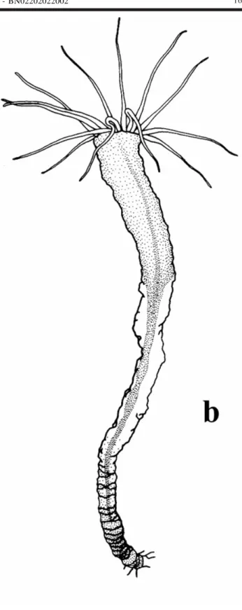
In this work we observed a total number of 268 stephanoscyphistomae, 141 and 127, respectively, from the control and experimental series. The following aspects were observed: (a) presence of particles closing the periderm tube; (b) particles pushed out of the periderm tube by the scyphistoma; (c) contracted living tissues within the perid-erm tube, but not contracting further upon gentle squeez-ing with delicate forceps; (d) contracted livsqueez-ing tissues within the periderm tube, but still contracting further upon gentle squeezing with delicate forceps; (e) contracted living tis-sues within the periderm tube, but oral disk with recogniz-able tentacles; (f) expanded polyp and oral disc with n
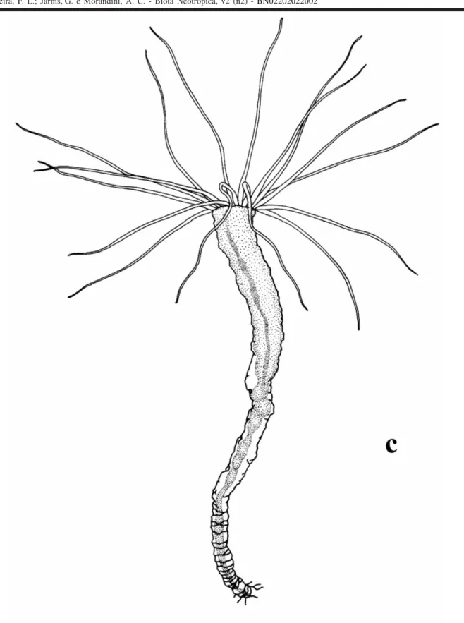
ten-tacles; (g) polyp with a simplecolumn, i.e. without mesen-teries or oral disc and tentacles; (h) polyp column with me-senteries, but without oral disc and tentacles; (i) strobila-tion and closed periderm operculum; (j) ephyrae; (k) half opened operculum; (l) removed operculum; (m) segmenta-tion sensu Silveira & Morandini (1998a); (n) planuloids (origi-nated by strobilation or segmentation) within the periderm tube; (o) planuloid inside the polyp column; (p) free planuloids (see Plate I).
The majority of the stephanoscyphistomae did not present any perceptible amount of zooxanthellae, except for the finding of 4 polyps with much zooxanthellae in the gastrodermis (2 polyps from the exposed-control different series and 2 polyps from the exposed and buried-experi-mental single series). Two calcareous substrata retrieved from the sea on March 29, 2001, showed, on one, the over-growth by the sponge Cliona aff. dioryssa De Laubenfels, 1950 and on the other the growth of coralline incrusting algae responsible for the absence of 3 and 5 stephanoscyphistomae, respectively, for the buried-con-trol and exposed-experimental series. Two polyps died during our observations: one in the buried-experimental series from the sample on January 22, 2001, due to a large
injury at the periderm pedal disk, at day 10 of cultivation; one in the buried-control series from the sample February 22, 2001, attacked by an unidentified ciliate protist (fast mul-tiplying within the periderm tube), at day 8 of cultivation.
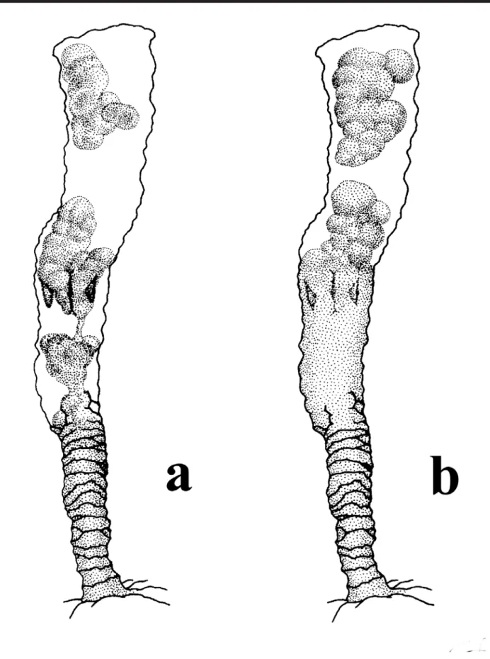
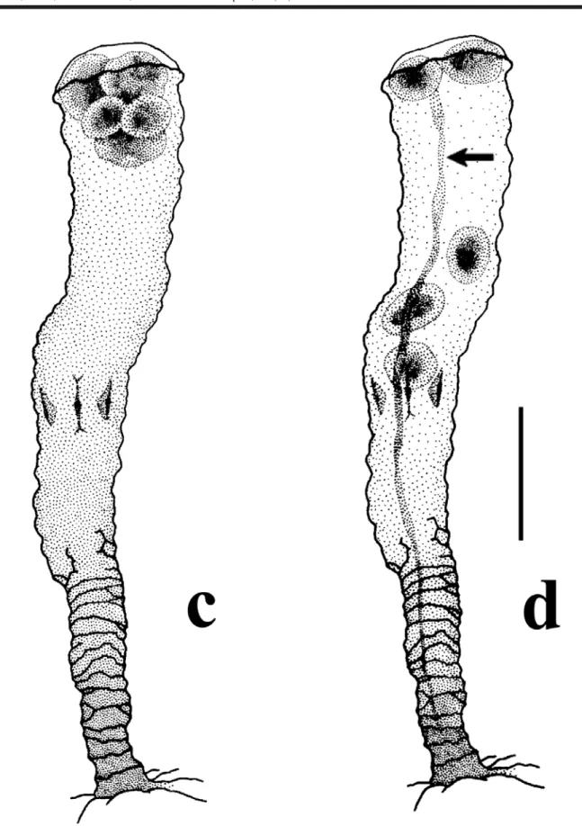
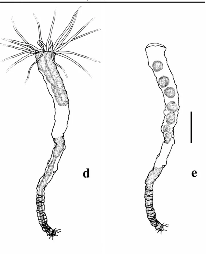
Apart from expected observations we could obtain with traditional rearing experiments in the laboratory,for Nausithoe aurea we found some different aspects as a) the rejection mechanism of particles within the periderm tube, b) segmentation and c) dormancy. During the rejec-tion of particles within the periderm tube the stephanoscyphistoma pushed it by stretching column, this requiring that part of the particles (when there were plenty) or all (when there are few) beingested by the distal region of the column and later spit out through the tube aperture. Segmentation was usually with the production of a perid-erm operculum and rarely under a plug of particles in the tube aperture, as Silveira & Morandini (1998a) had described for the colonial coronate Linuche unguiculata. Often after segmentation, part of the resulting planuloids remained in-side the gastrovascular cavity of the regenerating stephanoscyphistoma and, either left the polyp, with the opening of the operculum, or were resorbed (Figs 2 and 3). Segmentation may perhaps occur by an initial transverse fission halfway along the column of a normal polyp, andthe closing of the tube with an operculum (Fig. 4). After seg-mentation, with the opening of the operculum the residual tissues under it, and not in the form of planuloids, are in-gested by the stephanoscyphistoma (Fig. 5). We recog-nized dormancy as an undifferentiated polyp column, which could stay inside a blocked tube over a longer period.
For a summary analysis of our results, in terms of a general picture of the aspects related to the closing of the periderm tube, either by particles (mainly sand grains), by an operculum or particles + operculum, we combined all data in Table 2 for the control series and in Table 3 for the experimental series. In these tables we grouped our obser-vations considering: a) the expected (= known) aspects of the biology of N. aurea, i.e. strobilation producing detached ephyrae or planuloids and/or regeneration of a normal feed-ing polyp (sensu Silveira & Morandini, 1997: 242, Tab. VI; 237; 244); b) the different (= novel) aspects of the biology of N. aurea, i.e. asegmentation or dormancy mechanism as observed for the colonial L. unguiculata (sensu Silveira & Morandini, 1998a and b). In the tables we note whether the results refer to the exposed or buried stephanoscyphistomae.
5 Silveira, F. L.; Jarms,G. e Morandini, A. C. - Biota Neotropica, v2 (n2) - BN02202022002
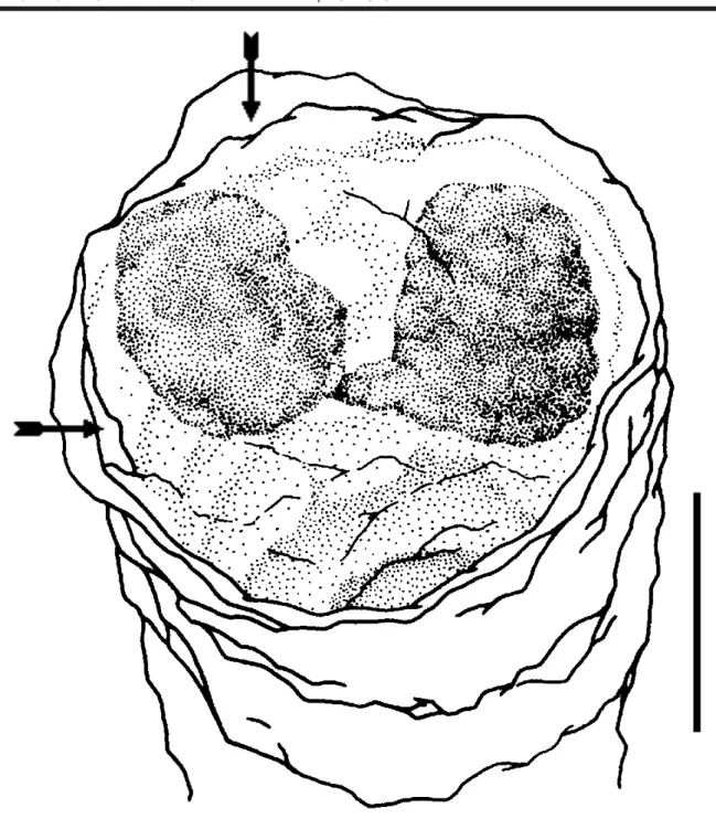
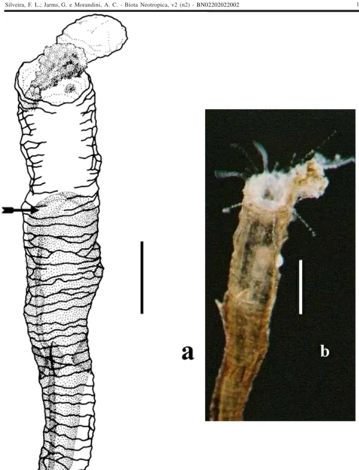
Plate I. Observed aspects of individual stephanoschyphistomae: (a) presence of particles closing the periderm tube (arrow);
6 Silveira, F. L.; Jarms,G. e Morandini, A. C. - Biota Neotropica, v2 (n2) - BN02202022002
Plate I. Observed aspects of individual stephanoschyphistomae: (c) contracted living tissues within the periderm tube, but not contracting further upon gentle squeeze with delicate forceps;
7 Silveira, F. L.; Jarms,G. e Morandini, A. C. - Biota Neotropica, v2 (n2) - BN02202022002
Plate I. Observed aspects of individual stephanoschyphistomae: (e) contracted living tissues within the periderm tube, but oral disk with recognizable tentacles;
8 Silveira, F. L.; Jarms,G. e Morandini, A. C. - Biota Neotropica, v2 (n2) - BN02202022002
Plate I. Observed aspects of individual stephanoschyphistomae: (g) polyp with simple column, i.e. without mesenteries or oral disc with tentacles (arrow points to particles);
9 Silveira, F. L.; Jarms,G. e Morandini, A. C. - Biota Neotropica, v2 (n2) - BN02202022002
Plate I. Observed aspects of individual stephanoschyphistomae: (i) strobilation under closed periderm operculum (arrow) (note conical head);
10 Silveira, F. L.; Jarms,G. e Morandini, A. C. - Biota Neotropica, v2 (n2) - BN02202022002
Plate I. Observed aspects of individual stephanoschyphistomae: (k) half opened operculum (arrow);
11 Silveira, F. L.; Jarms,G. e Morandini, A. C. - Biota Neotropica, v2 (n2) - BN02202022002
Plate I. Observed aspects of individual stephanoschyphistomae: (m) segmentation;
12 Silveira, F. L.; Jarms,G. e Morandini, A. C. - Biota Neotropica, v2 (n2) - BN02202022002
Plate I. Observed aspects of individual stephanoschyphistomae: (o) planuloid inside polyp column (arrow);
13 Silveira, F. L.; Jarms,G. e Morandini, A. C. - Biota Neotropica, v2 (n2) - BN02202022002
14 Silveira, F. L.; Jarms,G. e Morandini, A. C. - Biota Neotropica, v2 (n2) - BN02202022002
15 Silveira, F. L.; Jarms,G. e Morandini, A. C. - Biota Neotropica, v2 (n2) - BN02202022002
Figure 3. Stephanoscyphistoma periderm tube of Nausithoe aurea, distal end with operculum partially opened (arrows) (6 planuloids had already escaped through the aperture), and with remaining planuloids still becoming distinct (buried polyp, experiment series)
16 Silveira, F. L.; Jarms,G. e Morandini, A. C. - Biota Neotropica, v2 (n2) - BN02202022002
17 Silveira, F. L.; Jarms,G. e Morandini, A. C. - Biota Neotropica, v2 (n2) - BN02202022002
18 Silveira, F. L.; Jarms,G. e Morandini, A. C. - Biota Neotropica, v2 (n2) - BN02202022002
19 Silveira, F. L.; Jarms,G. e Morandini, A. C. - Biota Neotropica, v2 (n2) - BN02202022002
20 Silveira, F. L.; Jarms,G. e Morandini, A. C. - Biota Neotropica, v2 (n2) - BN02202022002
21 Silveira, F. L.; Jarms,G. e Morandini, A. C. - Biota Neotropica, v2 (n2) - BN02202022002
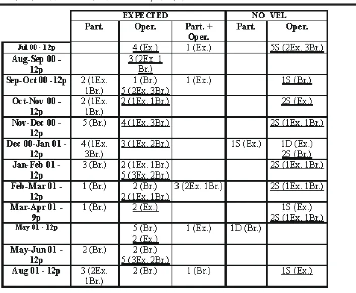
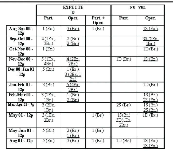
Table 3: Observations within the experimental series of the expected and novel aspects (dormancy or segmentation) for the biology
Nausithoe aurea related to the possibilities that the periderm tubes were closed by particles (Part.), or by an operculum (Oper.), or by particles and operculum (Part. + Oper.). Abbreviations: Br. = buried scyphistoma; D = dormancy; Ex. = exposed scyphistoma; np =
22 Silveira, F. L.; Jarms,G. e Morandini, A. C. - Biota Neotropica, v2 (n2) - BN02202022002
Examining Table 2, the polyps that showed expected aspects for the biology of N. aurea, we noticed that: 2 (1.41%) with particles — 1 buried in dormancy and 1 ex-posed in segmentation; 21 (14.8%) with an operculum, but 19 (13.47%) produced this in the laboratory and only 2 (1.41%) exposed presented it when sampled — 1 in dor-mancy and 1 in segmentation; NONE with particles + oper-culum. For these polyps the operculum had been correlated with segmentation.
Examining Table 3, the stephanoscyphistomae which showed expected aspects of the biology of N. aurea, we notice: 38(29.92%) with particles inside the tube — 14 (11.02%) exposed and 24 (18.89%) buried; 30 (23.62%)with an operculum, although 21 (16.53%) produced this in the laboratory and only 9 (7.08%) presented it when sampled; only 4 (3.15%) with particles + operculum. For all these pol-yps, the operculum had been correlated with strobilation. Among the polyps that showed novel aspects for the biol-ogy of N. aurea, we noticed: 8 (6.29%) with particles — 5 in dormancy (1 exposed and 4 buried) and 3 buried in segmen-tation; 16 (12.59%) with an operculum, although 10 (7.87%) produced in the laboratory and 6 (4.72%) presented it when sampled (2 exposed and 4 buried) — 3 in dormancy and 3 in segmentation; NONE with particles + operculum. For these polyps the operculum had been correlated with segmenta-tion.
Discussion
The fact that between 1/4 - 1/3 of all the stephanoscyphistomae under observation had oral discs and mesenteries suggests that in nature the condition of feeding polyps must prevail against every other described situations respectively at any time. All polyps were culti-vated under controlled homogeneous conditions and sub-jected to the same feeding regime, a favorable condition to induce strobilation for Nausithoe aurea. Nevertheless, we believe that the amount and the quality of food given to the stephanoscyphistomae did not reach a sufficient level to induce massive strobilation, even if this had far exceeded available food in nature. Werner (1979) had anticipated that for solitary coronates the surplus of energy stored by the polyps results in consecutive strobilation if there is not an annual regulation of species in waters with a high summer-winter range of temperature. Additionally, Morandini (1999) observed that cultivating 23 stephanoscyphistomae for 92 days, fed at 48 h intervals with the homogenate of the go-nad of the clam Perna perna (Linnaeus, 1767), 22 of them strobilated and 1 remained in the normal feeding condition; 17 stephanoscyphistomae strobilated during the first 25 days of cultivation; 3, 11 and 8 polyps strobilated, respec-tively, three, two and one time.
Survivorship of individual polyps of N. aurea over one year is the first record for any Coronatae polyp in
na-ture. The scyphistomae of Coronatae are known to show perennation under laboratory conditions (see Ortiz-Corp’s et al., 1987; Jarms, 1997; Silveira & Morandini, 1998a). The longest documented period of survival in the laboratory is 37 years(Atorella vanhoeffeni), the stephanoscyphistoma still beingalive and strobilating regularly (Jarms pers. obser. following initial culture observations by the late Dr. B. Werner). We think that stephanoscyphistomae, like the other polyps of Scyphozoa, arepotentially immortal. Neverthe-less, we observed that the fast growth of two encrusting organisms — sponge and coralline algae, resulted in death of some stephanoscyphistomae. We believe it to have been just by chancethe time in which we made these combined observations and consider them separate events. Never-theless, we believe that in environments with a high abun-dance of overgrowing or encrusting species, at least, soli-tary polyps cannot survive. This restricts the habitats also of N. aurea. In laboratory, without other animals or plants in the cultures, polyps of coronates grow continuously and there is no genetically defined maximum length. They only die if they are not able to reach a certain height in the tube after strobilation to get enough food, this being mostly the rim of the tube.
For the scyphistomae that showed expected aspects for the biology of the species we notice, comparatively, for the control and experimental series (deducing from Tables 2 and 3) that: 1) polyp tubes with particles occurred in higher frequency among the experimental series (29.92%) in rela-tion to the control series (16.31%) and, for both, among the buried scyphistomae. We assume a greater probability for particles to occur inside the tubes of buried scyphistomae. Thus, in the experimental series the exposed scyphistomae are more likely to have particles inside their tubes; 2) scyph-istomae with operculum occurred in higher frequency among the control series (36.17%) in relation to the experimental series (23.62%), but for both and in relative proportions (72.54% x 70%) the operculum was produced in the labora-tory and during strobilation; 3) scyphistomae with particles + operculum occurred in small numbers in both series. There-fore, it is suggested that the production of the operculum is under the control of the intrinsic regulative mechanism that causes strobilation, mainly the surplus of food.
23 Silveira, F. L.; Jarms,G. e Morandini, A. C. - Biota Neotropica, v2 (n2) - BN02202022002
dormancy. We believe that these findings suggest that these phenomena are more likely to happen among buried scyph-istomae; 2)stephanoscyphistomae with operculum occurred in slightest higher frequency among the control series (14.89%) in relation to the experimental series (12.59%), but for both, they occurred in the laboratory in higher rela-tive proportions among the control series compared with the experimental series (90.47% x 62.5%). We observed that for the control series the production of the operculum in the laboratory has always been correlated with segmenta-tion in 9 exposed and 10 buried scyphistomae, a close ratio of 1:1 operculated polyps just sampled, respectively, in dor-mancy and segmentation. We observed that for the experi-mental series the production of the operculum in the labo-ratory has always been correlated with segmentation in 7 exposed and 3 buried scyphistomae, a close ratio of some 2:1 polyps just sampled, respectively, in segmentation (1 exposed and 2 buried polyps) and dormancy (1 exposed and 2 buried polyps); 3) Wedid not observe any polyp with particles + operculum in segmentation or dormancy. In this series the production of an operculum is related to tation and dormancy. So this again suggests that segmen-tation must be derived from normal strobilation.
The description of the reduced metagenesis, caused by unfavorable conditions in the planktonicstage in some species of the Nausithoidae (Jarms, 1997), gave rise to the idea that coronates in general have the capacity to save all living tissue, andtransform it to the energy saving sessile stage — the polyp. Observations that polyps can survive up to three years without food, but under the cover of a periderm operculum, enforce this hypothesis (Jarms unpubl. data). The results of this paper strongly support this con-cept. Obviously, N. aurea is able to react with transforma-tion of the phenotype if ecological impacts do so require. Regulation mechanisms are still unknown.
Silveira & Morandini (1998a) defined segmentation in Linuche unguiculata as a mechanism leading to rejuve-nation of the polyps and production of planuloids. The seg-mentation observed for N. aurea is similar,and is also a further argument to support planuloid formation as an ex-planation for polyp-stage philopatry (Silveira & Morandini, 1997). The differences noticed are inpart due to the further development of planuloids. Distinct mobile planuloids have been seen inside the gastrovascular cavity of feeding scy-phistomae of L. unguiculata, but there was no evidence that they were re-absorbed as they could easily get out (Silveira & Morandini unpubl. data). Distinct mobile planuloids were seldom seen after segmentation in L.unguiculata, and and whenever present seemed to be dispersive stages of asexual reproduction and propagation (Silveira & Morandini, 1998a). Often, we had observed dis-tinct mobile planuloids inside the gastrovascular cavity of regenerating polyps of N. aurea, following segmentation, and these werein part entirely re-absorbed. Morandini (1999:
Tabs III-X) has noticed planuloids inside 13 scyphistomae that strobilated and produced planuloids, and that in 8 of these a few planuloids were entirely re-absorbed. Addition-ally, we noticed that the regenerating oral disc of the scyph-istoma, after segmentation and opening of the operculum, ingests residual tissues. By inadvertent removal of the oper-culum, we observed a polyp of N. aurea beginning segmen-tation that quickly regenerated the oral disc, with many long tentacles, but with a very slender and stretched column. The stephanoscyphistoma started segmentation by first di-viding the column in half and later producing distinct planuloids inside an operculated tube. We suggest that the transverse fission observed in the scyphistoma was deter-mined by the incapacity of the slender column to convey food for absorption in the basal part as observed for Nausithoe planulophora and Thecoscyphus zibrowii by Bumann & Jarms (1997).
Silveira & Morandini (1998b) compared the transfor-mation of polyp tissues inside two closed periderm tubes of L. unguiculata cysts, and to dormancy according to Cáceres (1997). We also compared the quiescent tissues of N. aurea inside closed tubes with cysts just sampled towards the end of the period of our observations. The significance of our findings goes together with the discussion in Silveira & Morandini (1998b) that for the Scyphozoa, mainly resting stages are reported as podocysts, or, eventually, planulocysts. Nevertheless, we point out that for N. aurea, dormancy seems to be related not only with operculate polyp, but alternatively with periderm tube plugged with fine and firmed particles from the surrounding sediments.
A recent study on the evolution of Cnidaria life cycles, using data from the small sub-unit of of the ribo-some to derive a phylogenetic hypothesis for “Medusozoa”, suggests that Coronatae is the sister group of “semaeostomes + rhizostomes” (Collins, 2002), a probable relationship also supported by some morphological charac-ters — the lappets, and the process of strobilation. Silveira and Morandini (1998a) have presented the hypothesis for L. unguiculata that segmentation was an advanced trait derived from a peculiar strobilation, with the addition of the operculum, although there is no operculum, either of tissue or periderm, during strobilation of the species. The obser-vation of segmentation in an operculated strobilating spe-cies as N. aurea supports the idea of linking this asexual phenomenon with strobilation.
24 Silveira, F. L.; Jarms,G. e Morandini, A. C. - Biota Neotropica, v2 (n2) - BN02202022002
days. If there are too many stones on the top of the soft body these are partly ingested. The stretching polyp pushes out the free particles, and the ingested ones are later spat out, while transported by ciliary currents. If there is too much pressure on the tube aperture from a more overlying sediment, the polyps can ingest some particles but are not able to push out either the ingested or the rest – the tube staying blocked. We believe this is the reason for shedding an operculum and undergoing dormancy.
With the results of our experiments in nature, com-bined with those following cultivation, we were able to show that the plasticity of the life cycle enables N. aurea to sur-vive under changing conditions in the São Sebastião Chan-nel. The trend to save all living tissue is attested by the possibility of a modification among normal metagenesis with ephyrae, the development of planuloids, and the trans-formation of polyp tissue by segmentation. If all these strat-egies fail, N. aurea can even undergo dormancy for a longer time.
Acknowledgements
This work was supported by Fundação de Amparo à Pesquisa do Estado de São Paulo (FAPESP 99/12433-0) and CAPES/USP PROAP/2000. ACM received financial support from FAPESP (97/03325-3, 97/08137-0 and 99/05374-7). We thank an anonymous reviewer from FAPESP for sugges-tions to improve the experimental design at the start of the work and for later comments on the project. We are indebted to MSc Helena Krieg Boscolo for help in the fieldwork and to Dr. Márcio Custódio (CEBIMar USP) for the identifica-tion of the sponge. We thank CEBIMar USP for providing the required facilities to obtain and examine the material throughout the work.
References
BUMANN, D. & G. JARMS, 1997. Localization of digestion activities in polyps of Nausithoe planulophora and Thecoscyphus zibrowii (Coronatae, Scyphozoa, Cnidaria). Helgoländer Meeresunter., 51: 477-485. CÁCERES, C.E., 1997. Dormancy in invertebrates. Invert.
Biol., 116(4): 371-383.
COLLINS, A.G., 2002. Phylogeny of the Medusozoa and the evolution of cnidarian life cycles. J. Evol. Biol., 15: 418-432.
HOLST, S. 2002. Können Polypen der Coronatae (Cnidaria, Scyphozoa) Fremdkörper aus ihren Röhren entfernen?. Diplomarbeit, Zoologisches Institut und Museum, Fachbereich Biologie der Universität Hamburg, Hamburg, 99p.
JARMS, G., 1990. Neubeschreibung dreier Arten der Gattung
Nausithoe (Coronata, Scyphozoa) sowie
Wiederbeschreibung der Art Nausithoe marginata Kölliker, 1853. Mitt. hamb. zool. Mus. Inst., 87: 7-39. JARMS, G., 1997. The polyps of Coronatae (Scyphozoa), a
review and some new results. In: den HARTOG, J.C. (ed.), Proceedings of the 6th International Conference
on Coelenterate Biology 1995. Nationaal Natuurhistorisch Museum, Leiden, p. 271-278. JARMS, G., 2001. The life cycle of Nausithoe hagenbecki
sp. nov. (Scyphozoa, Coronatae). Mitt. hamb. zool. Mus. Inst., 98: 13-22.
JARMS, G.; U. BÅMSTEDT; H. TIEMANN; M.B. MARTINUSSEN & J.H. FOSSÅ, 1999. The holopelagic life cycle of the deep-sea medusa Periphylla periphylla (Scyphozoa, Coronatae). Sarsia, 84: 55-65.
JARMS, G.; A.C. MORANDINI & F.L. da SILVEIRA, 2002. Polyps of the families Atorellidae and Nausithoidae (Scyphozoa: Coronatae). Biota Neotropica, 2(1): 11p. KAWAGUTI, S. & A. MATSUNO, 1981. A new species of
the Coronatae, Scyphozoa, from the Japan sea; Atorella japonica n. sp. Bull. Kawasaki Parmed. Coll., 1: 15-21. KOMAI, T., 1936 On another form of Stephanoscyphus
found in the waters of Japan. Mem. Coll. Sci. Kyoto (Ser. B), 11(3): 175-83.
MORANDINI, A.C., 1999. Gametogênese e desenvolvimento embrionário de Nausithoe aurea (Scyphozoa, Coronatae) do Canal de São Sebastião – SP. MSc Dissertation, Instituto de Biociências, Universidade de São Paulo. i-iv, 136 p.
MORANDINI, A.C. & F.L. da SILVEIRA, 2001a. New ob-servations and new record of Nausithoe aurea (Scyphozoa, Coronatae). Pap. Av. Zool., 41(27): 519-527.
MORANDINI, A.C. & F.L. da SILVEIRA, 2001b. Sexual re-production of Nausithoe aurea (Scyphozoa, Coronatae). Gametogenesis, egg release, embryonic development, and gastrulation. Sci. Mar., 65(2): 139-149.
ORTIZ-CORP’S, E.; C.E. CUTRESS & B.M. CUTRESS, 1987. Life history of the coronate scyphozoan Linuche unguiculata (
KOMAI, T., 1936 On another form of Stephanoscyphus found in the waters of Japan. Mem. Coll. Sci. Kyoto (Ser. B), 11(3): 175-83.
MORANDINI, A.C., 1999. Gametogênese e desenvolvimento embrionário de Nausithoe aurea (Scyphozoa, Coronatae) do Canal de São Sebastião – SP. MSc Dissertation, Instituto de Biociências, Universidade de São Paulo. i-iv, 136 p.
25 Silveira, F. L.; Jarms,G. e Morandini, A. C. - Biota Neotropica, v2 (n2) - BN02202022002
Title: Experiments in nature and laboratory observations
with Nausithoe aurea ( SCYPHOZOA: CORONATAE )
support the concept of perennation by tissue saving and confirm dormancy
Authors: Fábio Lang da Silveira, Gerhard Jarms, André Carrara Morandini
Biota Neotropica, Vol. 2 (number 2): 2003
h t t p : / / w w w. b i o t a n e o t r o p i c a . o rg . b r / v 2 n 2 / p t / a b s t r a c t ? a r t i c l e + B N 0 2 2 0 2 0 2 2 0 0 2
Date Received 07/19/2002 - Revised 09/23/2002 Accepted 10/02/2002
ISSN 1676-0611
MORANDINI, A.C. & F.L. da SILVEIRA, 2001b. Sexual re-production of Nausithoe aurea (Scyphozoa, Coronatae). Gametogenesis, egg release, embryonic development, and gastrulation. Sci. Mar., 65(2): 139-149.
ORTIZ-CORP’S, E.; C.E. CUTRESS & B.M. CUTRESS, 1987. Life history of the coronate scyphozoan Linuche unguiculata (Swartz, 1788). Carib. Jour. Sci., 23: 432-443. SILVEIRA, F.L. da. & A.C. MORANDINI, 1997. Nausithoe aurea n. sp. (Scyphozoa: Coronatae: Nausithoidae), a species with two pathways of reproduction after strobi-lation: sexual and asexual. Cont. Zool., 66(4): 235-246. SILVEIRA, F.L. da. & A.C. MORANDINI, 1998a. Asexual
reproduction in Linuche unguiculata (Swartz, 1788) (Scyphozoa: Coronatae) by planuloid formation through strobilation and segmentation. Proc. biol. Soc. Wash., 111(4): 781-794.
SILVEIRA, F.L. da. & A.C. MORANDINI, 1998b. New ob-servations on dormancy mechanisms in Linuche unguiculata (Swartz, 1788) (Scyphozoa: Coronatae). Bol. Mus. Nac., N.S., Zool., 393: 1-7.
SÖTJE, I. & G. JARMS, 1999. Description of Thecoscyphus zibrowii Werner, 1984 (Scyphozoa, Coronatae) with re-marks on the ontogeny. Mitt. Hamb. Zool. Mus. Inst., 96:5-13.
WERNER, B. 1966. Stephanoscyphus (Scyphozoa, Coronatae) und seine direkte Abstammung von den fossilen Conulata. Helgoländer wiss. Meeresunters., 13: 317-347.
WERNER, B., 1971. Stephanoscyphus planulophorus n.spec., eine neuer Scyphopolyp mit einem neun Entwichlungsmodus. Helgoländer wiss. Meeresunters., 22: 120-140.
WERNER, B., 1973. New investigations on systematics and evolution of the class Scyphozoa and the Phylum Cnidaria. Publs Seto mar. biol. Lab., 20: 35-61.
WERNER, B. 1974. Stephanoscyphus eumedusoides n. sp. (Syphozoa, Coronatae), ein Höhlenpolyp mit einem neuen Entwicklungsmodus. Helgoländer wiss.Meeresunters., 26: 434-463.