A m icro m e tho d fo r quantitatio n o f
debrisoquine and 4-hydroxydebrisoquine
in urine by liquid chro m ato graphy
1Instituto do Coração, Faculdade de Medicina, Universidade de São Paulo, São Paulo, SP, Brasil
2Laboratório de Toxicologia, Faculdade de Ciências Farmacêuticas de Ribeirão Preto, Universidade de São Paulo, Ribeirão Preto, SP, Brasil
3Laboratório de Farmacologia Terapêutica, Unidade 2, Faculdade de Ciências Farmacêuticas, Universidade de São Paulo, São Paulo, SP, Brasil
4Leiden-Amsterdam Center for Drug Research, Leiden, Holland V.A. Pereira3, J.O . Auler Jr.1,
M.J. Carmona1, F.H. Mateus2, V.L. Lanchote2, D.D. Breimer4 and S.R.C.J. Santos1
Abstract
We describe a new simple, selective and sensitive micromethod based on HPLC and fluorescence detection to measure debrisoquine (D) and 4-hydroxydebrisoquine (4-OHD) in urine for the investigation of xenobiotic metabolism by debrisoquine hydroxylase (CYP2D6). Four hundred µl of urine was required for the analysis of D and 4-OHD. Peaks were eluted at 8.3 min (4-OHD), 14.0 min (D) and 16.6 min for the internal standard, metoprolol (20 µg/ml). The 5-µm CN-reverse-phase column (Shimpack, 250 x 4.6 mm) was eluted with a mobile phase consisting of 0.25 M acetate buffer, pH 5.0, and acetonitrile (9:1, v/v) at 0.7 ml/min with detection at lexcitation = 210 nm and lemission
= 290 nm. The method, validated on the basis of measurements of spiked urine, presented 3 ng/ml (D) and 6 ng/ml (4-OHD) sensitivity, 390-6240 ng/ml (D) and 750-12000 ng/ml (4-OHD) linearity, and 5.7/ 8.2% (D) and 5.3/8.2% (4-OHD) intra/interassay precision. The method was validated using urine of a healthy Caucasian volunteer who received one 10-mg tablet of Declinax®, po, in the morning after an
overnight fast. Urine samples (diuresis of 4 or 6 h) were collected from zero to 24 h. The urinary excretion of D and 4-OHD, Fel (0-24 h), i.e., fraction of dose administered and excreted into urine, was 6.4% and 31.9%, respectively. The hydroxylation capacity index reported as metabolic ratio was 0.18 (D/4-OHD) for the person investigated and can be compared to reference limits of >12.5 for poor metabolizers (PM) and <12.5 for extensive metabolizers (EM). In parallel, the recovery ratio (RR), another hydroxylation capacity index, was 0.85 (4-OHD:SD + 4-OHD) versus reference limits of RR <0.12 for PM and RR >0.12 for EM. The healthy volunteer was considered to be an extensive metabolizer on the basis of the debrisoquine test.
Co rre spo nde nce
S.R.C.J. Santos
Laboratório de Pesquisa Instituto do Coração, HC, FM, USP Av. Dr. Eneas C. de Aguiar, 44 05403-000 São Paulo, SP Brasil
Research supported by FAPESP
(No. 97/14466-7).
Received April 23, 1999 Accepted February 3, 2000
Ke y wo rds
·Debrisoquine
·4-Hydroxydebrisoquine ·HPLC-F
·Urinary excretion kinetics ·Debrisoquine test validation ·CYP2D6
A series of drugs commonly prescribed for the treatment of cardiac patients have been identified as substrates of the CYP superfamilies I, II or III. Debrisoquine (D) hydroxylase, CYP2D6, an enzyme of
after a single po dose of Declinax®
, a known antihypertensive agent commercialized by Roche (Produtos Roche Químicos e Farma-cêuticos S.A., São Paulo, SP, Brazil) which contains 10 mg debrisoquine sulfate per tab-let. The measurement of CYP2D6 activity involves urine collection and simultaneous analysis and determination of D and 4-hydroxydebrisoquine (4-OHD). The high sensitivity required, of the order of nano-grams per milliliter of urine, requires the use of specific and sensitive methods.
Several sophisticated and high cost chro-matographic methods involving gas chroma-tography with a flame ionization detector (2), a nitrogen-phosphorous flame ioniza-tion detector (2,3) and a mass spectrometer detector (4) have been proposed but a series of difficulties were detected in their applica-tion to routine phenotyping. Liquid chroma-tography techniques using an ultraviolet de-tector have been reported (3,5-8). However, the low sensitivity of UV detection requires pre- or post-column derivatization proce-dures.
Since only a fluorescence detector can provide sufficient sensitivity without requir-ing derivatization, the objective of the pres-ent study was to develop a simple, rapid and sensitive fluorimetric method for the simul-taneous determination of D and 4-OHD in urine for routine population phenotyping.
All reagents and organic solvents, analyt-ical or chromatography grade, were pur-chased from Sigma Chemical Company (St. Louis, MO, USA), EM Science (Gibbstown, NJ, USA), Merck (Darmstadt, Germany) and Grupo Química (Penha, RJ, Brazil). Sodium chloride, sodium hydroxide and sodium ace-tate (Sigma), acetonitrile and AXO142-1 (EM Science), dichlormethane and LiChrosolv 6044 (Merck), and isopropanol (UV-HPLC 03008) (Grupo Química) were necessary for the procedure. The nitrogen (ONU1066) used for solvent evaporation in the organic ex-tracts and the helium (ONU1046) used to degas the mobile phase of chromatography,
99.99% purity, were purchased from IBG Indústria Brasileira de Gases Ltda. (Jundiaí, SP, Brazil). A type HA 45 membrane was used for buffer filtration and a type FHLP45 membrane was used for filtration of the mo-bile phase and of the organic extracts (Milli-pore Corporation, Bedford, MA, USA).
The reference standard, debrisoquine sul-fate, was donated by Roche and the 4-hydroxydebrisoquine standard was kindly provided by Professor Sompon Wanwimol-ruk (Department of Pharmacy and MRC Toxicology Research Unit, University of Otago Medical School, Dunedin, New Zealand).
Ultrapure water was obtained using the
MILLIQ® MILLIRO® systems (Millipore
Corporation) and was used to prepare the buffer solution of the mobile phase of chro-matography and to clean and regenerate the liquid chromatography apparatus (Shimadzu Corporation, Tokyo, Japan).
Debrisoquine was determined in urine samples by high performance liquid chro-matography using a Shimadzu LC-10AS ap-paratus equipped with an RF-10AXL fluo-rescence detector and connected to a model
7125 Rheodyne®
injector with a 50 µl loop,
a Nova-Pak®
CN Guard-PakTM
HPLC pre-column insert (Waters Corporation, Milford,
CT, USA) and a Shimpack®
CLC-CN Shimadzu column, 250 x 4.6 mm ID, 5 µm. The mobile phase consisted of 0.25 M ace-tate buffer, pH 5.0, and acetonitrile (9:1, v/v, final pH = 5.4) in an isocratic elution system at a flow rate of 0.7 ml/min. The column effluent was monitored at 210 nm (lexcitation)
and 290 nm (lemission). The chromatograms
were obtained with a Shimadzu C-R6A Chromatopac® printer-plotter, which
provid-ed the elution diagrams and peak integra-tion.
Fifty µl metoprolol in methanol (20 µg/ ml), the internal standard, was added to a dry clean extraction tube, followed by evapora-tion in a water bath at 37o
used in duplicate both for calibration with reference standards and for the samples. The biological sample was purified by the addi-tion of 80 mg sodium chloride/assay, urine was alkalinized to pH 9.0 by the addition of 50 µl 0.4 M sodium hydroxide (20 µmol) and extraction was performed with a mixture of dichloromethane:isopropanol (6:4, v/v) in a vortex type tube shaker for 1 min. After extraction and centrifugation at 3000 rpm for 10 min, the aqueous phase was separated from the organic phase by aspiration and discarded. The tube containing the remain-ing organic phase was then immersed in liquid nitrogen for 10 s. The organic extract was then transferred to a clean and dry coni-cal tube. The extracts were concentrated to
dryness in a water bath at 37o
C under a nitrogen flow. The dried extract was then dissolved in 100 µl of the mobile phase and 50 µl was injected into the liquid chromatog-raphy apparatus.
Calibration curves were constructed by the addition of 100 µl of D (150 µg/ml stock methanol solution) and 100 µl of 4-OHD (300 µg/ml stock aqueous solution) to a volumetric flask. Volume was completed with drug-free human urine up to 5 ml. The spiked urine was diluted serially to obtain concentrations of D of 390 to 3120 ng/ml and of 4-OHD in the 750- to 6000-ng/ml range. The standard calibration curve was prepared in duplicate and contained the in-ternal standard (metoprolol, 1 µg/assay).
We determined the absolute recovery of D (85 ± 8%) and 4-OHD (80 ± 7%) by comparing the injection of purified urine extracts with direct injection of standards (N = 8 replicates for D and 4-OHD). Relative recoveries calculated on the basis of the internal standard were 83 ± 5% (D) and 95 ± 4% (4-OHD). The detection limit was 3 ng/ ml (7.9%) and 6 ng/ml (8.0%) for D and 4-OHD, respectively. The quantification lim-its determined on the basis of the analysis of urine aliquots (10 replicates) were 12 ng/ml D (7.7%) and 23 ng/ml 4-OHD (7.6%). The
precision of the analytical procedure meas-ured by quantitative analysis of the two com-pounds under study in urine aliquots (5 rep-licates) on the same day (intraday precision)
was 5.7/5.3% for D/4-OHD, and 8.2%
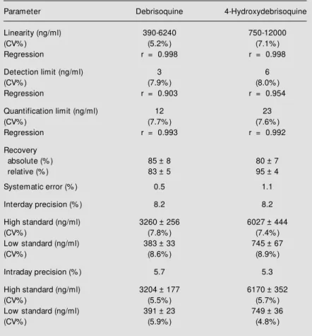
for both D and 4-OHD in 5 replicates on five consecutive days (interday precision) (Table 1).
We evaluated the debrisoquine activity of a healthy adult volunteer, a 50-year-old Caucasian female, height 172 cm, weight 60
kg, and body surface area 1.70 m2
, with normal hepatic, renal, endocrine and cardiac functions. The volunteer received detailed information about the procedures to be per-formed and gave written inper-formed consent to participate in the study. The study proto-col was approved by the Ethics Committee of the School Hospital under number 1414/ 98/109. The volunteer then received a single po dose of Declinax®
, one tablet containing 10 mg debrisoquine sulfate, in the morning after an overnight fast. Urine was then col-lected from 0 to 4 h during the 0-24 h interval after administration of the CYP2D6 marker drug for validation of the analytical method described earlier.
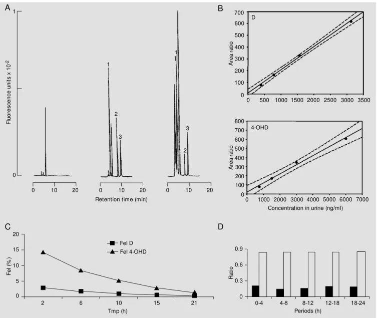
The chromatographic pattern of the puri-fied urine extracts is illustrated in Figure 1A. Chromatography was carried out for about 20 min.
The high selectivity of the chromato-graphic system utilized, together with the use of the fluorescence detector (Figure 1A), provided high sensitivity despite the small urine volume used. The linearity obtained for D and 4-OHD (Figure 1B) guaranteed good precision, with high reproducibility of the results. The confidence limits of the ana-lytical method of simultaneous determina-tion of D and of its 4-OHD in urine are listed in Table 1.
F
lu
o
re
s
c
e
n
c
e
u
n
it
s
x
1
0
-2
1
A
re
a
r
a
ti
o
700
600
500
400
300
200
100
0
0 500 1000 1500 2000 2500 3000 3500
A
re
a
r
a
ti
o
800 700
600 500
400 300
0
0 1000 2000 3000 4000 5000 6000 7000
200 100
Concentration in urine (ng/ml)
F
e
l
(%
)
20
10
5
0 15
2 6 10 15 21
Fel D Fel 4-OHD
Tmp (h)
R
a
ti
o
0.9
0.3
0 0.6
0-4 4-8 8-12 12-18 18-24
Periods (h) 0
0 10 20 0 10 20 0 10 20
Figure 1 - A, Chromatographic pattern for the simultaneous determination of D and 4-OHD in urine by HPLC-F. Peaks: 1 - 4-OHD: 8.3 min, 300 ng; 2 - D:
14.0 min, 160 ng; 3 - internal standard (metoprolol): 16.6 min, 8 µg. B, Calibration curves for D (upper) and 4-OHD (low er) peak area ratio versus
concentration in urine. C, Exponential curve decay. Urinary excretion of debrisoquine (Fel D, squares) and of 4-hydroxydebrisoquine (Fel 4-OHD,
triangles) versus time of midpoint (Tmp) after administration of the CYP2D6 marker drug to a healthy Caucasian volunteer. D, Ability to hydroxylate
debrisoquine up to 24 h: metabolic ratio (closed bars) and recovery ratio (open bars) of diuresis (4- or 6-h intervals) for the healthy Caucasian volunteer investigated.
D
4-OHD
A B
C D
The exponential decay of D and 4-OHD forms in urine is illustrated in Figure 1C by plotting the fraction of dose administered and excreted into urine (Fel) versus time of midpoint (Tmp) (9-11). It is important to emphasize that D and 4-OHD were excreted at a 1:4 molar ratio during the 24-h period of investigation. The elimination rate constant
for D in urine (Ku), formation rate constant for 4-OHD (Kf) and their respective half-lives (t(1/2)ß) were estimated by Fel versus Tmp, using a semilogarithmic plot, as fol-lows: 0.133/h (Ku), 0.119/h (Kf) and 5.2 h (D), 5.8 h (4-OHD) for the biological half-lives.
On the basis of the data for accumulated
1
2
3
1
3
2
fraction of dose administered and excreted into urine versus time, the following per-centages were obtained for the D:4-OHD ratio: 2.9:14.2% (0-4 h), 4.6:22.6% (0-8 h), 5.6:27.8% (0-12 h), 6.2:30.6% (0-18 h), and 6.4:31.9% (0-24 h). It is interesting to ob-serve that the total eliminated fraction (D + 4-OHD) ranged from 20 to 40% during the study period, as follows: 17.1% (0-4 h), 27.2% (0-8 h), 33.4% (0-12 h), 36.8% (0-18 h), and 38.3% (0-24 h).
The capacity for D hydroxylation meas-ured on the basis of the recovery ratio (RR) that corresponds to the 4-OHD excretion in relation to the total eliminated (D + 4-OHD) yielded a value of 0.85 for the recovery ratio versus reference values of RR >0.12 and RR <0.12 described previously for extensive metabolizers (EM) and poor metabolizers, (PM), respectively, for the 0-8-h period (12). The patient studied is an EM since the value obtained was 0.85>0.12. It should also be pointed out that the ratios were constant at all intervals from zero to 24 h as illustrated in Figure 1D.
When the capacity for D hydroxylation was measured on the basis of the metabolic ratio (MR) which corresponds to the excre-tion of D in relaexcre-tion to the metabolite elimi-nated, 4-OHD (1), the ratio obtained for the subject investigated was 0.18 versus refer-ence values of MR >12.5 for PM and MR <12.5 for EM (Figure 1D). Therefore, this calculation confirmed the result obtained previously, since this was an EM.
The debrisoquine test based on 24-h urine collection (4 to 6 h diuresis) designed for this study protocol indicates that MR and RR, indexes of hydroxylation capacity, remained unchanged throughout the investigation. These findings indicate that urine collection can be simplified by reducing it to 8 h (0-4, 4-8 h diuresis), allowing not only population phenotyping of hospitalized patients and outpatients, but also estimation of the kinetic parameters such as half-life and rate con-stants.
On the basis of the confidence intervals obtained in the present study and their valida-tion by the applicavalida-tion of the debrisoquine test to a healthy Caucasian volunteer (explained in detail above), we consider the proposed mi-cromethod for simultaneous analysis to be sufficient for application to routine population phenotyping as an index of the capacity for hydroxylation through the measurement of CYP2D6 activity. This methodology has per-mitted the application of this test in studies currently carried out on high-risk surgical pa-tients, guaranteeing the dose adjustment and the success of pharmacological therapy. Fi-nally, MR and RR proved to be good indexes of the hydroxylation capacity of the enzyme, CYP2D6 for population phenotyping purposes.
Table 1 - Confidence limits of the analytical method for simultaneous determination of debrisoquine and its hydroxylated metabolite by HPLC-F of urine.
(D:4-OHD) spiked blank of urine: low standard (390:750 ng/ml), high standard (3120:6000 ng/ml). CV: Coefficient of variation; r: coefficient of linear correlation.
Parameter Debrisoquine 4-Hydroxydebrisoquine
Linearity (ng/ml) 390-6240 750-12000
(CV% ) (5.2% ) (7.1% )
Regression r = 0.998 r = 0.998
Detection limit (ng/ml) 3 6
(CV% ) (7.9% ) (8.0% )
Regression r = 0.903 r = 0.954
Quantification limit (ng/ml) 12 23
(CV% ) (7.7% ) (7.6% )
Regression r = 0.993 r = 0.992
Recovery
absolute (% ) 85 ± 8 80 ± 7
relative (% ) 83 ± 5 95 ± 4
Systematic error (% ) 0.5 1.1
Interday precision (% ) 8.2 8.2
High standard (ng/ml) 3260 ± 256 6027 ± 444
(CV% ) (7.8% ) (7.4% )
Low standard (ng/ml) 383 ± 33 745 ± 67
(CV% ) (8.6% ) (8.9% )
Intraday precision (% ) 5.7 5.3
High standard (ng/ml) 3204 ± 177 6170 ± 352
(CV% ) (5.5% ) (5.7% )
Low standard (ng/ml) 391 ± 23 749 ± 36
Ackno wle dgm e nts
We are indebted to Roche (Produtos Roche Químicos e Farmacêuticos S.A.) for the reference standard, pure debrisoquine sulfate, and to Professor Sompon
Wanwi-molruk (Department of Pharmacy and MRC Toxicology Research Unit, University of Otago Medical School, Dunedin, New Zealand) for the 4-hydroxydebrisoquine stan-dard.
Re fe re nce s
1. Evans DAP, M ahgoub A, Sloan TP, Idle JR & Smith RL (1980). A family and popula-tion study of the genetic polymorphism of debrisoquine oxidation in a w hite British
population. Journal of M edical Genetics,
17: 102-105.
2. Lennard M S, Silas JH, Smith AJ & Tucker GT (1977). Determination of debrisoquine and its 4-hydroxy metabolite in biological fluids by gas chromatography w ith flame-ionization and nitrogen-selective
detec-tion. Journal of Chromatography, 133:
161-166.
3. Chan K (1988). Comparison of gas chro-matographic and high-performance liquid chromatographic assays for the determi-nation of debrisoquine and its 4-hydroxy
metabolite in human fluids. Journal of
Chromatography. B, Biomedical Applica-tions, 425: 311-321.
4. Daumas L, Sabot JF, Vermeulen E, Clapot P, Allegre F & Pinatel H (1991). Determi-nation of debrisoquine and metabolites in human urine by gas
chromatography-mass spectrometry. Journal of
Chroma-tography. B, Biomedical Applications, 570: 89-97.
5. Duche JC, Barre J & Tillement JP (1987). Rapid liquid chromatographic determina-tion of debrisoquine and its hydroxy me-tabolite in human urine to define
hydroxy-lation phenotypes. Journal of
Chromatog-raphy. B, Biomedical Applications, 423: 340-343.
6. Johnson KA, Kolatkar V & Straka RJ (1990). Improved selectivity of a high-per-formance liquid chromatography assay for debrisoquine and its 4-hydroxy
metabo-lite from urine. Therapeutic Drug M
onitor-ing, 12: 478-480.
7. M oncrieff J (1988). Assay of debrisoquine and 4-hydroxydebrisoquine in urine by re-versed-phase high-perform ance liquid chrom atography using on-line sam ple clean-up on a standard isocratic
chromato-graph. Journal of Chromatography. B,
Bio-medical Applications, 428: 178-182. 8. Wanw imolruk S & Ferry DG (1990). Rapid
high-performance liquid chromatographic method for the analysis of debrisoquine and 4-hydroxydebrisoquine in urine w
ith-out derivatization. Journal of Liquid
Chro-matography, 13: 961-968.
9. Gibaldi M (1991). Biopharmaceutics and
Clinical Pharmacokinetics. 4th edn. Lea & Febiger, Philadelphia, 203-233.
10. Shargel L & Yu ABC (1999). Applied
Bio-pharmaceutics & Pharmacokinetics. 4th edn. Appleton & Lange, Stamford, 325-352.
11. Ritschel WA (1986). Handbook and Basic
Pharmacokinetics. 2nd edn. Drug Intelli-gence Publications, Hamilton, 284-295. 12. Frye RF & Branch RA (1996). Improved
high-performance liquid chromatographic determ ination of debrisoquine and 4-hydroxydebrisoquine in human urine
fol-low ing direct injection. Journal of