Histo patho lo gical analysis o f rat
m e se nte ry as a m e tho d fo r e valuating
ne utro phil m igratio n: diffe re ntial e ffe cts
o f de xam e thaso ne and pe rtussis to xin
1Departamento de Fisiologia e Farmacologia, Unidade de Pesquisa Clínica,
and 2Departamento de Morfologia, Faculdade de Medicina,
Universidade Federal do Ceará, Fortaleza, CE, Brasil G.A.C. Brito2,
J.L.A.A. Falcão1,
S.N.R. Saraiva2
A.A.M. Lima1,
C.A. Flores1 and
R.A. Ribeiro1
Abstract
In the present study, histopathological analysis of rat mesentery was used to quantify the effect of two anti-inflammatory agents, dexameth-asone (Dex) and pertussis toxin (Ptx), on leukocyte migration. The intravenous injection of Dex (1 mg/kg) and Ptx (1,200 ng) 1 h prior to the intraperitoneal injection of the inflammatory stimuli lipopolysac-charide (LPS) or formyl-methionyl-leucyl-phenylalanine (fMLP) sig-nificantly reduced the neutrophil diapedesis (LPS: Ptx = 0.86 ± 0.19 and Dex = 0.35 ± 0.13 vs saline (S) = 2.85 ± 0.59; fMLP: Ptx = 0.43 ± 0.09 and Dex 0.01 ± 0.01 vs S = 1.08 ± 0.15 neutrophil diapedesis/ field) and infiltration (LPS: Ptx = 6.29 ± 1.4 and Dex = 3.06 ± 0.76 vs S = 15.94 ± 3.97; fMLP: Ptx = 3.85 ± 0.56 and Dex = 0.40 ± 0.16 vs S = 7.15 ± 1.17 neutrophils/field) induced by the two agonists in the rat mesentery. The inhibitory effect of Dex and Ptx was clearly visible in the fields nearest the venule (up to 200 µm), demonstrating that these anti-inflammatory agents act preferentially in the transmigration of neutrophils from the vascular lumen into the interstitial space, but not in cell movement in response to a haptotactic gradient. The mesentery of rats pretreated with Dex showed a decreased number of neutrophils within the venules (LPS: Dex = 1.50 ± 0.38 vs S = 4.20 ± 1.01; fMLP: Dex = 0.25 ± 0.11 vs S = 2.20 ± 0.34 neutrophils in the lumen/field), suggesting that this inhibitor may be acting at a step that precedes neutrophil arrival in the inflamed tissue. In contrast to that observed with Dex treatment, the number of neutrophils found in mesenteric venules was significantly elevated in animals pretreated with Ptx (LPS: Ptx = 9.85 ± 2.25 vs S = 4.20 ± 1.01; fMLP: Ptx = 4.66 ± 1.24 vs S = 2.20 ± 0.34 neutrophils in the lumen/field). This discrepancy shows that Ptx and Dex act via different mechanisms and suggests that Ptx prevents locomotion of neutrophils from the vascular lumen to the interstitial space. In conclusion, the method described here is useful for quantifying the inflammatory and anti-inflammatory effect of different substances. The advantage of this histopathological ap-proach is that it provides additional information about the steps involved in leucocyte migration.
Co rre spo nde nce
R.A. Ribeiro
Departamento de Fisiologia e Farmacologia, CCS, UFCE Rua Coronel Nunes de Melo, 1127 60430-270 Fortaleza, CE Brasil
Fax: + 55-85-243-9333 E-mail: gerly@ fortalnet.com.br
Research supported by CNPq (No. 523808/96-8) and FUNCAP (No. SO S 092/94).
Received August 26, 1997 Accepted June 22, 1998
Ke y wo rds
•Neutrophil migration
•Pertussis toxin
•LPS
•Dexamethasone
•Mesentery
Intro ductio n
The migration of specific leukocyte sub-sets from the microvascular lumen to ex-travascular tissue is a characteristic feature of the inflammatory response. Such leuko-cytes function as the primary line of host defense in the destruction of microorgan-isms and in the initiation of tissue repair. This process is initiated by the extravascular generation of chemoattractants (1,2) and by the formation of weak reversible adhesive interactions between leukocytes and vascu-lar endothelial cells. This binding is medi-ated by selectins, and although of relatively low affinity, is sufficient to serve as a bio-logical brake which quickly decelerates leu-kocytes by making them roll on endothelial cells. During rolling, leukocytes may be acti-vated by chemoattractants, with a conse-quent greatly increased affinity of their ß2 integrin adhesion receptors for ligands on activated endothelium. A chemotactic signal present outside the venule induces leuko-cytes to squeeze between the endothelial cells of the venule and to migrate to the site of inflammation (3,4).
Although leukocytes are necessary for host defense against infection, an excessive infil-tration of these cells into the inflammatory site can cause tissue damage. Thus, efforts have been made to understand this process, and to develop substances that can interfere with spe-cific steps of leukocyte recruitment.
The mesentery is a translucent tissue that allows direct viewing of the microcirculation without microtomy (5). This tissue can be easily inflamed by the injection of various stimuli into the peritoneal cavity, and has been extensively used as a model for anti-inflam-matory drug screening. The easiest means of quantifying leukocyte infiltration is to perform total and differential cell counts in peritoneal exudates (6). However, using this procedure, little information is obtained about the mecha-nisms involved in leukocyte recruitment into the inflamed mesentery. The technique of
in-travital microscopy was introduced to over-come this deficiency and to allow the gather-ing of important information on the contribu-tion of adhesion molecules to the mechanisms that mediate the passage of leukocytes across the vessel wall in vivo (3,7). The principal disadvantages of this method are its level of sophistication and elevated cost, since it re-quires a good recording setup (microscope, camera, video monitor) and computer facili-ties (software, printer).
The objetive of the present study was to develop a simpler and less expensive method for the analysis and quantification of neutro-phil migration into inflamed peritoneal cavi-ties based on histopathological features of the rat mesentery. Throughout this study, we used lipopolysaccharide (LPS) and formyl-methionyl-leucyl-phenylalanine (fMLP) as the pro-inflammatory stimuli and dexameth-asone (Dex) (8) and pertussis toxin (Ptx) from Bordetella pertussis (9) as inhibitors of neutrophil migration. We have recently dem-onstrated that the inhibitory activity of per-tussis toxin depends on the ADP-ribosylating activity of its A protomer on G proteins (10).
Mate rial and Me tho ds
Animals
Male Wistar rats (150-200 g) from the animal colony of the Federal University of Ceará were used throughout the study. The animals received water and food ad libitum.
D rugs and to xins
Pertussis toxin was purified from the su-pernatants of B. pertussis 165 cultures by hydroxylapatite chromatography and fetuin affinity chromatography (11). Ptx was a gen-erous gift from Dr. Erik Hewlett (University of Virginia, VA, USA).
Expe rim e ntal pro to co ls
LPS (200 ng/cavity) or fMLP (44 ng/ cavity) was injected intraperitoneally (ip; 1 ml) into rats pretreated intravenously (iv) 1 h earlier with Ptx (1,200 ng), Dex (1 mg/kg) or saline (0.2 ml).
The animals were sacrificed 2, 3 or 4 h after the ip injection of LPS and 4 h after fMLP, and the peritoneal cells harvested by washing the cavities with 10 ml of PBS containing 5 U heparin/ml. Total and differ-ential cell counts were performed as de-scribed elsewhere (6). The results are re-ported as number of neutrophils/ml. After harvesting the exudate, the peritoneal cavity was opened, the mesentery removed and fixed in 10% neutral buffered formalin. Af-ter 48 h, the tissue was sectioned into small pieces and mounted on albumin-coated glass slides. Twenty-four hours later the histologi-cal sections were stained with hematoxylin and eosin. Quantitative histological analysis was performed by light microscopy using a 1000X-immersion objective. The extent of neutrophil infiltration was quantified in five consecutive microscopic fields on both sides of the luminal and extraluminal sides. The number of neutrophils inside the vessel and the number of cells in diapedesis (neutro-phils passing between the venule endothelial cells, i.e., transmigration) were obtained in five fields along the venule, in mesentery of animals sacrificed 4 h after the inflammatory stimuli. The results are reported as the mean ± SEM of the number of neutrophils/field.
Statistical analysis
The statistical significance of the
differ-ences among groups was determined by ANOVA followed by the Tukey test. Re-gression analysis was performed to deter-mine the 95% confidence interval and the linear correlation coefficient (r). P values less than 5% were considered to indicate statistical significance.
Re sults
LPS- and fMLP-induce d ne utro phil infiltratio n
into rat me se nte ric tissue
Ptx (1,200 ng/rat) and Dex (0.2 mg/rat) injected iv 1 h before LPS (200 ng) or fMLP (44 ng) significantly (P<0.05) reduced the mesenteric neutrophil infiltration induced by both inflammatory stimuli (Figure 1A and C). The reduced neutrophil infiltration could be seen in the first 200 µm close to the venule (P<0.05; Figure 1B and D). The ef-fect of Dex or Ptx on LPS-induced neutro-phil infiltration is illustrated in Figure 2. There was a clear reduction in the number of neutrophils in the mesentery of animals pre-treated with Ptx (C and D) or Dex (E and F) when compared to controls (A and B). A similar response was observed with fMLP (data not shown). Regression analysis showed that there was a positive correlation (r = 0.77) between the number of infiltrated neu-trophils in the mesentery and the number of these cells present in the peritoneal washes 4 h after the injection of LPS. The 95% confi-dence interval of the correlation coefficient for the population was different from zero, indicating that there was a linear correlation between the methods used (Figure 3). When the animals were sacrificed 2 or 3 h after the injection of LPS, the correlation coefficient between the methods was r = 0.74 and r = 0.68, respectively (data not shown).
Time co urse o f ne utro phil infiltratio n
induce d by LPS into rat me se nte ric tissue
administered iv 1 h before LPS (200 ng) or fMLP (44 ng) significantly reduced (P<0.05) the number of neutrophils in diapedesis across the mesenteric venule wall (Figure 5A and B).
Ne utro phils in the lume n o f the me se nte ric
ve nule
The number of neutrophils inside the mesenteric venules was significantly creased (P<0.05) by Ptx (1,200 ng/rat) in-jected iv 1 h before LPS (200 ng) or fMLP (44 ng) challenge (Figure 6A and B). On the other hand, Dex (0.2 mg/rat) administered iv 1 h before LPS or fMLP (in the same doses) significantly reduced (P<0.05) the number of neutrophils inside the mesenteric venules when compared to saline-treated rats (Figure 6A and B).
D iscussio n
The histopathological method presented here proved to be useful for quantifying leukocyte migration and the effect of anti-inflammatory agents on this phenomenon. Our results confirmed the inhibitory effect of Dex (12) and Ptx (10) on neutrophil mi-gration induced by inflammatory stimuli. Rats treated with these agents showed a signifi-cant reduction in the neutrophil infiltration into inflamed mesentery induced by fMLP, a direct chemotactic agent, and LPS, a sub-stance that induces neutrophil recruitment by an indirect mechanism dependent on peri-toneal resident cells (13).
Dexamethasone reduced the neutrophil infiltration induced by both chemotactic stimuli. The inhibitory effect of Dex and Ptx was particularly visible in the fields nearest the venule (within a distance of up to 200 µm), thus demonstrating that these anti-in-flammatory agents act preferentially on the migration of neutrophils from the vascular lumen into the interstitial space, rather than on the movement of cells in response to a
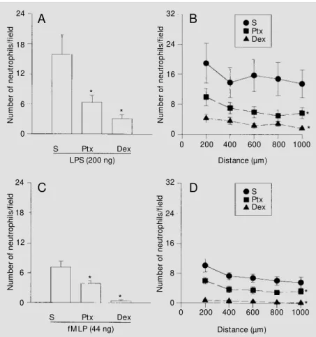
Figure 1 - Pertussis toxin (Ptx) and dexamethasone (Dex) inhibit LPS- or fM LP-induced neutrophil migration into the rat mesentery. Ptx (1,200 ng/rat) and Dex (0.2 mg/rat) w ere injected iv 1 h before the ip administration of LPS (200 ng/cavity; A and B) or fM LP (44 ng/ cavity; C and D). The rats w ere sacrificed 4 h after injection of the inflammatory stimuli. The neutrophils w ere counted in five consecutive fields on both sides of a venule perpendicular axis. The results are reported as the mean ± SEM of the total number of neutrophils/field (A and C) or number of neutrophils/field as a function of the distance from the venule (B and D). Six rats w ere used per treatment. * P<0.05 compared to the group that received saline (S) iv at all points. (ANOVA and Tukey test).
N u m b e r o f n e u tr o p h ils /f ie ld 24 N u m b e r o f n e u tr o p h ils /f ie ld 32 18 6 0 12 24 16 8 0 * * * *
S Ptx Dex
200
0 400 600 800 1000
Distance (µm)
200
0 400 600 800 1000
Distance (µm) fM LP (44 ng)
LPS (200 ng)
S Ptx Dex S Ptx Dex * * N u m b e r o f n e u tr o p h ils /f ie ld 32 24 16 8 0 N u m b e r o f n e u tr o p h ils /f ie ld 24 18 12 6 0 * *
A
B
S Ptx Dex
D
C
injected iv 1 h before LPS (200 ng) signifi-cantly (P<0.05) reduced the mesenteric neu-trophil infiltration in the 2nd, 3rd and 4th h after the inflammatory stimuli (Figure 4). The peak of neutrophil infiltration in the mesentery was seen at the 3rd h (Figure 4), whereas the highest counts of neutrophil in peritoneal wash were obtained 4 h after the inflammatory stimuli (data not shown).
Ne utro phil diape de sis induce d by LPS and
fMLP in me se nte ric ve nule s
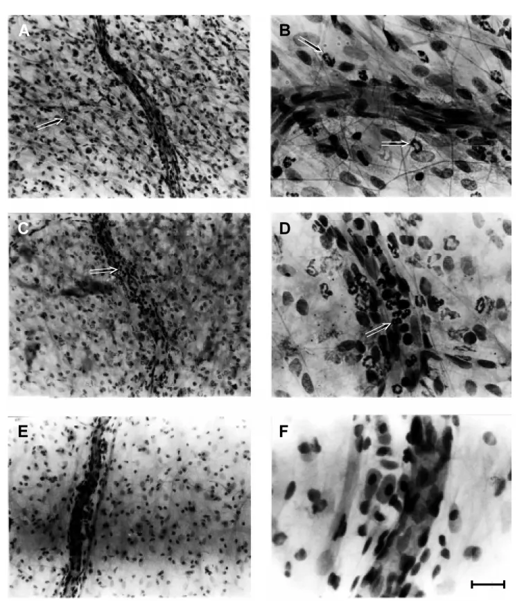
Figure 2 - Histological aspect of inflamed rat mesentery 4 h after the ip injection of LPS (200 ng/cavity) and iv pretreated (1 h earlier) w ith sterile saline (A and B), pertussis toxin (Ptx; 1,200 ng/animal; C and D) or dexamethasone (Dex; 0.2 mg/animal; E and F). The neutrophil infil trate in the Ptx- and Dex-treated rats w as clearly less intense than that observed in the saline-Dex-treated group. In Ptx-Dex-treated rats, the neutrophils w ere retained in the lumen of the blood vessels. Arrow s are pointing to neutrophils. Hematoxylin and eosin stain; bar = 100 µm for A, C, and E, and bar = 25 µm for B, D, and F.
A
B
D
C
E
F
⇒
⇒
⇒
⇒
haptotactic gradient. This hypothesis is sup-ported by two observations: a) at distances of 200 µm from the venule, the curves for cell migration in the control and treated groups were almost parallel, and b) the num-ber of neutrophils undergoing diapedesis in the mesenteric venules was significantly re-duced in both Ptx- and Dex-treated rats.
It has been proposed by Katori et al. (12) that at least five sequential steps are in-volved in the extravasation of leukocytes from the vascular lumen into the interstitial space following the administration of che-moattractants: 1) rolling, 2) adhesion to the inner surface of the venules, 3) passage through endothelial gaps, 4) accumulation in
the space between the endothelial cells and the basement membrane of pericytes, and 5) penetration of the pericyte basement mem-brane to reach the interstitial space. All of these steps can be triggered or modulated by chemotactic mediators, cytokines and inter-actions between leukocytes and endothelial cells through the expression of cell adhesion molecules (14). Dexamethasone is a potent anti-inflammatory agent with a well-known inhibitory action on neutrophil migration. Like other glucocorticoids, Dex induces the synthesis of lipocortin that inhibits the en-zyme phospholipase A2, thereby preventing the generation of prostaglandins, leukotri-enes and PAF which can contribute to the recruitment of leukocytes to the inflamma-tory site (15,16). In addition, glucocorticoids inhibit the synthesis and activity of cyto-kines with chemotactic properties, including IL-1 (17,18) and TNF-α (19,20), and their ability to induce the expression of adhesion molecules such as E-selectin, ICAM-1 (21) and VCAM-1 (22).
Our results show that the mesentery of rats pretreated with Dex had a lower number of neutrophils within the venule. However, this was not related to a possible neutropenic effect of the corticoid (data not shown). Indeed, acute treatment with corticoids causes neutrophilia (23). Thus, we suggest that Dex acts at a step that precedes neutrophil arrival in the inflamed tissue by inhibiting produc-tion and/or activity of chemotactic agents released by stimulated resident cells. Inhibi-tion of chemotactic activity would explain the inhibitory effect of Dex on fMLP since this agent acts independently on the release of other chemotactic factors from peritoneal resident cells (24). In agreement with others (25), we believe that the inhibition of neutro-phil-endothelium adhesion is the main mech-anism for the anti-inflammatory effect of glucocorticoids. Such an action can explain the reductions in diapedesis and neutrophil infiltration observed in Dex-treated rats. On the other hand, some in vivo studies have
N
e
u
tr
o
p
h
ils
x
1
0
6/m
l
7
5
2 3
0 1 4 6
0 5 10 15 20 25 30
Neutrophils/field Figure 3 - The correlation
be-t w een be-t he hisbe-t opabe-t hological method and conventional leuko-cyte counts in peritoneal exu-date 4 h after the injection of LPS. The straight line represents the correlation index betw een these methods (r = 0.77). The dashed lines represent the con-fidence interval (P<0.05).
S Ptx Dex
N
u
m
b
e
r
o
f
n
e
u
tr
o
p
h
ils
/f
ie
ld
24
18
6 12
0
1 2 3 4 5
Time (h) Figure 4 - Time course of
shown that Dex does not inhibit neutrophil rolling or adhesion to venular endothelium and does not inhibit neutrophil penetration through endothelial cell junctions (12,26).
Ptx reduced the number of cells in dia-pedesis and the neutrophil infiltration into inflamed mesentery. In contrast to Dex, the number of neutrophils in the lumen of mes-enteric venules was significantly increased in rats pretreated with this toxin. This diver-gence indicates that Ptx and Dex act via different mechanisms. The retention of neu-trophils in the lumen of mesenteric blood
vessels suggests that Ptx prevents neutrophil locomotion from the vascular lumen into the interstitial space. It has been demonstrated that the lectin domains of two Ptx subunits share amino acid sequence similarities with the lectin domains of the eukaryotic selectin family (27). During the inflammatory pro-cess, selectins appear on endothelial cells and promote the rolling of leukocytes by reversible binding to carbohydrates (14). Thus, it is possible that Ptx decreases the leukocyte recruitment to the sites of inflamed tissue by competitively blocking common
Figure 5 - Pertussis toxin (Ptx) and dexamethasone (Dex) re-duced the number of neutrophils in diapedesis in rat mesenteric tissue follow ing exposure to LPS or fM LP. Ptx (1,200 ng/rat) and Dex (0.2 mg/rat) w ere in-jected iv 1 h before the ip ad-ministration of LPS (200 ng/cav-ity; A) or fM LP (44 ng/cavng/cav-ity; B). The rats w ere sacrificed 4 h af-ter injection of the inflammatory stimuli. The neutrophils in dia-pedesis w ere counted in five consecutive microscopy fields along the venule. The results are reported as the mean ± SEM of the number of neutrophils/field. Six rats w ere used per treat-ment. * P<0.05 compared to the group that received saline (S) iv (ANOVA and Tukey test).
Figure 6 - Pertussis toxin (Ptx) and dexamethasone (Dex) dif-ferentially affect the number of neutrophils inside the venule. Ptx (1,200 ng/rat) and Dex (0.2 mg/rat) w ere injected iv 1 h be-fore the ip administration of LPS (200 ng/cavity; A) or fM LP (44 ng/cavity; B). The rats w ere sac-rificed 4 h after injection of the inflammatory stimuli. The neu-trophils w ere counted in five consecutive fields along the venule. The results are reported as the mean ± SEM of the num-ber of neutrophils in the lumen/ field. Six rats w ere used per treatment. * P<0.05 compared to the group that received sa-line (S) iv (ANOVA and Tukey test). N e u tr o p h il d ia p e d e s is /f ie ld 4 3 2 1 0 A * * * * 4 3 2 1 0 B
S Ptx Dex
LPS (200 ng)
S Ptx Dex
fM LP (44 ng)
N e u tr o p h il d ia p e d e s is /f ie ld N e u tr o p h ils i n t h e l u m e n /f ie ld 16 12 8 4 0 A * * * * 16 12 8 4 0 B
S Ptx Dex
LPS (200 ng)
S Ptx Dex
fM LP (44 ng)
leukocyte and/or endothelial selectin carbo-hydrate binding sites (28). We have demon-strated that the in vivo anti-inflammatory effect of Ptx is dependent on its ability to inhibit the action of Gi protein (10,29). Fur-thermore, it has been shown that a Ptx-sensi-tive G protein is involved in cellular move-ment (2) as well as in other effects triggered by inflammatory mediators, including an oxidative burst, the release of arachidonate-derived metabolites, and cell degranulation (30).
In conclusion, the histopathological pro-cedure presented here proved to be an ad-equate means of quantifying the inflamma-tory and anti-inflammainflamma-tory actions of sev-eral substances. The method showed a sig-nificant (P<0.05) correlation with the more conventional approach of leukocyte counts in peritoneal exudates. The two methods could not be strictly compared because of differences in the time course of neutrophil vascular migration to the mesentery (peak at the 3rd h) and the arrival of these cells into peritoneal cavities (peak at the 4th h). The principal advantage of this method com-pared to simple cell counting is the possibil-ity of obtaining additional information about
the steps involved in leukocyte migration. Such information may contribute to our un-derstanding of the mechanisms underlying the action of different anti-inflammatory sub-stances. For example, using this method it was possible to show that Ptx and Dex act via different mechanisms suggesting that Ptx prevents the locomotion of neutrophil from the vascular lumen into the interstitial space, whereas Dex acts at a step that precedes neutrophil arrival in the inflamed tissue, prob-ably inhibiting production and/or activity of chemotactic agents. The methodology de-scribed here is not as practical as leukocyte counts in peritoneal exudates, but provides a good alternative to intravital microscopy (31,32), because it is simpler and less expen-sive than the later technique, does not need prolonged training and can be carried out using the facilities of a conventional histol-ogy laboratory.
Ackno wle dgm e nts
We thank Erik L. Hewlett for kindly pro-viding the toxins and Stephen Hyslop for language review.
Re fe re nce s
1. Springer TA (1994). Traffic signals for lym-phocyte recirculation and leukocyte emi-gration: the multistep paradigm. Cell, 76: 301-304.
2. Ben-Baruch A, M ichiel DF & Oppenheim JJ (1995). Signals and receptors involved in recruitment of inflammatory cells. Jour-nal of Biological Chemistry, 270: 11703-11706.
3. Nourshargh S, Larkin SW, Das A & Wil-liams TJ (1995). Interleukin-1-induced leu-kocyte extravasation across rat mesenter-ic mmesenter-icrovessels is mediated by platelet-activating factor. Blood, 85: 2553-2558. 4. Frenette PS & Wagner DD (1996).
Adhe-sion molecules - Part II: Blood vessels and blood cells. New England Journal of M edi-cine, 335: 43-45.
5. Gabe M (1968). Techniques Histologi-ques. M ason, Paris.
6. Souza GEP & Ferreira SH (1985). Block-ade by anti-macrophage serum of the mi-gration of PM N neutrophils into the in-flamed peritoneal cavity. Agents and Ac-tions, 17: 97-103.
7. Arndt H, Palitzsch K-D, Anderson DC, Rusche J, Grisham M B & Granger DN (1995). Leukocyte-endothelial cell adhe-sion in a model of intestinal inflammation. Gut, 37: 374-379.
8. Flow er RJ (1989). Glucocorticoids and the inhibit ion of phospholipase A2. In:
Schleimer RP, Clamor HN & Oronsky AL (Editors), Anti-Inflammatory Steroid Ac-tion. Basic and Clinical Aspects. Academ-ic Press, New York, 46-66.
9. Imagaw a T, Kanoh M , Sonoda S & Utsumi S (1980). Polymorphonuclear leukocyte-inhibitory factor of Bordetella pertussis. III. Inhibition of Arthus reaction and
peri-toneal infiltration of PM N. M icrobiology and Immunology, 24: 895-905.
10. Brito GCA, Souza M HLP, M elo-Filho AA, Hew lett EL, Lima AA, Flores CA & Ribeiro RA (1997). Role of pertussis toxin A sub-unit in neutrophil migration and vascular permeability. Infection and Immunity, 65: 1114-1118.
11. Sindt KA, Hew let t EL, Redpat h GT, Rappuoli R, Gray LS & Vandenberg SR (1994). Pertussis toxin activates platelets through an interaction w ith platelet glyco-protein Ib. Infection and Immunity, 62: 3108-3114.
12. Katori M , Oda T & Nagai K (1990). A site of action of dexamethasone on leukocyte extravasation in microcirculation. Agents and Actions, 29: 24-26.
mechanisms of neutrophil emigration. Dif-ferent mechanisms of inflammation in rabbits induced by interleukin-1, tumor ne-crosis factor alpha or endotoxins versus leukocyte chemoattractants. American Journal of Pathology, 135: 227-237. 14. M cEver RP (1992). Leukocyte-endothelial
cell interactions. Current Biology, 4: 840-849.
15. Flow er RJ & Blackw ell GJ (1979). Anti-inflammatory steroids induce synthesis of a phospholipase A2 inhibitor w hich
pre-vents prostaglandin generation. Nature, 278: 456-459.
16. Hirata F, Notsu Y, Iw ata M , Parente L, DiRosa M & Flow er RJ (1982). Identifica-tion of several species of phospholipase inhibitory protein(s) by radioimmunoassay for lipomodulin. Biochemical and Bio-physical Research Communications, 109: 223-230.
17. Staruch M J & Wood DD (1985). Reduc-tion of serum interleukin-1 like activity af-ter treatment w ith dexamethasone. Jour-nal of Leukocyte Biology, 37: 193-207. 18. Bochner BS, Rutledge BK & Schleimer
RP (1987). Interleukin 1 production by hu-man lung tissue. II: inhibition by anti-in-flammatory steroids. Journal of Immunol-ogy, 139: 2303-2307.
19. Beutler B, Krochin N, M ilsark IW, Luedke C & Cerami A (1986). Control of cachectin (tumor necrosis factor) synthesis: mecha-nisms of endotoxin resistance. Science, 232: 977-980.
20. Schw iebert LA, Beck LA, Stellato C, Bickel CA, Bochner BS & Schleimer RP (1996).
Glucocorticoids inhibition of cytokine pro-duction: Relevance to antiallergic actions. Journal of Allergy and Clinical Immunol-ogy, 97: 143-152.
21. Cronstein BN & Weissmann G (1993). The adhesion molecules of inflammation. Ar-thritis and Rheumatism, 36: 147-157. 22. Tessier PA, Cattaruzzi P & M cColl SR
(1996). Inhibition of lymphocyte adhesion to cytokine-activated synovial fibroblasts by glucocorticoids involves the attenua-tion of vascular cell adhesion molecule 1 and intercellular adhesion molecule 1 gene expression. Arthritis and Rheuma-tism, 39: 226-234.
23. Dale DC, Fauci AS & Wolff SM (1974). Alternate-day prednisone leukocyte kinet-ics and susceptibility to infections. New England Journal of M edicine, 291: 1154-1158.
24. Ribeiro RA, Souza-Filho M VP, Souza M HLP, Oliveira SHP, Costa CHS, Cunha FQ & Ferreira SH (1996). Role of resident mast cells and macrophages in the neu-trophil migration induced by LTB4, fM LP and C5a des Arg. International Archives of Allergy and Immunology, 112: 27-29. 25. Allison Jr F, Smith M R & Wood WB
(1955). Studies on the pathogenesis of acute inflammation. II. The actions of cor-tisone on the inflammatory response to thermal injury. Journal of Experimental M edicine, 102: 669-679.
26. Oda T & Katori M (1992). Inhibition site of dexamethasone on extravasation of poly-morphonuclear leukocytes in the hamster cheek pouch microcirculation. Journal of
Leukocyte Biology, 52: 1337-1342. 27. Saukkonen K, Burnette WN, M ar V, M asur
HR & Tuomanen E (1992). Pertussis toxin has eukaryotic-like carbohydrate recogni-tion domains. Proceedings of the National Academy of Sciences, USA, 89: 118-122. 28. Rodzinsky E, Jones T, Burnette W N, Burroughs M & Tuomanen E (1993). Anti-inf lam m at ory ef f ect s in experim ent al meningitis of prokaryotic peptides that mimic selectin. Journal of Infectious Dis-eases, 168: 1422-1428.
29. Thomazzi SM , Souza M HLP, M elo-Filho AA, Hew lett EL, Lima AAM & Ribeiro RA (1995). Pertussis toxin from Bordetella pertussis blocks neutrophil migration and neutrophil-dependent edema in response to inflammation. Brazilian Journal of M edi-cal and Biologiedi-cal Research, 28: 120-124. 30. Nourshargh S & Williams TJ (1990). Evi-dence that a receptor-operated event on the neutrophil mediates neutrophil accu-mulation in vivo. Pretreatment of 111In-neutrophils w ith pertussis toxin in vitro inhibits their accumulation in vivo. Journal of Immunology, 145: 2633-2638. 31. Granger DN, Benoit JN, Suzuky M &
Grisham M B (1989). Leukocyte adher-ence to venular endothelium during is-chemia/reperfusion. American Journal of Physiology, 257: G683-G688.