Reactive oxygen species and
angiotensin II signaling in vascular cells
-implications in cardiovascular disease
Multidisciplinary Research Group on Hypertension,
Canadian Institute of Health Research, Clinical Research Institute of Montreal, University of Montreal, Quebec, Canada
R.M. Touyz
Abstract
Diseases such as hypertension, atherosclerosis, hyperlipidemia, and diabetes are associated with vascular functional and structural changes including endothelial dysfunction, altered contractility and vascular remodeling. Cellular events underlying these processes involve changes in vascular smooth muscle cell (VSMC) growth, apoptosis/anoikis, cell migration, inflammation, and fibrosis. Many factors influence cellular changes, of which angiotensin II (Ang II) appears to be amongst the most important. The physiological and pathophysiologi-cal actions of Ang II are mediated primarily via the Ang II type 1 receptor. Growing evidence indicates that Ang II induces its pleiotro-pic vascular effects through NADPH-driven generation of reactive oxygen species (ROS). ROS function as important intracellular and intercellular second messengers to modulate many downstream sig-naling molecules, such as protein tyrosine phosphatases, protein tyrosine kinases, transcription factors, mitogen-activated protein ki-nases, and ion channels. Induction of these signaling cascades leads to VSMC growth and migration, regulation of endothelial function, expression of pro-inflammatory mediators, and modification of extra-cellular matrix. In addition, ROS increase intraextra-cellular free Ca2+ concentration ([Ca2+]
i), a major determinant of vascular reactivity. ROS influence signaling molecules by altering the intracellular redox state and by oxidative modification of proteins. In physiological conditions, these events play an important role in maintaining vascular function and integrity. Under pathological conditions ROS contribute to vascular dysfunction and remodeling through oxidative damage. The present review focuses on the biology of ROS in Ang II signaling in vascular cells and discusses how oxidative stress contributes to vascular damage in cardiovascular disease.
Correspondence
R.M. Touyz
Clinical Research Institute of Montreal
110 Pine Ave. West Montreal, H2W 1R7, Quebec Canada
Fax: 514-987-5523 E-mail: touyzr@ircm.qc.ca
Presented at the V International Symposium on Vasoactive Peptides, Ouro Preto, MG, Brazil, February 12-14, 2004.
Research supported by Canadian Institutes of Health Research, Heart and Stroke Foundation of Canada, Canadian Hypertension Society, and fonds de la Recherche en Santé du Quebec.
Received April 22, 2004 Accepted June 3, 2004
Key words
•Vascular smooth muscle cells •Remodeling •Inflammation •Signal transduction •Reactive oxygen species
Introduction
Angiotensin II (Ang II), originally de-scribed as a potent vasoconstrictor, is now recognized as a multifunctional hormone in-fluencing many cellular processes important
blood pressure and fluid homeostasis in physiological conditions. In pathological con-ditions, through its vasoconstrictor, mito-genic, pro-inflammatory, and pro-fibrotic actions, Ang II contributes to altered vascu-lar tone, endothelial dysfunction, structural remodeling, and vascular inflammation, char-acteristic features of vascular damage in hy-pertension, atherosclerosis, vasculitis, and diabetes (2-5).
The subcellular mechanisms and signal-ing pathways whereby Ang II mediates its physiological and pathophysiological vas-cular effects are complex (2). Growing evi-dence indicates that production of reactive oxygen species (ROS) and activation of re-duction-oxidation (redox)-dependent signal-ing cascades are critically and centrally in-volved in Ang II-induced actions (3,5). All vascular cell types, including endothelial cells, smooth muscle cells, adventitial fibro-blasts, and resident macrophages, produce ROS (6-10). Of particular importance in the vasculature are superoxide (•••••O
2-) and hy-drogen peroxide (H2O2), since these ROS act as inter- and intra-cellular signaling mol-ecules. The major source of ROS in the vascular wall is non-phagocytic NADPH oxidase, which is regulated by vasoactive agents (Ang II, ET-1, thrombin, serotonin),
cytokines (IL-1, TNFα), growth factors
(PDGF, IGF-1, EGF) and mechanical forces (cyclic stretch, laminar and oscillatory shear stress) (5). The best characterized system in vascular cells is Ang II-stimulated NADPH oxidase-mediated generation of •••••O
2-, which appears to be upregulated in hypertension, atherosclerosis and diabetes (5).
The present review focuses on recent progress in mechanisms whereby Ang II gen-erates ROS in vascular cells, how ROS influ-ence signaling events and cellular function and what the implications are in vascular func-tion and remodeling in cardiovascular dis-eases. Emerging concepts on mechanisms of signal transduction by ROS that involve per-turbations in cellular redox state and oxidative
modifications of proteins are also discussed.
Reactive oxygen species, redox signaling and oxidative stress
ROS are formed as intermediates in redox processes, leading from oxygen to water (11). The univalent reduction of oxygen, in the presence of a free electron (e), yields •••••O
2-, H2O2 and •••••OH (Figure 1). Superoxide has an unpaired electron, which imparts high reactiv-ity and renders it unstable and short-lived. It is water soluble and membrane impermeable, but can cross cell membranes via anion chan-nels (12,13). In physiological conditions in aqueous solutions at a neutral pH, •••••O
2 -dismutatesyielding H2O2. However, when pro-duced in excess, a significant amount of •••••O
2
-reacts with NO to produce ONOO- (14).
Hydrogen peroxide is produced mainly from dismutation of •••••O
2-. This reaction can be spontaneous or can be catalyzed by superox-ide dismutase (SOD), of which there are three isoforms, CuZnSOD, MnSOD and extracellu-lar SOD (EC-SOD) (11). The SOD-catalyzed dismutation is favored when the concentration of •••••O
2- is low and when the concentration of SOD is high, which occurs under physiologi-cal conditions. Unlike •••••O
2-, H2O2 is not a free radical and is a much more stable molecule. Hydrogen peroxide is lipid soluble, crosses cell membranes and has a longer half-life than
• • • • •O
2-. In biological systems, it is scavenged by catalase and by glutathione peroxidase (13). Hydrogen peroxide can also be reduced to generate the highly reactive •••••OH in the pres-ence of metal-containing molecules such as Fe2+ (11). Hydroxyl radical is extremely reac-tive and, unlike •••••O
2- and H2O2, which travel some distance from their site of generation,
• • • •
•OH induces local damage where it is formed.
In the vasculature, •••••O
conditions, the rate of ROS production is bal-anced by the rate of elimination.
ROS share several features with classical second messengers and have been impli-cated as important signaling molecules. Simi-lar to second messengers, production of ROS is tightly regulated by extracellular stimuli. ROS are small molecules that can diffuse locally, their existence is transient and they act on specific downstream effectors to in-fluence cell activity and function (17). Re-dox signaling involves at least one reaction in which oxidation of a signaling molecule by a ROS occurs and which is reversible (18). Physiologic generation of ROS has
been implicated in a varietyof biological
responses from transcriptional activation to cell proliferation. “Redox regulation” refers to the biological responses maintaining cell homeostasis against oxidative excess. Under pathological conditions, a disequilibrium between ROS generation and antioxidant protection results in increased bioavailabil-ity of ROS leading to a state of oxidative stress (19,20). Hence oxidative events in which ROS play specific roles in signaling cascades and which are non-damaging are referred to as redox-signaling processes (17,18). On the other hand, an oxidative burden in which ROS cause injury and where repair or cell death are non-specific responses with respect to the involvement of oxidants is termed oxidative stress. The pathogenic outcome of oxidative stress is oxidative dam-age (13), a major cause of vascular injury in cardiovascular disease.
Ang II-induced production of reactive oxygen species in vascular cells
Vascular NADPH oxidase
Ang II elicits its actions via two distinct receptors, the Ang II type 1 (AT1) and Ang II type 2 receptors (AT2) (2). Most known physi-ological and pathophysiphysi-ological effects of Ang II are mediated via AT1 receptors, which
couple to multiple interacting signal trans-duction cascades, leading to diverse biologi-cal actions. These signaling processes are multiphasic with distinct temporal charac-teristics and have been well described in recent reviews (2,21).
Exciting new research in the field of
vas-cular biology is the demonstration that AT1
receptor activation stimulates non-phagocytic NADPH oxidase and generation of •••••O
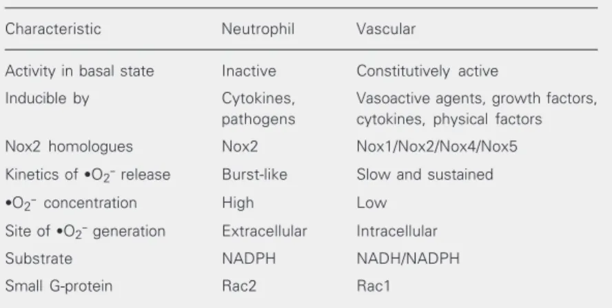
2- in various vascular cell types, including vascu-lar smooth muscle cells (VSMC) (5,6), en-dothelial cells (7) and fibroblasts (8). Vascu-lar NADPH oxidase is simiVascu-lar, but not iden-tical, to neutrophil NADPH oxidase, as sum-marized in Table 1. The prototypical phago-cytic NADPH oxidases are multimeric pro-tein complexes comprising membrane-bound
Table 1. Characteristics of neutrophil and vascular NADPH oxidase.
Characteristic Neutrophil Vascular
Activity in basal state Inactive Constitutively active
Inducible by Cytokines, Vasoactive agents, growth factors, pathogens cytokines, physical factors
Nox2 homologues Nox2 Nox1/Nox2/Nox4/Nox5
Kinetics of ·O2- release Burst-like Slow and sustained
·O2- concentration High Low
Site of ·O2- generation Extracellular Intracellular
Substrate NADPH NADH/NADPH
Small G-protein Rac2 Rac1
Glutathione
H2O + O2
O2
2
Peroxidase
Thioredoxin Catalase SOD
2·O2
-+ NO
·OH ONOO
-H2O2
Fe2+
e
-+ Fe3+
Oxidation
flavocytochrome b558 (formed by gp91phox (Nox2) and p22phox), up to three cytoplas-mic subunits, p47phox, p67phox and p40phox and a regulatory G-protein (Rac1 or Rac2) (9). All neutrophil subunits have been demonstrated, to varying degrees, in vascular cells (6-9) (Figure 2). In addition, Nox2 homologues, Nox1, a 563-amino acid protein that shares 55% homology with gp91phox, and Nox4, a 578-amino acid pro-tein with 39% homology to gp91phox, have been implicated to play a role in vascular cell
• • • • •O
2- production (22,23). Both Nox1 and
Nox4 are expressed in vascular cells and are regulated by factors that stimulate ROS gen-eration, such as Ang II and PDGF (24,25). Nox1 was initially suggested to be a subunit-independent low capacity •••••O
2- generating enzyme involved in the regulation of mito-genesis (22). However, recent data indicate that Nox1 requires p47phox and p67phox and that it is regulated by NoxO1 (Nox orga-nizer 1, a p47phox homologue) and NoxA1 (Nox activator 1, a p67phox homologue)
(26). Nox4 has recently been implicated to be the major catalytic component in endo-thelial cells (27). Although the renin-angio-tensin system has been demonstrated to
up-regulate vascular Nox1 and Nox4 in vitro
and in vivo (28), the physiological signifi-cance of these processes in the cardiovascu-lar system awaits ccardiovascu-larification.
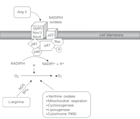
Activation of NADPH oxidase is a multi-step process initiated by serine phosphoryla-tion of p47phox, which triggers complex for-mation of cytoplasmic subunits followed by translocation to the membrane where, together with Rac, it associates with cytochrome b558 to assemble the active oxidase (9) (Figure 2). Of the many vasoactive factors that stimulate this process, Ang II appears to be one of the most important in the vasculature (5,6,29). Mechanisms linking Ang II to the enzyme and upstream signaling molecules modulating NADPH oxidase in VSMCs have not been fully elucidated, but PLD, PKC, c-Src, EGFR transactivation, PI3K, and Rac may be in-volved (30-32). In its activated state, NADPH oxidase accepts electrons from its substrate NADPH and donates these to molecular oxy-gen. In this way, a one-electron reduction of oxygen to •••••O
2- is catalyzed at the expense of NADPH according to the following reaction: 2O2 + NADPH - NADPH oxidase → 2•••••O
2- + NADP+ + H+.
Other enzymatic sources
Nitric oxide synthase (NOS), the enzyme primarily responsible for NO production, can also generate •••••O
2- in conditions of sub-strate (arginine) or co-factor (tetrahydrobi-opterin) (BH4) deficiency (33). These find-ings have led to the concept of “NOS uncou-pling”, where the activity of the enzyme for NO production is decreased in association with an increase in NOS-dependent •••••O
2 -formation. Ang II may play a role in these processes in pathological conditions (34). eNOS uncoupling has been demonstrated in atherosclerosis (35), diabetes (36),
hyperho-Xanthine oxidase Mitochondrial respiration Cyclooxygenase Lipoxygenase Cytochrome P450 NOS
BH 4
NAD(P)H NAD(P)+ + H+
Ang II
NAD(P)H oxidase
L-arginine
p67 p47
p40
Rac p22 Gp91/ Nox1/ Nox4
p
Cell Membrane
e
-O2 O2
-Figure 2. Generation of O2- and H2O2 from O2 in vascular cells. Many enzyme systems,
mocystinemia (37), and hypertension (38), all of which are associated with activation of the renin-angiotensin system. Other enzy-matic sources capable of generating ROS in the vasculature are xanthine oxidase, cyto-chrome P450, mitochondrial respiratory chain enzymes, and phagocyte-derived mye-loperoxidase (4-6). However, the contribu-tion of these enzymes to vascular generacontribu-tion of ROS is relatively minor compared with NADPH oxidase.
Signaling molecules targeted by reactive oxygen species
Observations that ROS could function as second messengers were first made in the 1970s when it was demonstrated that exog-enous H2O2 mimics the action of insulin and that insulin and growth factors stimulate
cel-lular H2O2 production (39). Accumulating
evidence indicates that endogenous ROS participate in signaling cascades in many cell types (17,18).
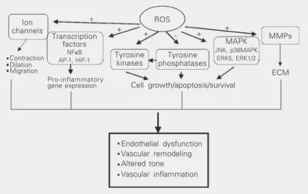
ROS appear to be important participants in Ang II signaling in vascular cells. This is based on the findings that 1) Ang II is ca-pable of generating ROS in vascular cells, 2) antioxidants and inhibitors of ROS-generat-ing systems abolish agonist-mediated sig-naling pathways, and 3) exogenous addition of oxidants activate the same signaling cas-cades as Ang II. Major targets of ROS in-clude transcription factors, protein tyrosine phosphatases (PTP), protein tyrosine kinases (PTK), mitogen-activated protein (MAP) ki-nases, ion channels, phospholipases, and transcription factors (40), all of which are regulated by Ang II (Figure 3).
Transcription factors
Transcription factors were the first sig-naling proteins identified as redox-sensitive. The DNA binding activity is regulated through specific cysteine motifs that need to
be reduced for activity. Nuclear factor κB
(NFκB), which is activated by Ang II in
vascular cells, is the prototype of redox-sensitive transcription factors. NFκB is
se-questered in the cytoplasm in a complex with its inhibitor IκB. ROS influence NFκB
activity by oxidative modification of
cys-teine residues, by IκB degradation and by
oxidative enhancement of upstream signal cascades (40). NFκB regulates transcription
of many genes involved in vascular inflam-mation and growth, including interleukins, adhesion molecules and proto-oncogenes (41). Other Ang II-activated redox-sensitive transcription factors include activator pro-tein-1 (AP-1) and hypoxia-inducible factor-1 (HIF-1). AP-1 is a transcription factor com-plex formed by homo- or heterodimerization of members of the c-Jun and c-Fos families of proteins and influences vascular cell dif-ferentiation and growth. ROS regulate AP-1 activity through numerous mechanisms and targets, including the reversible
S-glutathi-Contraction Dilation Migration
Ion channels
Transcription factors
NFκB AP-1, HIF-1
ROS
MMPs
ECM MAPK
JNK, p38MAPK ERK5, ERK1/2
Tyrosine kinases
Tyrosine phosphatases
Cell growth/apoptosis/survival
Endothelial dysfunction Vascular remodeling Altered tone Vascular inflammation
Figure 3. Redox-dependent signaling pathways by Ang II in vascular smooth muscle cells. Intracellular reactive oxygen species (ROS) modify the activity of tyrosine kinases, such as Src, Ras, JAK2, Pyk2, PI3K, and EGFR, as well as mitogen-activated protein kinases (MAPK), particularly p38MAPK, JNK and ERK5. ROS may inhibit protein tyrosine phos-phatase activity, further contributing to protein tyrosine kinase activation. ROS also influ-ence gene and protein expression by activating transcription factors, such as NFκB, activator protein-1 (AP-1) and hypoxia-inducible factor-1 (HIF-1). ROS stimulate ion chan-nels, such as plasma membrane Ca2+ and K+ channels, leading to changes in cation
concentration. Activation of these redox-sensitive pathways results in numerous cellular responses which, if uncontrolled, could contribute to hypertensive vascular damage. -, inhibitory effect; +, stimulatory effect; ECM, extracellular matrix; MMPs, matrix metallo-proteinases.
olation of a single conserved cysteine resi-due, the reversible redox regulation by thioredoxin and the nuclear protein Ref1 (42) and through regulation by the c-Jun N-terminal kinase (JNK) cascade. JNK phos-phorylates serine residues 63 and 73 of the
NH2-terminal transactivation of c-Jun,
re-quired for functional activation of AP-1 (43).
Protein tyrosine phosphatases and protein tyrosine kinases
Currently the best-established direct mo-lecular targets of ROS are PTPs. Protein-tyrosine phosphorylation is a major mechan-ism for post-translational modification of proteins and plays a critical role in regulat-ing cell proliferation, differentiation, migra-tion, and transformation. The level of ty-rosine phosphorylation in cells is controlled by the tightly regulated balance between PTK and PTPs (44). By dephosphorylating PTK substrate proteins, PTPs counteract effects of PTK activity. Hence PTPs may be consid-ered as negative regulators and terminators of a signaling process initiated by PTK acti-vation. Exposure of cells to low doses of oxidants or thiol-directed agents induces an increase in tyrosine phosphorylation due to
PTP inactivation.
Protein tyrosine phosphatases. PTPs are a large, structurally diverse family of recep-tor and non-receprecep-tor enzymes that are criti-cal regulators of multiple signaling path-ways (44). Because of their particular struc-ture, PTPs are susceptible to oxidation and inactivation by ROS. All PTPs possess a conserved 230-amino acid domain that con-tains a reactive and redox-regulated cysteine, which catalyzes the hydrolysis of protein phosphotyrosine residues by the formation of a cysteinyl-phosphate intermediate (45). This cysteine forms thiol phosphate, an in-termediate in the dephosphorylation reac-tion of PTPs. Oxidareac-tion of this cysteine resi-due to sulfenic acid by H2O2 renders the PTP completely inactive (45) (Figure 4). Since the oxidation of PTP is reversible, PTPs exist in two forms: an active state with a reduced cysteine or an inactive state with an oxidized cysteine. Activation and inactivation of PTPs are regulated by extracellular signals, in-cluding Ang II (46) and EGFand H2O2 plays a major role as a secondary messenger in this process (45) (Figure 4). Lee and colleagues (47) demonstrated that EGF-induced PTP1B inactivation is dependent on reversible oxi-dation of cysteine residues by H2O2. Recent studies suggest that PTP1B may be more efficiently regulated by •••••O
2- than by H2O2 (48). Peroxynitrite rapidly and irreversibly inhibits PTPs, supporting the role of this ROS in oxidative damage.
Besides soluble phosphatases, receptor PTP (RPTP) can also be modulated by oxidative stress (49). A model has been proposed in which oxidative stress induces a conforma-tional change in RPTPα-D2, leading to
stabili-zation of RPTPα dimers, and thus to inhibition
of RPTPα activity (49). In addition, the
inacti-vation of PTPs is involved in oxidative stress-induced activation of several PTK such as the EGFR, insulin receptor, Lck and Fyn (41). This is particularly important with respect to Ang II, which mediates many of its signaling events in vascular cells through EGFR
trans-PTP-cysteine-SOH
Oxidized Inactive
Reduction
PTP-cysteine-S-Reduced Active
PTK
MAPK MAPK
PTK H2O
H2O2
Oxidation
Cell growth, Apoptosis, Survival Figure 4. Protein tyrosine
phos-phatases (PTPs) are susceptible to oxidation and inactivation by reactive oxygen species (ROS). All PTPs possess a redox-regu-lated cysteine, which catalyzes the hydrolysis of protein phos-photyrosine residues by the for-mation of a cysteinyl-phosphate intermediate. Oxidation of this cysteine residue to sulfenic acid by H2O2 renders the PTP
activation (2). H2O2 has also been shown to regulate MAP kinases through inhibition of PTP activity of CD45, SHP-1 and HePTP (50). Thus, activation of vascular MAP kinases by Ang II may be mediated, in part, through redox-dependent inactivation of PTPs.
Protein tyrosine kinases. Receptor- and non-receptor tyrosine kinases are also
tar-gets of ROS (40,41,47). Exogenous H2O2
induces tyrosine phosphorylation and acti-vation of PDGFR and EGFR, probably due to ROS-mediated inhibition of dephospho-rylation of PDGFR and EGFR by inactiva-tion of membrane-associated PTPs. Oxygen intermediates, which are produced in re-sponse to tyrosine kinase receptor activa-tion, are also involved in transactivation of PDGFR and EGFR by Ang II. This mechan-ism involves c-Src and Ras (32). In patho-logical conditions associated with oxidative stress, ROS may directly activate cell sur-face receptors, thereby amplifying the pro-cess of •••••O
2-generation. Non-receptor
ty-rosine kinases such as Src, JAK2, STAT, p21Ras, Pyk2, and Akt, all of which are stimulated in response to Ang II and which have been implicated in cardiovascular re-modeling and vascular damage, are regu-lated by ROS.
MAP kinases.MAP kinases are a family of ubiquitous proline-directed, protein-serine/ threonine kinases, which participate in signal transduction classically associated with cell differentiation, cell growth and cell death (51). Of the major mammalian MAP kinases, ERK1/ 2, p38 MAP kinase and JNK are the best characterized. ERK1/2, phosphorylated by MEK1/2 (MAP/ERK kinase), is a key growth signaling kinase, whereas JNK and p38 MAP kinase, phosphorylated by MEK4/7 and MEK3/6, respectively, influence cell survival, apoptosis, differentiation, and inflammation. ERK5, regulated by MEK5, is involved in protein synthesis, cell cycle progression and cell growth. All MAP kinases are regulated, to varying degrees, by Ang II in vascular cells (2). Enhanced activation of vascular MAP
kinases has been demonstrated in hyperten-sion, atherosclerosis and diabetes and seems to be a major mechanism contributing to vas-cular damage associated with these conditions (51,52). MAP kinases are regulated by phos-phorylation cascades and are strongly acti-vated by ROS or by a mild oxidative shift of the intracellular thiol/disulfide redox state. Most studies have examined effects of exogenous H2O2 to activate MAP kinases (53). There are relatively few reports of endogenous ROS regulating the MAP kinase cascade. In VSMCs, intracellular ROS are critical for Ang II-in-duced activation of p38MAPK, JNK and ERK5, whereas phosphorylation of ERK1/2 appears to be redox-insensitive (54). How-ever, serotonin-mediated ERK1/2 activation in smooth muscle cells is redox-sensitive, but in fibroblasts, it is not (40). Thus, redox-regu-lation of MAP kinases may be ligand- and cell-specific. Although MAP kinases are regulated by oxygen free radicals, they are probably not direct substrates of •••••O
2-and H2O2.
Mechanisms whereby MAP kinases are activated by ROS are unclear, but MAP ki-nase phosphatases (MKP) are possible tar-gets. Similar to PTPs, MKPs share a con-served essential redox-sensitive cysteine that confers catalytic activity. Inhibition of MKPs by ROS, through oxidative modification, re-sults in activation of MAP kinases (41). In fact, decreased phosphatase activity has been linked to increased vascular ERK1/2 activa-tion in hypertension (55). Other processes by which ROS influence MAP kinases may be through upstream activators, such as Src tyrosine kinases, the small GTPase Ras and PKC (17,18).
Calcium transport systems. In addition to influencing signaling pathways associ-ated with cell growth and inflammation, ROS
modulate intracellular Ca2+ concentration
([Ca2+]
i), a major determinant of vascular
contraction. Superoxide and H2O2 increase [Ca2+]
Ca2+ mobilization, increased Ca2+ influx and
decreased activation of Ca2+-ATPase (57).
Plasma membrane K+ channels in VSMCs
that control a hyperpolarization-elicited re-laxation are opened by mechanisms associ-ated with thiol oxidation by ROS (40). Re-cent studies reported that contractile re-sponses to H2O2 are exaggerated in arteries from spontaneously hypertensive rats (SHR) compared with their normotensive counter-parts (57). Findings from our laboratory dem-onstrated that H2O2-induced [Ca2+]i transients are increased in VSMCs SHR (58). These data suggest that, in addition to impaired endothelium-dependent vasodilation (due to increased quenching of NO by •••••O
2-),
redox-sensitive Ca2+ changes could contribute to
altered vascular tone.
Processes whereby ROS influence signaling molecules
Two major processes have been identified whereby ROS influence signaling molecules: 1) oxidative modification of proteins and 2) changes in intracellular redox state (41).
Modification of proteins by oxidation. ROS can influence protein function and struc-ture by various mechanisms: by altering im-portant amino acid residues, by inducing protein dimerization and by interacting with metal complexes such as Fe-S moieties (41). Oxidative modification of amino acids within the functional domain of proteins occurs through many ways. The best characterized change involves cysteine residues. The sulf-hydryl group (-SH) of a single cysteine resi-due may be oxidized to form sulfenic (-SOH), sulfinic (-SO2H), sulfonic (-SO3H), or S-glutathionylated (-SSG) derivatives. These changes alter the activity of the enzyme if the cysteine is within the catalytic domain or the ability of a transcription factor to bind DNA is located within its DNA binding motif (40,41). PTPs are directly inactivated by ROS-induced reversible oxidation of the cata-lytic site Cys215. Other mechanisms by which
ROS can influence proteins are by intramo-lecular disulfide bridge formation, where two or more cysteine residues within the same protein are oxidized, by protein dimer-ization through inter-molecular disulfide link-ages, by diotyrosine formation and through metal-catalyzed oxidation by ROS.
Change in intracellular redox state. The intracellular compartment is generally main-tained in a reduced state by the redox buffer-ing capacity of intracellular thiols, particu-larly glutathione (GSH) and thioredoxin (TRX). These thiol redox systems counter-act intracellular oxidative stress by reducing H2O2 and lipid peroxides. GSH peroxidases, which are selenoproteins, are located in the cytosol and mitochondria and use GSH to reduce H2O2 to produce GSSG: H2O2 + 2GSH
-glutathione peroxidase →→ 2H2O + GSSG.
As antioxidants, glutathione-dependent enzymes are particularly important because the intracellular concentrations are relatively high with glutathione in the millimolar range and thioredoxin in the micromolar range.
In addition to their antioxidant potential, GSH and TRX participate directly in redox signaling (59). GSH regulates signaling by modulating the levels of total GSH and the ratio of oxidized to reduced (GSH) forms. GSH can translocate to the nucleus where it regulates DNA binding of transcription fac-tors (41). TRX is secreted by cells and was originally cloned as a cytokine-like factor. It is a low molecular weight (12 kDa) multi-functional protein with two redox-active cys-teines within a conserved active site. TRX regulates activity of proteins by directly bind-ing to them and by translocatbind-ing to the nucleus to regulate gene expression through Ref1. Binding and activation of Ref1 by TRX in-duces DNA binding of the Jun-Fos complex to the AP-1 site to mediate transcription
(41). NFκB and HIF-1 are also regulated by
and Ang II, there is evidence that the TRX system is modulated by the renin angio-tensin system, since ACE inhibition improves severity of myocarditis via redox regulation mechanisms involving TRX. In addition, in SHR, a model of Ang II-dependent hyper-tension, vascular TRX expression is impaired (60).
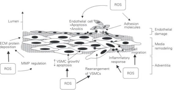
Reactive oxygen species as mediators of vascular damage
Under physiological conditions, vascu-lar production of ROS and the consequent activation of redox-dependent signaling path-ways and induction of redox-sensitive genes are tightly regulated. However, in pathologi-cal conditions, such as in hypertension, ath-erosclerosis, hyperlipidemia, hyperhomocys-teinemia, and diabetes, where generation of ROS is increased and the renin angiotensin system may be upregulated, these redox-sensitive events may contribute to cellular processes involved in vascular dysfunction and structural remodeling (3-5).
Increased bioavailability of vascular ROS leads to VSMC growth, migration, collagen deposition, and altered MMP activity,
im-portant factors in arterial remodeling in car-diovascular disease (3-5) (Figure 5). In en-dothelial cells, oxidative excess induces ap-optosis and aniokis (cell shedding), leading to endothelial cell loss and resultant im-paired endothelial function. In addition, oxi-dative stress stimulates activation of
trans-cription factors (e.g., NFκB and AP-1) and
pro-inflammatory genes (cytokines, interleu-kins), upregulation of adhesion molecules (e.g., ICAM, VCAM, PECAM), stimulation of chemokine production (e.g., MCP-1) and recruitment of inflammatory cells (mono-cytes, macrophages), critical processes in-volved in vascular inflammation and injury (5,52). Increased vascular •••••O
2- and H2O2 also impair endothelium-dependent relax-ation, increase contractile reactivity and al-ter vascular tone. These effects may be
me-diated directly by elevating cytosolic Ca2+
concentration or indirectly by reducing con-centrations of the vasodilator NO••••• (56).
Conclusions
Evidence is growing in support of ROS acting as signaling molecules in various cell types. The present review focuses on
redox-Inflammatory
Endothelial damage Lumen
response ECM protein
deposition
Endothelial cell Apoptosis Anoikis
ROS
Adhesion molecules
Media remodeling
Adventitia Cell
Rearrangement of VSMCs
migration
ROS
ROS ROS
MMP regulation VSMC growth/apoptosis
sensitive pathways whereby Ang II mediates vascular changes associated with cardiovas-cular diseases. Although the processes un-derlying Ang II-generated ROS in the vascu-lature are becoming clearer, there is still a paucity of knowledge of how reactive oxy-gen intermediates function as second mes-sengers in response to Ang II and how these redox-sensitive processes lead to vascular
remodeling, endothelial dysfunction and in-flammation. Future investigation in the field of redox signaling should elucidate how low levels of ROS act as signaling molecules in signal transduction cascades that regulate vascular function and maintain vascular in-tegrity and what factors tip the balance so that high level oxidants act as damaging stress signals to induce vascular injury.
References
1. Wolf G & Wenzel UO (2004). Angiotensin II and cell cycle regula-tion. Hypertension, 43: 693-698.
2. Touyz RM & Schiffrin EL (2000). Signal transduction mechanisms mediating the physiological and pathophysiological actions of angio-tensin II in vascular smooth muscle cells. Pharmacological Re-views, 52: 639-672.
3. Touyz RM (2000). Oxidative stress and vascular damage in hyper-tension. Current Hypertension Reports, 2: 98-105.
4. Wilcox CS (2002). Reactive oxygen species: roles in blood pressure and kidney function. Current Hypertension Reports, 4: 160-166. 5. Griendling KK, Sorescu D & Ushio-Fukai M (2000). NADPH oxidase.
Role in cardiovascular biology and disease. Circulation Research, 86: 494-501.
6. Touyz RM, Chen X, He G, Quinn MT & Schiffrin EL (2002). Expres-sion of a gp91phox-containing leukocyte-type NADPH oxidase in human vascular smooth muscle cells - modulation by Ang II. Circula-tion Research, 90: 1205-1213.
7. Ushio-Fukai M, Tang Y, Fukai T, Dikalov SI, Ma Y, Fujimoto M, Quinn MT, Pagano PJ, Johnson C & Alexander RW (2002). Novel role of gp91(phox)-containing NAD(P)H oxidase in vascular endothelial growth factor-induced signaling and angiogenesis. Circulation Re-search, 91: 1160-1167.
8. Rey FE & Pagano PJ (2002). The reactive adventitia: fibroblast oxidase in vascular function. Arteriosclerosis, Thrombosis, and Vas-cular Biology, 22: 1962-1971.
9. Babior BM, Lambeth JD & Nauseef W (2002). The neutrophil NADPH oxidase. Archives of Biochemistry and Biophysics, 397: 342-344.
10. Lassegue B & Clempus RE (2003). Vascular NAD(P)H oxidases: specific features, expression and regulation. American Journal of Physiology, 285: R277-R297.
11. Fridovich I (1997). Superoxide anion radical, superoxide dismutases, and related matters. Journal of Biological Chemistry, 272: 18515-18517.
12. Han D, Antunes F, Canali R, Rettori D & Cadenas E (2003). Voltage-dependent anion channels control the release of the superoxide anion from mitochondria to cytosol. Journal of Biological Chemistry, 278: 5557-5563.
13. Schafer FQ & Buettner GR (2001). Redox environment of the cell as viewed through the redox state of the glutathione disulfide/glutathi-one couple. Free Radical Biology and Medicine, 30: 1191-1212. 14. Darley-Usmar V, Wiseman H & Halliwell B (1995). Nitric oxide and
oxygen radicals, a question of balance. FEBS Letters, 369: 13-15. 15. Channon KM & Guzik TJ (2002). Mechanisms of superoxide
produc-tion in human blood vessels: relaproduc-tionship to endothelial dysfuncproduc-tion, clinical and genetic risk factors. Journal of Physiology and Pharma-cology, 53: 515-524.
16. Halliwell B (1999). Antioxidant defence mechanisms: from the be-ginning to the end (of the bebe-ginning). Free Radical Research, 31: 261-272.
17. Forman HJ, Torres M & Fukuto J (2002). Redox signaling. Molecular and Cellular Biochemistry, 234-235: 49-62.
18. Forman HJ & Torres M (2002). Reactive oxygen species and cell signaling: respiratory burst in macrophage signaling. American Jour-nal of Respiratory and Critical Care Medicine, 166 (Part 2): S4-S8. 19. Zalba G, San Jose G, Moreno MU, Fortuno MA, Fortuno A,
Beau-mont FJ & Diez J (2001). Oxidative stress in arterial hypertension: role of NAD(P)H oxidase. Hypertension, 38: 1395-1399.
20. Landmesser U & Harrison DG (2001). Oxidative stress and vascular damage in hypertension. Coronary Artery Disease, 12: 455-461. 21. Saito Y & Berk BC (2001). Transactivation: a novel signaling
path-way from angiotensin II to tyrosine kinase receptors. Journal of Molecular and Cellular Cardiology, 33: 3-7.
22. Suh Y, Arnold RS, Lassegue B, Shi J, Xu X, Sorescu D, Chung AB, Griendling KK & Lambeth JD (1999). Cell transformation by the superoxide-generating oxidase mox 1. Nature, 401: 79-82. 23. Cheng G, Cao Z, Xu X, van Meir EG & Lambeth JD (2001). Homologs
of gp91phox: cloning and tissue expression of Nox3, Nox4, and Nox5. Gene, 269: 131-140.
24. Sorescu D, Weiss D, Lassegue B et al. (2002). Superoxide produc-tion and expression of nox family proteins in human atherosclero-sis. Circulation, 105: 1429-1435.
25. Bengtsson SH, Gulluyan LM, Dusting GJ & Drummond GR (2003). Novel isoforms of NADPH oxidase in vascular physiology and patho-physiology. Clinical and Experimental Pharmacology and Physiolo-gy, 30: 849-854.
26. Banfi B, Clark RA, Steger K & Krause K-H (2003). Two novel proteins activate superoxide generation by the NADPH oxidase Nox1. Jour-nal of Biological Chemistry, 278: 3510-3513.
27. Ago T, Kitazono T, Ooboshi H, Iyama T, Han YH, Takada J, Wakisaka M, Ibayashi S, Utsumi H & Iida M (2004). Nox4 as the major catalytic component of an endothelial NAD(P)H oxidase. Circulation, 109: 227-233.
HH (2001). Upregulation of the vascular NAD(P)H-oxidase isoforms Nox1 and Nox4 by the renin-angiotensin system in vitro and in vivo.
Free Radical Biology and Medicine, 31: 1456-1464.
29. Berry C, Hamilton CA, Brosnan MJ, Magill FG, Berg G, McMurray JJV & Dominiczak AF (2000). An investigation into the sources of superoxide production in human blood vessels: Ang II increases superoxide production in human internal mammary arteries. Circu-lation, 101: 2206-2212.
30. Touyz RM & Schiffrin EL (2001). Increased generation of superoxide by angiotensin II is mediated via PLD-dependent, NADPH oxidase-sensitive pathways in vascular smooth muscle cells from hyperten-sive patients. Journal of Hypertension, 19: 1245-1254.
31. Touyz RM, Yao G & Schiffrin EL (2003). c-Src induces phosphoryla-tion and translocaphosphoryla-tion of p47phox: Role in superoxide generaphosphoryla-tion by Ang II in human vascular smooth muscle cells. Arteriosclerosis, Thrombosis, and Vascular Biology, 23: 981-987.
32. Seshiah PN, Weber DS, Rocic P, Valppu L, Taniyama Y & Griendling KK (2002). Angiotensin II stimulation of NAD(P)H oxidase activity. Upstream mediators. Circulation Research, 91: 406-413.
33. Cosentino F, Barker JE, Brand MP, Heales SJ, Werner ER, Tippins JR, West N, Channon KM, Volpe M & Luscher TF (2001). Reactive oxygen species mediate endothelium-dependent relaxations in tet-rahydrobiopterin-deficient mice. Arteriosclerosis, Thrombosis, and Vascular Biology, 21: 496-502.
34. Mollnau H, Wendt M, Szocs K et al. (2002). Effects of angiotensin II infusion on the expression and function of NAD(P)H oxidase and components of nitric oxide/cGMP signaling. Circulation Research, 90: E58-E65.
35. Vasquez-Vivar J, Duquaine D, Whitsett J, Kalyanaraman B & Rajagopalan S (2002). Altered tetrahydrobiopterin metabolism in atherosclerosis: implications for use of oxidized tetrahydrobiopterin analogues and thiol antioxidants. Arteriosclerosis, Thrombosis, and Vascular Biology, 22: 1655-1661.
36. Bagi Z & Koller A (2003). Lack of nitric oxide mediation of flow-dependent arteriolar dilation in type I diabetes is restored by sepiapterin. Journal of Vascular Research, 40: 47-57.
37. Virdis A, Iglarz M, Neves MF, Amiri F, Touyz RM, Rozen R & Schiffrin EL (2003). Effect of hyperhomocystinemia and hyperten-sion on endothelial function in methylenetetrahydrofolate reduc-tase-deficient mice. Arteriosclerosis, Thrombosis, and Vascular Bi-ology, 23: 1352-1357.
38. Landmesser U, Dikalov S, Price SR, McCann L, Fukai T, Holland SM, Mitch WE & Harrison DG (2003). Oxidation of tetrahydrobiopterin leads to uncoupling of endothelial cell nitric oxide synthase in hyper-tension. Journal of Clinical Investigation, 111: 1201-1209. 39. Mukherjee SP, Lane RH & Lynn WS (1978). Endogenous hydrogen
peroxide and peroxidative metabolism in adipocytes in response to insulin and sulfhydryl reagents. Biochemical Pharmacology, 27: 2589-2594.
40. Droge W (2001). Free radicals in the physiological control of cell function. Physiological Reviews, 82: 47-95.
41. Thannickal VJ & Fanburg BL (2000). . . Reactive oxygen species in cell signaling. American Journal of Physiology, 279: L1005-L1028. 42. Xanthoudakis S & Curran T (1992). Identification and
characteriza-tion of Ref-1, a nuclear protein that facilitates AP-1 DNA-binding activity. EMBO Journal, 11: 653-665.
43. Karin M, Liu Z & Zandi E (1997). AP-1 function and regulation.
Current Opinion in Cell Biology, 9: 240-246.
44. Anderson JN, Mortensen OH, Peters GH, Drake PG, Iversen LF, Olsen OH, Jansen PG, Andersen HS, Tonks NK & Moller NP (2001). Structural and evolutionary relationships among protein tyrosine
phosphatase domains. Molecular and Cellular Biology, 21: 7117-7136.
45. Denu JM & Tanner KG (1998). Specific and reversible inactivation of protein tyrosine phosphatases by hydrogen peroxide: evidence for a sulfenic acid intermediate and implications for redox regulation.
Biochemistry, 7: 5633-5642.
46. Guillemot L, Levy A, Zhao ZJ, Bereziat G & Rothhut B (2000). The protein-tyrosine phosphatase SHP-2 is required during angiotensin II-mediated activation of cyclin D1 promoter in CHO-AT1A cells.
Journal of Biological Chemistry, 275: 26349-26358.
47. Lee SR, Kwon KS, Kim SR & Rhee SG (1998). Reversible inactivation of protein-tyrosine phosphatase 1B in A431 cells stimulated with epidermal growth factor. Journal of Biological Chemistry, 273: 15366-15372.
48. Barrett WC, DeGnore JP, Konig S, Fales HM, Keng YF, Zhang ZY, Yim MB & Chock PB (1999). Regulation of PTPB1 via glutathionyla-tion of the active site cysteine 215. Biochemistry, 38: 6699-6705. 49. Blanchetot C, Tertoolen LGJ & Hertog JD (2002). Regulation of
receptor protein tyrosine phosphatase α by oxidative stress. EMBO
Journal, 21: 493-503.
50. Lee K & Esselman WJ (2002). Inhibition of PTPS by H2O2 regulates
the activation of distinct MAPK pathways. Free Radical Biology and Medicine, 33: 1121-1132.
51. Torres M & Forman HJ (2003). Redox signaling and the MAP kinase pathways. Biofactors, 17: 287-296.
52. Touyz RM, Deschepper C, Park JB, He G, Chen X, Neves MF, Virdis A & Schiffrin EL (2002). Inhibition of mitogen-activated protein/ extracellular signal-regulated kinase improves endothelial function and attenuates Ang II-induced contractility of mesenteric resistance arteries from spontaneously hypertensive rats. Journal of Hyperten-sion, 20: 1127-1134.
53. Baas AS & Berk BC (1995). Differential activation of mitogen-acti-vated protein kinases by H2O2 and O2- in vascular smooth muscle
cells. Circulation Research, 77: 29-36.
54. Touyz RM, Cruzado M, Tabet F, Yao G, Salomon S & Schiffrin EL (2003). Redox-dependent MAP kinase signaling by Ang II in vascular smooth muscle cells - role of receptor tyrosine kinase transactiva-tion. Canadian Journal of Physiology and Pharmacology, 81: 159-167. 55. Begum N, Ragolia L, Rienzie J, McCarthy M & Duddy N (1998). Regulation of mitogen-activated protein kinase phosphatase-1 in-duction by insulin in vascular smooth muscle cells. Evaluation of the role of the nitric oxide signaling pathway and potential defects in hypertension. Journal of Biological Chemistry, 273: 25164-25170. 56. Lounsbury KM, Hu Q & Ziegelstein RC (2000). Calcium signaling
and oxidant stress in the vasculature. Free Radical Biology and Medicine, 28: 1362-1369.
57. Gao YJ & Lee RM (2001). Hydrogen peroxide induces a greater contraction in mesenteric arteries of spontaneously hypertensive rats through thromboxane A(2) production. British Journal of Phar-macology, 134: 1639-1646.
58. Tabet F, Schiffrin EL & Touyz RM (2004). Differential calcium regula-tion by hydrogen peroxide and superoxide in vascular smooth muscle cells from SHR. Journal of Cardiovascular Pharmacology (in press).
59. Nakamura H (2004). Thioredoxin as a key molecule in redox signal-ing. Antioxidants and Redox Signalling, 6: 15-17.