Bio physical characte ristics o f gap
junctio ns in vascular wall ce lls:
implicatio ns fo r vascular bio lo gy
and dise ase
Departments of 1Physiology and Biophysics, Institute for Molecular Cardiology, and 2Surgery, SUNY at Stony Brook, Stony Brook, NY, USA
Departments of 3Urology, and 4Physiology and Biophysics, Institute for Smooth Muscle
Biology, Albert Einstein College of Medicine, Bronx, NY, USA P.R. Brink1,
J. Ricotta2 and
G.J. Christ3,4
Abstract
The role gap junction channels play in the normal and abnormal functioning of the vascular wall is the subject of much research. The biophysical properties of gap junctions are an essential component in understanding how gap junctions function to allow coordinated relax-ation and contraction of vascular smooth muscle. This study reviews the properties thus far elucidated and relates those properties to tissue function. We ask how biophysical and structural properties such as gating, permselectivity, subconductive states and channel type (het-eromeric vs homotypic vs heterotypic) might affect vascular smooth muscle tone.
Co rre spo nde nce
P.R. Brink
Department of Physiology and Biophysics
SUNY at Stony Brook Stony Brook, NY 11794 USA
Presented at the Meeting “Gap Junctions in the Nervous and Cardiovascular Systems: Clinical Implications”, Rio de Janeiro, RJ, Brazil, June 6-11, 1998. Research supported by NIH grant GM55263.
Received August 13, 1999 Accepted November 23, 1999
Ke y words
·Vascular smooth muscle
·Gap junctions
Intro ductio n
In vascular smooth muscle the role that gap junctions play is integral among a num-ber of other processes and parameters influ-encing tissue function (1-4). The ultimate tissue function, however, is coordinated re-laxation and contraction of the many cells within the wall of any particular vessel. Ulti-mately, the major factors which influence such coordinated tissue behavior are neu-ronal innervation density, cell to cell com-munication and cellular excitability (1,5-7). As vascular smooth muscle cells do not tend to generate action potentials, innervation density and gap junction-mediated commu-nication become paramount as potential regu-latory sites. The former parameter is the
Re sults and D iscussion
Ge ne ric gap junction channe l type s in
vascular tissue s
The subunit protein of any gap junction channel is the connexin and in vascular smooth muscle connexin43 (Cx43) is the most abundant, followed by connexin40 (Cx40), connexin45 (Cx45) and possibly connexin37 (Cx37) (1,10-12). Endothelial cells are also able to co-express Cx43, Cx40 and Cx37, the latter being the most abundant
in situ (see 1 for review).
The putative presence of multiple con-nexin subtypes in a given vascular smooth muscle cell has potentially important physi-ological implications. For example, there are three types of gap junction channels possible when more than one connexin is being ex-pressed in the same cell. They are as follows: 1) a homotypic gap junction channel com-posed of 12 identical connexin subunits, 2) a heterotypic gap junction channel where all the connexins of one cell are identical but different from the connexins of the adjacent cell, and 3) the heteromeric gap junction channel where the connexins in any one cell are not identical within a hemichannel. In the case of vascular smooth muscle cells the dominant connexin is Cx43. Therefore, we will focus our discussion on properties of homotypic gap junction channels composed of Cx43, Cx40, Cx45 and Cx37. Further we will compare homotypic Cx43 and Cx37 to the in vitro documentation of Cx43-Cx37
heterotypics and heteromerics. The relevant properties to be described are gating and permselectivity.
Homotypic gap junction me mbrane voltage
de pe nde nce
In general, all mammalian homotypic gap junction channels display symmetric voltage dependence. Because of its ubiquity we will first characterize Cx43 homotypic gap
junc-tion channels and subsequently compare their behavior to Cx37, Cx40 and Cx45 whenever possible. In this regard, the instantaneous junctional current for Cx43 is linear over a large voltage range. Transjunctional voltage steps of sufficient amplitude and duration cause junctional currents to decline with time, resulting in a steady-state conductance which is a fraction of the instantaneous con-ductance. The time course of the voltage-dependent behavior is of the order of hun-dreds of milliseconds to seconds (13). Cx43 shows relatively symmetric voltage depend-ence but has different voltage sensitivity relative to Cx37. That is, the point on the Vj
axis where junctional conductance is 50% of the maximum (i.e., Vo) is approximately
25-30 mV for Cx37 and 70-85 mV for Cx43 (12,14,15). Cx40 has a Vo in the 40 mV
range, while the Vo for Cx45 has been
reported to be 20 mV (16). Such observa-tions emphasize that, with the possible ex-ception of Cx45, the voltage sensitivity of connexins in vascular wall cells is not likely to effect dynamic changes in intercellular coupling.
process is modified or driven in the presence of physiologically important ligands/intra-cellular messengers, such as cAMP or IP3, it
could well be a regulatory point with great dynamic range. This latter postulate has yet to be experimentally tested, let alone shown to be functional under physiological condi-tions. Mode shifting has not been character-ized for Cx37 but has been demonstrated in A7r5 cells which co-express Cx40 and Cx43 (He DS, personal communication). Thus, voltage-dependent gating, a common prop-erty of all the vertebrate connexins thus far studied, seems to be an unlikely tool for cells to utilize in order to affect dynamic alter-ations in cell to cell coupling mediated by gap junctions. Whether ligand gating, linked with processes such as mode shifting, might be able to provide dynamic behaviors that are physiologically relevant to the vascular wall cell has yet to be determined.
Homotypic gap junction channe l
co nductance
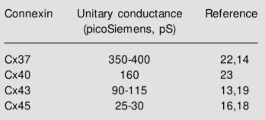
The unitary conductance for Cx43 is 90-120 pS (13,17- 20). Table 1 lists the unitary conductances for all four connexin types found in the vascular system. The different unitary conductance values imply that dif-ferent permselectivity properties may exist for distinct connexins. What becomes im-portant then, is determining if differential conductance values translate into vessel-spe-cific behaviors of the vascular wall, such as, for example, propagated vasodilation or con-striction (21).
Subconductive state s: are the y
physiologically re le vant?
All gap junction channels thus far stud-ied have displayed subconductive states. One common feature is the predominance of sub-conductive states with large transjunctional voltages, the classic example of this being Cx37 (22) and Cx43 (24). The observation
that the permselective properties of the sub-conductive states are very different from the main state conductance has given rise to much speculation. For example, if the sub-conductive state is the dominant one in terms of dwell time, then it would necessarily fol-low that the permeation rates of solutes seek-ing transit from cell to cell would be quite dependent on the nature of the subconducting state(s). However, before even addressing the permselective properties, the first ques-tion answered must be how much of the open time is dominated by subconductive states. If the dwell time in any given subconductance state is significant (10-20% or greater?), then the permeation characteristics of the distinct subconducting states become of interest. For Cx43, Christ and Brink (24) have deter-mined that the occupancy time of the sub-conductive state(s), for a transjunctional volt-age of 40 mV, is less than 2% for a channel whose open probability is on the order of 70-90%. In this case, even if the permselectivity of the subconductive state is dramatically different from the main state it would seem hardly relevant to macroscopic tissue behav-ior with such a low occupancy time. Con-versely, if gap junction channels are driven into subconductive states by interaction with ligands, or alterations in second messenger levels, such that for the majority of the open time the conductive and permeation rates are dictated by the subconductive state, then this property of gap junction channels would have very telling affects on tissue function.
Table 1 - Unitary conductance of the four connexin types found in the vascular system.
Connexin Unitary conductance Reference
(picoSiemens, pS)
Cx37 350-400 22,14
Cx40 160 23
Cx43 90-115 13,19
Channe l pe rmse le ctivity: its role in tissue
physio lo gy
The homotypic Cx43 gap junction chan-nel has been studied the most with respect to permselectivity. A recent study by Wang and Veenstra (20) delineated the permeability of Cx43 to monovalent cations. The analysis revealed that homotypic Cx43 is non-selec-tive, and based on comparison of unitary conductance, follows an Eisenmann series I or II sequence (25), i.e., Rb = Cs>K>Na> Li>TMA>TEA. The anion series also shows a proportionality between mobility and uni-tary conductance. Hence Cx43 appears to be very poorly selective and falls into the cat-egory of Porin OmpF (26-28) which itself is permeable to both cations and anions. How-ever, it should be noted that the porins have no sequence homology with the connexins. Cx40 follows the same cation sequence, but has a very different anion sequence as that described for Cx43 and Cx37. In fact, uni-tary conductance was slightly decreased with increased ionic mobility, indicating that Cx40 is much less anion permeable and/or that the cation-anion environment surrounding or within the channel affects the selectivity filter(s). If this latter case is in fact true, then more detailed analysis will clearly be neces-sary.
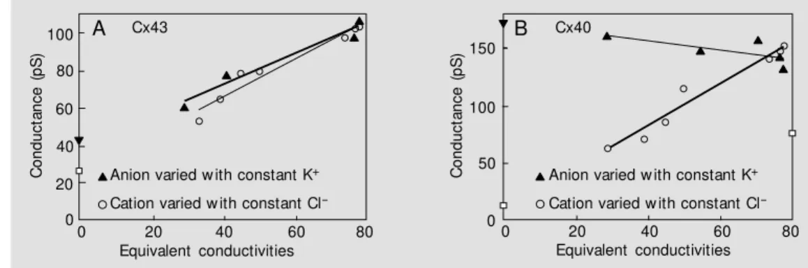
Figure 1A shows two data sets taken from Wang and Veenstra (20) for Cx43 where unitary conductance is plotted against the cation mobility of the major cation in the pipette solution when Cl is the major anion (circles). Unitary conductance was also plot-ted against anion mobility (triangles) where the major cation is K. The solid lines repre-sent linear regression fits of the data. The thicker of the two lines represents the anion data where the Y-intercept denotes the re-sidual cation conductance of 41 pS. The thinner line represents the cation data where the Y-intercept is the residual anion conduc-tance of 27 pS. Figure 1B represents the same analysis for Cx40. The circles repre-sent cation data for Cx40 and the triangles anion data. The homotypic Cx43 seems to be a highly non-selective channel where ionic mobility is the rate limiting factor for perme-ation. For homotypic Cx40 the data imply an interaction between highly mobile anion spe-cies and cations which hampers cation tran-sit through the channel. In fact, as the anion mobility declines the unitary conductance increases, indicating that some interaction between cation and anion in Cx40 channels could be occurring (23).
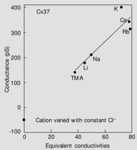
Data for Cx37 are shown in Figure 2 which reveals a potential hazard to the con-ventional interpretation for such data.
Spe-Figure 1 - A, Selectivity sequence of Cx43 for cations and anions taken from Wang and Veenstra (20). Unitary conductance is plot-ted against equivalent conductiv-ity (cm2/Ohm). The triangles
rep-resent unit ary conduct ances measured w ith different anions, w ith K+ as the cation. The circles
are unitary conductance meas-ured w ith different cations, w ith Cl- as the anion. The lines
repre-sent the linear regression fits to the data points. The thick solid line is the fit for the anions w hile
the thinner line is for the cations. The y-intercept for the anions represents the conductance due to K+ (41 pS) w hile the y-intercept for the cation data
represents the Cl- conductance (27 pS). B, Cx40 taken from Beblo and Veenstra (23). The analysis is the same as that for Cx43. With constant K+ as the
anion species, increases in size and decreases in mobility resulting in increased channel conductance imply a cation/anion interaction for the more mobile anions w hich reduces conductance. The y-intercepts again represent the cation conductance (171 pS) and anion conductance (8 pS), respectively.
C
o
n
d
u
c
ta
n
c
e
(
p
S
)
100
C
o
n
d
u
c
ta
n
c
e
(
p
S
) 150 80
60
40
20
0
0 20 40 60 80
Cx43
Anion varied w ith constant K+
Cation varied w ith constant Cl
-Equivalent conductivities
100
50
0
0 20 40 60 80
Equivalent conductivities Cx40
Anion varied w ith constant K+
Cation varied w ith constant Cl
cifically, note that the y-intercept is -49 pS, the theoretical Cl conductance. Such behav-ior predicts that the unitary conductance vs
cation/anion concentration will be non-lin-ear in the mM range. This has been shown to be the case for Cx37 (29) and is a ready explanation for the aberrant Y-intercept.
For some connexins the permselectivity properties might be dramatically altered by the ionic environment. Gap junction mem-brane conductance is known to be modu-lated by a number of molecules which also are able to diffuse through the gap junction channel (30). For example, hydrogen and calcium ions are two species which cause the channel to gate closed in sufficiently high concentration, but in turn both are able to diffuse through the channel at concentra-tion ranges normally found in cells (31,32). It could be that cation and anion species also affect gap junction channel gating as well as the permeation path. There is now evidence for the supposition that the concentration of monovalents affects the gating of Cx43 through a screening mechanism (29). But for Cx37 homotypic gap junction channels the monovalent cation species has no detectable effect. Rather, Mg2+ is effective in altering
the kinetics of the voltage dependence (33). However, with respect to Cx37, there are still no data available on the screening ef-fects and/or the binding of specific sites on the permeation path.
Another technical approach which has been utilized to document the poor selectiv-ity characteristics of gap junctions is the transfer of fluorescent probes. Few attempts have been made to quantify transfer rates, but dye transfer has been demonstrated for the majority of the mammalian connexins. In one study the transfer of carboxyfluorescein and dichlorofluorescein was shown for Cx45, Cx43, Cx40 and Cx37 (18). Millimolar a-mounts of dye were introduced into one cell of a pair via a whole cell patch. After 10 min, fluorescence was monitored. Cell pairs were scored either as having transferred probe to
the adjacent cell or not. The junctional con-ductance was also determined. For all the connexin types studied, the minimal con-ductance for observing dye transfer was in the 2-5 nS range. These data indicate that many, if not all, connexins are capable of passing both cations and anions of some size and charge. At issue is not the poor selectiv-ity but rather the permeation rates necessary to allow specific messenger molecules to activate/inhibit whole groups of cells. This latter point is very important to answer if we are going to be able to understand the role of gap junctions in non-excitable tissue rather than continue to have speculation as the sole basis of further investigation.
Are he te rotypic or he te rome ric channe l
forms important to function?
An interesting issue which has yet to be addressed with regard to permselectivity is whether or not heteromeric and/or hetero-typic biophysical properties are sufficiently different from homotypic channels. Suffi-ciently different, that is, to affect the cell to cell diffusion of critical substances able to trigger/alter cellular responses. In the vascu-lar wall this is of particuvascu-lar interest because of the connexin co-expression that occurs in endothelial and vascular smooth muscle cells.
C
o
n
d
u
c
ta
n
c
e
(
p
S
)
400
300
200
100
0
-100
Cx37
Cation varied w ith constant Cl
-K
Cs
Rb
Na Li
TM A
0 20 40 60 80
Equivalent conductivities
Figure 2 - M easured unitary con-ductance for Cx37 w ith various cations, w ith Cl- again
remain-ing constant. The data indicate that the Cl- conductance is a
A question which has yet to be answered is the gap junction types which are present in co-expressing cells in situ. Heterotypic and heteromeric channel types have been dem-onstrated in vitro, implying that the same types of channels could form in situ (14,34).
In this regard, it becomes obvious that permselectivity measurements of the hetero-typic, heteromeric and, to a lesser extent, homotypic channel types in situ are not
go-ing to be readily available in the near future. Dye transfer between cells co-expressing connexins has been documented in situ (35)
but quantitative analysis has not been forth-coming to allow comparison with cells ex-pressing a single connexin. An interesting observation made by Little et al. (35) was the lack of dye transfer from smooth muscle cells of the tunica media to endothelial cells in the tunica intima. The reverse transfer (from endothelial to smooth muscle cells) was demonstrable. This type of result might well be best explained as one of the two cell types representing an infinite sink, the clas-sic example of this phenomenon being that illustrated by Rae et al. (36).
In vitro studies of Cx43 and Cx37 have
shown that Cx43 and Cx37 form heterotypic and heteromeric channels along with homo-typic forms (14). The most notable param-eter which was different in cells co-express-ing Cx34 and Cx37 was a broadenco-express-ing of the Vo. The second feature was the occurrence
of channel conductances that could not be explained as heterotypic or homotypic. The different conductive states imply, as indi-cated before, different permselectivity prop-erties. This observation points to the issue already raised, i.e., that even if the permse-lectivity is different, is it different enough to affect tissue physiology? There is a second perplexing problem. In a given vascular tis-sue co-expressing distinct connexins, what population of channel types are there? What fraction of the total number of channels is homotypic, heterotypic or heteromeric? With-out this information it is impossible to
quan-tify the role that the three generic gap junc-tion types might play in normal and abnor-mal tissue physiology.
Conclusions and clinical re le vance
The major role played by intercellular communication through gap junctions in modulating vasomotor tone throughout the vascular tree has been discussed in detail elsewhere (37,38). The implication for this report is that it follows naturally that since gap junctions play such a critical role in maintaining circulatory homeostasis, they are logical molecular targets for vascular disease, and as such, for vascular therapy. However, it should be emphasized that there are currently no published reports that have either confirmed or denied a direct proximal role for gap junctions in vascular disease.
regula-tion of vascular tone is just beginning to be studied. There are many possibilities, and it is clear the real work has just begun.
On the other hand, theoretical studies suggest that the degree of coupling among vascular smooth muscle cells may already be such that 10-fold or greater increases in cou-pling would be required for physiologically relevant changes in vascular contractility (8,9). This is not to say that altered connexin expression would play no role in vascular pathology, but that intercellular communi-cation is so critical to vascular function, that there is significant plasticity or reserve to prevent it from becoming the focal point for tissue pathology. Of course all of the latter considerations are based on the known bio-physical characteristics of Cx43 in human vascular smooth muscle, i.e., long open times, high open probabilities, physiologically ir-relevant subconductance states, and lack of selectivity. In this regard, however, if there are indeed physiological or
pathophysiologi-cal alterations in second messenger mol-ecules/ions that result in dramatic changes in the documented biophysical behaviors of connexins, then the role of gap junctions could be drastically altered. Dynamic ligand gating of substate dwell times or permselec-tivity properties, for example, or the pres-ence of significant heteromeric or hetero-typic channel types, could have profound implications for normal vascular physiology and disease. Certainly the potential clinical correlates of altered intercellular communi-cation can be visualized, but have not yet been documented. On this basis, this would seem to be a very fruitful area of future research.
Ackno wle dgm e nts
The authors would like to acknowledge Dr. S.V. Ramanan, SUNY at Stony Brook, NY, USA, who kindly donated the data for Figure 2.
Re fe re nce s
1. Christ GJ, Spray DC, ElSabban M , M oore LK & Brink PR (1996). Gap junctions in vascular tissues: Evaluating the role of intercellular communication in the modu-lation of vasomotor tone. Circulation Re-search, 79: 631-646.
2. Segal SS & Kurjiaka DT (1995). Coordina-tion of blood flow control in the resis-tance vasculature of skeletal muscle. M edicine and Science in Sports and Exer-cise, 27: 1158-1164.
3. Beach JM , M cGahren ED & Duling BR (1998). Capillaries and arterioles are elec-trically coupled in hamster cheek pouch. American Journal of Physiology, 275 (Part 2): H1489-H1496.
4. Pacicca C, Schaad O & Beny J (1996). Electrotonic propagation of kinin-induced, endothelium -dependent hyperpolariza-tions in pig coronary smooth muscles. Journal of Vascular Research, 33: 380-385.
5. Bevan JA & Torok J (1970). M ovement of norepinephrine through the media of rab-bit aorta. Circulation Research, 27: 325-331.
6. Hirst GDS & Edw ards FR (1989). Sympa-thetic neuroeffector transmission in ar-teries and arterioles. Physiological Re-view s, 69: 546-604.
7. Burnstock G (1970). Structure of smooth muscle and its innervation. In: Bulbring E, Brading AF, Jones AW & Tomita T (Edi-tors), Smooth M uscle. Williams & Wilkins, Baltimore, M D, 1-69.
8. Christ GJ, Brink PR & Ramanan SV (1994). Dynamic gap junctional communication: a delimiting model for tissue responses. Biophysical Journal, 67: 1335-1344. 9. Ramanan SV, Brink PR & Christ GJ (1998).
Neuronal innervation, intracellular signal transduction and intercellular coupling: a model for syncytial tissue responses in the steady state. Journal of Theoretical Biology, 193: 69-84.
10. Beyer EC (1993). Gap junctions. Interna-tional Review of Cytology, 137: 1-37. 11. Brink PR (1998). Gap junctions in smooth
muscle. Acta Physiologica Scandinavica, 164: 349-356.
12. Brink PR, Valunias V & Christ GJ (2000). Homotypic, heterotypic and heteromeric
gap junctions. In: Perrachia C (Editor), Gap Junctions. Academic Press, New York. 13. Brink PR, Ramanan SV & Christ GJ (1996).
Human connexin43 gap junction channel gating evidence for mode shifts and/or heterogeneity. American Journal of Phys-iology, 271: C321-C331.
14. Brink PR, Cronin K, Banach K, Peterson E, Westphale EM , Seul KH, Ramanan SV & Beyer EC (1997). Evidence for hetero-meric gap junction channels formed from rat connexin43 and human connexin37. American Journal of Physiology, 273: C1386-C1396.
15. Valiunas V, Bukaukas F & Weingart R (1997). Conductances and selective per-m eability of connexin43 gap junction channels examined in neonatal rat heart cells. Cirulation Research, 80: 708-719. 16. M oreno AP, Liang JG, Beyer EC & Spray
DC (1995). Properties of gap junction channels formed of connexin 45 endog-enously expressed in human hepatoma (SKHep1) cells. American Journal of Phys-iology, 268: C356-C365.
Christ GJ & Spray DC (1993). Gap junc-tional communication betw een human corpus cavernosum smooth muscle cells in culture: Gating behavior and single channel events. Am erican Journal of Physiology, 264: C80-C92.
18. Campos de Carvalho AC, M oreno AP, Christ GJ, M elman A, Roy C, Hertzberg EL & Spray DC (1993). Gap junctions f orm ed of connexin43 int erconnect smooth muscle cells of the human corpus cavernosum. Journal of Urology, 149: 1568-1575.
19. Veenstra RD, Wang HZ, Beblo DA, Chilton M G, Harris AL, Beyer EC & Brink PR (1995). Selectivity of connexin-specific gap junctions does not correlate w ith channel conductance. Circulation Re-search, 77: 1156-1165.
20. Wang H-Z & Veenstra RD (1997). M onova-lent ion selectivity sequences of the rat connexin 43 gap junction channel. Journal of General Physiology, 109: 491-507. 21. Segal SS, Welsh DG & Kurjiaka DT (1999).
Spread of vasodilation and vasoconstric-tion along feed arteries and arterioles of hamster skeletal muscle. Journal of Phys-iology, 516: 283-291.
22. Veenst ra RD, W ang HZ, Beyer EC, Ramanan SV & Brink PR (1994). Con-nexin37 forms high conductance gap junc-tion channels w ith subconductance state activity and selective dye and ionic per-meabilities. Biophysical Journal, 66: 1915-1928.
23. Beblo DA & Veenstra RD (1997). M onova-lent cation permeation through the con-nexin 40 gap junction channel. Journal of General Physiology, 109: 509-522. 24. Christ GJ & Brink PR (1999). An analysis
of the presence and physiological rel-evance of subconducting states of con-nexin43-derived gap junction channels in cultured human corporal vascular smooth
muscle cells. Circulation Research, 84: 797-803.
25. Eisenmann G & Horn R (1983). Ionic se-lectivity revisited: the role of kinetic and equilibrium processes in ion permeation through channels. Journal of M embrane Biology, 331: 599-635.
26. Benz R (1986). Analysis and chemical modification of bacterial porins. In: M iller C (Editor), Ion Channel Reconstitution. Plenum Publishing Corp., New York. 27. Benz R, Kanko K & Langer P (1980). Pore
formation by the matrix protein (porin) of Escherichia coli in planar bilayer mem-branes. Annals of the New York Academy of Sciences, 258: 13-24.
28. Brink PR & Fan SF (1989). Patch clamp recordings form membranes w hich con-tain gap junction channels. Biophysical Journal, 56: 579-593.
29. Banach K, Ramanan SV & Brink PR (1998). Homotypic hCx37 and rCx43 and their heterotypic form. In: Werner R (Editor), Gap Junction. IOS Press, Amsterdam, 76-80.
30. Burt J & Spray DC (1988). Inotropic agents modulate gap junctional conductance be-tw een cardiac myocytes. American Jour-nal of Physiology, 254: H1206-H1210. 31. Spray DC, Harris AL & Bennett M VL
(1981). Equilibrium properties of a volt-age-dependent junctional conductance. Journal of General Physiology, 77: 77-93. 32. Loew enstein W (1981). Junctional inter-cellular communication: The cell to cell m em brane channel. Physiological Re-view s, 61: 829-913.
33. Ram anan SV, Brink PR, Varadaraj K, Peterson E, Schirrmacher K & Banach K (1999). A three-state m odel for con-nexin37 gating kinetics. Biophysical Jour-nal, 76: 2520-2529.
34. Stauffer KA (1995). The gap junction pro-teins B1-connexin and B2-connexin can
form heteromeric hemichannels. Journal of Biological Chemistry, 270: 6768-6772. 35. Little TL, Xia J & Duling BR (1995). Dye
tracer defines differential endothelial and smooth muscle coupling patterns w ithin the arteriolar w all. Circulation Research, 76: 498-504.
36. Rae J, Bartling C, Rae J & M athias RT (1996). Dye transfer betw een cells of the lens. Journal of M embrane Biology, 150: 89-103.
37. Christ GJ, Brink PR, Xhao W, M oss J, Gondra J, Roy C & Spray DC (1993). Gap junctions modulate tissue contractility and
a1-adrenergic agonist efficacy in isolated
rat aorta. Journal of Pharmacology and Experimental Therapeutics, 266: 1054-1065.
38. Christ GJ (1995). M odulation of a1
-adre-nergic contractility in isolated vascular tis-sues by heptanol: a functional demonstra-tion of the potential importance of inter-cellular communication to vascular re-sponse generation. Life Sciences, 56: 709-721.
39. Watts SW, Tsai M L, Loch-Caruso R & Webb RC (1994). Gap junctional commu-nication and vascular smooth muscle re-activity: use of tetraethylammonium chlo-ride. Journal of Vascular Research, 31: 307-313.
40. Watts SW & Webb RC (1996). Vascular gap junct ional com m unicat ion is in-creased in mineralocorticoid-salt hyper-tension (see comments). Hypertension, 28: 888-893.