D iagno stic inve stigatio n o f
ve ntilato r-asso ciate d pne um o nia
using bro ncho alve o lar lavage :
co m parative study with a
po stm o rte m lung bio psy
1Disciplina de Pneumologia, Departamento de Clínica Médica,
Faculdade de Ciências Médicas, Hospital de Clínicas, Universidade Estadual de Campinas, Campinas, SP, Brasil
2Departamento de Patologia Clínica, Faculdade de Ciências Médicas,
Hospital de Clínicas, Universidade Estadual de Campinas, Campinas, SP, Brasil
3Disciplina de Fisiologia e Metabologia Cirúrgica, Departamento de Cirurgia,
Unidade de Terapia Intensiva, Faculdade de Ciências Médicas,
Hospital de Clínicas, Universidade Estadual de Campinas, Campinas, SP, Brasil A.B. Balthazar1,
A. Von Nowakonski2,
E.M. De Capitani1,
P.V. Bottini2,
R.G.G. Terzi3
and S. Araújo3
Abstract
The purpose of the present study was to validate the quantitative culture and cellularity of bronchoalveolar lavage (BAL) for the diag-nosis of ventilator-associated pneumonia (VAP). A prospective vali-dation test trial was carried out between 1992 and 1997 in a general adult intensive care unit of a teaching hospital. Thirty-seven patients on mechanical ventilation with suspected VAP who died at most three days after a BAL diagnostic procedure were submitted to a postmor-tem lung biopsy. BAL effluent was submitted to Gram staining, quantitative culture and cellularity count. Postmortem lung tissue quantitative culture and histopathological findings were considered to be the gold standard exams for VAP diagnosis. According to these criteria, 20 patients (54%) were diagnosed as having VAP and 17 (46%) as not having the condition. Quantitative culture of BAL effluent showed 90% sensitivity (18/20), 94.1% specificity (16/17), 94.7% positive predictive value and 88.8% negative predictive value. Fever and leukocytosis were useless for VAP diagnosis. Gram stain-ing of BAL effluent was negative in 94.1% of the patients without VAP (16/17). Regarding the total cellularity of BAL, a cut-off point of 400,000 cells/ml showed a specificity of 94.1% (16/17), and a cut-off point of 50% of BAL neutrophils showed a sensitivity of 90% (19/20). In conclusion, BAL quantitative culture, Gram staining and cellularity might be useful in the diagnostic investigation of VAP.
Co rre spo nde nce
A.B. Balthazar
Departamento de Clínica Médica FCM, UNICAMP
Caixa Postal 6166 13083-970 Campinas, SP Brasil
Fax: + 55-19-287-6436 E-mail: alipio@ unicamp.br
Publication supported by FAPESP.
Received August 16, 2000 Accepted May 29, 2001
Ke y wo rds
·Nosocomial pneumonia
·Mechanical ventilation
·Diagnostic techniques
·Bronchoalveolar lavage
·Lung biopsy
Intro ductio n
Nosocomial pneumonia is defined as the development of pneumonia at least 48 h after hospitalization and not incubating at the time of admission. Nowadays, it is the most com-mon nosocomial infection, and the first one in morbidity and mortality (1-3). Although patients submitted to mechanical ventilation (MV) do not represent the majority of pa-tients with nosocomial pneumonia, they are those at higher risk of acquiring it (3). Ven-tilator-associated pneumonia (VAP) is de-fined as pneumonia occurring in a patient on MV at least for 48 h (4,5). The mortality rate associated with VAP can vary from 33 to 71%, depending on the virulence of the mi-croorganisms and the severity of the clinical condition (5). However, it is hard to know which is the contribution of VAP to global mortality of infected patients since they al-ready suffer from threatening diseases (1,6,7). VAP is often caused by Gram-negative bacilli, resulting from the aspiration of oropharyngeal cavity-contaminated secre-tions (8). Colonization of the oropharynx by Gram-negative bacilli is usually associated with chronic diseases, recent use of antibiot-ics and endotracheal intubation (8,9). The relationship between continuous aspiration of colonized material around the endotra-cheal cuff and the development of pneumo-nia has been well established (2,8). At some hospitals, Staphylococcus aureus is the most frequently isolated microorganism,especially the methicillin-resistant strain (10).
The correct diagnosis of VAP remains a challenge due to the low accuracy of current clinical criteria, such as the presence of fe-ver, leukocytosis, purulent tracheal secre-tions or pulmonary radiographic opacities (1,11). Many patients on MV have severe background diseases, increased oropharyn-geal colonization, and several reasons to present fever and leukocytosis (1,9,12). Pu-rulent sputum might follow endotracheal in-tubation and leakage of secretions around
the endotracheal tube. Radiographic nary opacities can be secondary to pulmo-nary edema, pulmopulmo-nary thromboembolism, atelectasis and acute respiratory distress syn-drome (ARDS), among others. Thus, a high rate of uncertainty turns the current clinical criteria inadequate for the correct diagnosis of VAP (13). Therefore, the main objective of the present study was to validate the quan-titative culture and cellularity of bronchoal-veolar lavage (BAL) in the diagnosis of VAP by comparing them to the gold standard exam, i.e., quantitative culture of a postmor-tem lung tissue biopsy plus histopathologi-cal findings compatible with pneumonia.
Patie nts and Me tho ds
Patie nt se le ctio n
res-piratory tract decontamination.
The main reason for MV, use of antibiot-ics at the time of the BAL procedure and at the time of death, blood leukocyte counts, presence of fever and type of thoracic radio-graphic findings were analyzed as predictor variables. Histopathological findings com-patible with pneumonia and positive lung tissue microbiological culture coincident with BAL culture were considered as the out-come variables.
The study protocol was approved by the local Ethics Committee and informed consent for a postmortem lung biopsy was obtained from the patients relatives or guardians.
Me tho ds
Bronchoalveolar lavage. BAL was per-formed by fiberoptic bronchoscopy after se-dation with midazolam plus fentanyl or so-dium thiopental. In some cases, a short ac-tion neuromuscular blocking agent was also used. No local anesthetics were routinely used. During the procedure, ventilatory pa-rameters were adjusted to FiO2 = 1.0,
in-spiratory peak flow of 60 l/min or less, and peak pressure alarm at a level that allowed adequate ventilation, i.e., tidal volume as close as possible to that prior to the proce-dure, and all patients were kept under rigor-ous cardiopulmonary monitoring (systemic arterial pressure, heart rate and rhythm, and pulse oximetry). Fiberoptic bronchoscopy and BAL technique were performed as rec-ommended by Meduri and Chastre (14). Twenty-milliliter saline aliquots were in-jected each time, for a total of 200 ml (10 aliquots). The lung segment was chosen based on simple chest radiographic images, tho-racic computed tomography scan when avail-able, and by visualization of purulent secre-tion from a specific segmental bronchus dur-ing bronchoscopy. The first 40 ml of fluid obtained during the procedure was named bronchial aliquot, and the last 160 ml was named alveolar aliquot. An Olympus BF
type 20D apparatus was used. Cleaning and disinfecting were done using mechanical methods, initially brushing the internal and external parts of the equipment and washing it with saline. The equipment was then im-mersed in a soap solution (Endozime AW plus®
), rinsed with clean water, immersed again in a 2% glutaraldehyde solution (Glutaron®
) for 30 min, and finally rinsed with sterile water.
BAL processing. The bronchial aliquot was processed for a direct search of fungi (Grocott staining) and mycobacteria (Ziehl-Neelsen staining). Five milliliters of the 40-ml alveolar aliquot was selected for micro-biological examination, including quantita-tive culture, Gram staining, and culture for mycobacteria and fungi. Gram and acid-fast staining, as well as a direct search for fungi, were performed after cytocentrifugation. Quantitative culture was performed as rec-ommended by Baselski et al. (4) and was considered positive when bacterial growth was 104
colony-forming units per ml (104
cfu/ml) or more. The remaining part of the alveolar aliquot was homogenized and vor-texed for 30 to 60 s and submitted to specific cell counting using a Neubauer chamber, and the results were expressed as number of cells per ml (4). Differential counts were made after cytocentrifugation (10 min at 800 rpm), and staining by basic blue of May-Grünwald following room temperature dry-ing, using a sample of 50 to 500 µl inversely proportional to the number of cells present in the sample. For differential counts, 300 to 500 cells were counted under a simple light microscope at 100 times magnification, and the results were expressed as percentages of lymphocytes, monocytes, neutrophils, mac-rophages, epithelial cells, etc. Epithelial cells were used as markers of contamination, and when they were present at rates of 2% or more, the BAL sample was excluded from the study.
extend-ing from the median clavicular line to the median axillary line. The segments chosen for biopsy were from regions where BAL was performed. Two fragments were taken, one measuring approximately 1 x 1 x 1 cm and the other 7 x 4 x 4 cm. The smaller fragment was placed in 0.9% saline and sent to the microbiology laboratory for quantita-tive culture and the larger one was placed in 10% formalin and sent to the anatomic pa-thology laboratory.
Lung biopsy fragment processing. The dilution technique was used for lung tissue culture and results exceeding 104 cfu/ml were
considered positive. The interpretation of the histopathological results was based on the criteria described by Katzenstein and Askin (15). Definite diagnosis of pneumonia was established only when histopathologi-cal findings were in accordance with the quantitative culture results. Otherwise, the case was excluded from analysis.
D ata analysis
To validate the diagnostic tests, we used as gold standard for a definite diagnosis of pneumonia a compatible pattern obtained by histological examination plus a positive quan-titative culture of lung tissue as described above. Sensitivity, specificity, positive and negative predictive values, prevalence of pneumonia in the sample and accuracy of the test were calculated by the method of Sackett et al. (16), using a cut-off value that maxi-mized the true-positive rate, in the case of continuous variables. Cell counts are pre-sented as a box plot graph using Minitab 10.1 software.
Re sults
We studied 37 patients, 26 males (70.3%) and 11 females (29.7%) aged on average 37.5 years. Acute respiratory failure was due to clinical causes in 17 patients and to surgi-cal pathologies in 11 and was secondary to trauma in 9. The radiographic thoracic find-ings showed localized heterogeneous opaci-ties in 6 patients, localized homogenous opacities in 10 and diffuse heterogeneous opacities in 21. All patients were taking antibiotics at the time of BAL.
A definite diagnosis of pneumonia was made in 20 patients (54%). Patients without a histopathological diagnosis of pneumonia (17/37, 45.9%) presented the following iso-lated or associated pathological findings: ARDS (nine cases), interstitial fibrosis (three cases), diffuse alveolar damage (three cases), diffuse alveolar edema (two cases), pulmo-nary embolism (three cases), and pulmopulmo-nary embolism of neoplastic origin (one case).
Considering quantitative culture of lung tissue biopsy and histopathological diagno-sis of pneumonia as the diagnostic gold stan-dard, we found that quantitative culture of BAL showed 90% sensitivity (18/20) and 94.1% specificity (16/17), with 94.7% posi-tive predicposi-tive value, and 88.8% negaposi-tive
Gold standard: histopathological pattern plus quantitative culture
Pneumonia (+) Pneumonia (-)
1 18
(+)
(-) 2 16 18
a b
c d
20 17 37
19
Sensitivity = a/a + c = 18/20 = 90% Specificity = d/b + d = 16/17 = 94.1%
Positive predictive value = a/a + b = 18/19 = 94.7% Negative predictive value = d/c + d = 16/18 = 88.8% Prevalence of pneumonia = 20/37 = 54%
Accuracy = a + d/a + b + c + d = 24/37 = 64.8%
Figure 1. Summary of data concerning validation of bronchoalveolar lavage (BAL) results as a diagnostic test for pneumonia. The gold standard parameters w ere open lung biopsy histopathological pattern for pneumonia plus quantitative culture of lung tissue.
predictive value, with pneumonia prevalence expected to be 54% (Figure 1).
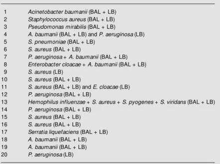
Table 1 shows the types of bacteria iso-lated from BAL and from the lung biopsy.
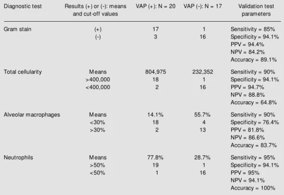
Table 2 shows the index of agreement between Gram stain and the results of BAL quantitative culture, mean cell counts in BAL (total cells, alveolar macrophages and neu-trophils), and sensitivity and specificity of these findings according to pre-established cut-off values.
Figure 2 shows the distribution of BAL cellularity in a box plot graph and the param-eters for test validity analysis using a cut-off value of 400,000 cells/ml.
Sensitivity and specificity were 50 and 76.4% for fever (>38o
C) and 60 and 76.4% for leukocytosis (>10,000 total leukocytes), re-spectively. The complementary association of these two parameters was not additive. The results obtained for Gram staining, total cellu-larity, macrophages and neutrophil differen-tiation counts in BAL for the analysis of test validity are presented in Table 2.
BAL procedure complications were ob-served in seven cases (18.8%): hemorrhage in four, hypoxemia in two and association of hemorrhage and hypoxemia in one case. All complications were of mild intensity, none of them resulting in the interruption of the procedure.
D iscussio n
VAP continues to be a diagnostic chal-lenge. Only a few studies on humans using postmortem lung biopsy to assess the fre-quency of VAP have been published (17-25).
The wide variation in VAP prevalence in these different studies is due to the size of the specimens examined, the use or not of anti-biotics prior to lung biopsy, as well as to the type of antimicrobial regimen used. In any case, the gold standard for VAP diagnosis is still a matter of debate in the specialized literature. Rouby et al. (18) have considered
that histological examination of every pul-monary segment could give a most realistic view of the prevalence of VAP. In their study, they performed unilateral pneumonec-tomy for histological assessment. Marquette et al. (20) found a 67% frequency of VAP when performing bilateral pneumonectomy for histological examination, which must explain their high prevalence index. Using small lung fragments of 1 x 1 x 1 cm, Chastre et al. (17)found only a 23% rate of histologi-cally defined VAP, probably due to the tiny specimen examined. However, other studies also using tiny lung fragments found higher frequencies of VAP, such as 55% (21), 60% (19), and 54% (present study). Whether our own prevalence was underestimated due to the size of the fragment examined is a matter of speculation, considering the 67% index found by Marquette et al. (20) using bilateral pneumonectomy. In this respect, further stud-ies are needed to provide new information later on.
Regarding a diagnosis of a disease other
Table 1. M icroorganisms isolated from bronchoalveolar lavage and a lung biopsy in 20 cases of ventilator-associated pneumonia.
Patients Bacteria found and type of examination used
1 Acinetobacter baumanii (BAL + LB) 2 Staphylococcus aureus (BAL + LB) 3 Pseudomonas mirabilis (BAL + LB)
4 A. baumanii (BAL + LB) and P. aeruginosa (LB) 5 S. pneumoniae (BAL + LB)
6 S. aureus (BAL + LB)
7 P. aeruginosa + A. baumanii (BAL + LB) 8 Enterobacter cloacae + A. baumanii (BAL + LB) 9 S. aureus (LB)
10 S. aureus (BAL + LB)
11 S. aureus (BAL + LB) and E. cloacae (LB) 12 P. aeruginosa (BAL + LB)
13 Hemophilus influenzae + S. aureus + S. pyogenes + S. viridans (BAL + LB) 14 P. aeruginosa (BAL + LB)
15 S. aureus (BAL + LB) 16 S. aureus (BAL + LB)
17 Serratia liquefaciens (BAL + LB) 18 A. baumanii (BAL + LB) 19 A. baumanii (BAL + LB) 20 P. aeruginosa (LB)
opsy procedure is probably one of the most important reasons for this variation.
In our study, BAL permitted the isolation of 25 of 27 bacterial species found in the culture of the lung fragment in patients with VAP, accounting for 92.5% of them. Five of 20 VAP patients (25%) had polymicrobial pneumonia. Association of germs in lung biopsy culture occurred only in four patients (see Table 1). Most published studies have pointed out the polymicrobial status of noso-comial pneumonia (26,27). Bartlett et al. (26) found polymicrobial pneumonia in 54% of 159 cases. Similarly, Fagon et al. (1) found a 40% rate in 52 cases, and Torres et al.(27) found a 13% rate in 78 cases.
The germ most frequently isolated from our VAP patients was Staphylococcus aureus
(from lung fragments in eight cases and from BAL in eight cases), followed by Acineto-bacter baumanii (five in biopsy examination than VAP, the most common respiratory
histopathological finding in our patients was ARDS, followed by diffuse alveolar damage and pulmonary fibrosis. Torres et al. (19) found a 43% rate of alveolar hemorrhage and a 29% rate of diffuse alveolar damage in their patients. Marquette et al. (20) found that the most common diagnosis was diffuse alveolar damage, followed by pulmonary fibrosis. In a study on 83 patients, Rouby et al. (18) detected 28 with diffuse alveolar damage and 13 with pulmonary fibrosis.
Only a few studies have assessed the sensitivity and specificity of BAL examina-tion using a postmortem lung biopsy as the gold standard for the diagnosis of VAP.
A possible reason for the high discrep-ancy in the reported frequency of VAP could be the wide variation in test validation for the use of BAL in its diagnosis. The use of broad spectrum antibiotics prior to the
bi-Table 2. Validation test for the other bronchoalveolar lavage variables. The gold standard parameters w ere open lung biopsy histopathological pattern for pneumonia plus quantitative culture of a lung tissue biopsy.
Diagnostic test Results (+) or (-): means VAP (+): N = 20 VAP (-): N = 17 Validation test
and cut-off values parameters
Gram stain (+) 17 1 Sensitivity = 85%
(-) 3 16 Specificity = 94.1%
PPV = 94.4% NPV = 84.2% Accuracy = 89.1%
Total cellularity M eans 804,975 232,352 Sensitivity = 90%
>400,000 18 1 Specificity = 94.1%
<400,000 2 16 PPV = 94.7%
NPV = 88.8% Accuracy = 64.8%
Alveolar macrophages M eans 14.1% 55.7% Sensitivity = 90%
<30% 18 4 Specificity = 76.4%
>30% 2 13 PPV = 81.8%
NPV = 86.6% Accuracy = 83.7%
Neutrophils M eans 77.8% 28.7% Sensitivity = 95%
>50% 19 1 Specificity = 94.1%
<50% 1 16 PPV = 95%
NPV = 94.1% Accuracy = 100%
and five in BAL), and Pseudomonas aerugi-nosa (five in biopsy specimens and four in BAL). These results agree with many pub-lished studies, where the most frequently isolated microorganisms were Gram-nega-tive bacilli (1,17,26-32) and Staphylococcus aureus (mainly the methicillin-resistant strain) (1,33-37). When compared with other studies that used postmortem lung biopsy examination, our culture results also showed Gram-negative bacilli and Staphylococcus aureus as the most important causal agents, as also reported by Torres et al. (19), Papazian et al. (22), and Marquette et al. (20).
The Gram staining results presented in Table 1 reveal that patients without VAP showed a good correlation between culture results and negative direct Gram staining examination (16/17 patients = 94.1%). This is a very high specificity index, excluding in daily clinical practice a diagnosis of pneu-monia in most of the suspected cases when Gram staining is negative. Marquette et al. (20) found a specificity of 87.5% (negative Gram staining in seven of eight cases with negative tissue cultures), and Papazian et al. (22) found 100% specificity in their study. However, the sensitivity of Gram staining in our study was only 85% (17/20). When con-sidering the microorganism species, when each Gram stain is matched to each tissue culture result, coincidence occurs in only 55% (11/20). Low levels of sensitivity were also reported in the studies of Marquette et al. (20) (47.3%), and Papazian et al. (25) (56%).
In our study, despite the wide variation in total BAL cellularity (a mean of 804,975 cells/ml, ranging from 140,000 to 1,745,000 in VAP patients; a mean of 232,352 cells/ml, ranging from 52,500 to 1,050,000 in patients without VAP) (Figure 2), using a cut-off point of 400,000 cells/ml we can define a sensitivity of 90% (18/20) and a specificity of 94.1% (16/17). In the study of Marquette et al. (20), despite the fact that the average number of BAL cells was higher in patients
with pneumonia, there was no significant difference between groups. Papazian et al. (25) obtained the same results, showing that this parameter must be interpreted with cau-tion in clinical practice.
Relative neutrophil number in BAL was also higher in patients with pneumonia (95% of the cases had 50% or more neutrophils). The same results were obtained by Marquette et al. (20). Despite significant differences between groups in their study, they con-cluded that this parameter has a poor predic-tive value when the variation and range of values are too wide. Papazian et al. (25) found no differences in neutrophil numbers between groups. However, Kirtland et al. (23) found that 50% of neutrophils in BAL is a good cut-off value to exclude pneumonia (100% specificity). Their patients with pneu-monia showed a mean percentage of BAL neutrophils of 75% (50-96%) versus 46% (1-96%) in the group without pneumonia (23). In this respect, our results, associated with those of Kirtland et al. (23), should stimulate further studies on the role of BAL neutrophils in the diagnosis of VAP.
Since a large number of critically ill pa-tients cannot wait for the result of culture to initiate antibiotic therapy, the clinical and radiographic suspicion of VAP allied to BAL
T
o
ta
l
c
e
llu
la
ri
ty
o
f
B
A
L
4,000,000
3,000,000
2,000,000
1,000,000
0 400,000
With VAP Without VAP
findings, such as total cellularity and per-centage of neutrophils, should contribute to the diagnosis and institution of early therapy. It seems reasonable that additional studies should be carried out on the cellularity of BAL, since the early use of antibiotics in this kind of patients will inevitably compromise the results of BAL or lung tissue culture.
The number of BAL procedure compli-cations observed in our study was rather high (18.9%), but all of them were
self-limited, not interfering with the continuity of the procedure in any case.
In conclusion, our data indicate that BAL quantitative culture, Gram staining and cel-lularity might be useful tools in the diagnos-tic investigation of VAP. It is evident that further studies are needed to assess the role of better diagnostic procedures (invasive or not) in VAP, regarding endpoints such as related morbidity, overall mortality, and health care costs.
Re fe re nce s
1. Fagon JY, Chastre J, Domart Y, Trouillet JL, Pierre J, Darne C & Gibert C (1989). Nosocomial pneumonia in patients receiv-ing continuous mechanical ventilation: prospective analysis of 52 episodes w ith use of a protected specimen brush and quantitative culture techniques. American Review of Respiratory Disease, 139: 877-884.
2. Craven DE, Steger KA & Barber TW (1991). Preventing nosocomial pneumo-nia: state of the art and perspectives for the 1990s. American Journal of M edicine, 91 (Suppl 3B): 44S-53S.
3. Tablan OC, Anderson LJ, Arden NH, Breiman RF, Butler JC, M cNeil M M & Pearson M L (1997). Guidelines for pre-vention of nosocomial pneumonia. Part I: an overview of the prevention of nosoco-mial pneumonia, 1994. M orbidity and M ortality Weekly Report, 46: 1-43. 4. Baselski VS, El-Torky M , Coalson JJ &
Griffin JP (1992). The standardization of criteria for processing and interpreting laboratory specimens in patients w ith sus-pected ventilator-associated pneumonia. Chest, 102 (Suppl): 571S-579S.
5. Sterling TR, Ho EJ, Brehm WT & Kirkpa-trick M B (1996). Diagnosis and treatment of ventilator-associated pneumonia - im-pact on survival. A decision analysis. Chest, 110: 1025-1034.
6. Fagon JY, Chastre J, Hance AJ, M ontraers PH, Novara A & Gibert C (1993). Nosoco-mial pneumonia in ventilated patients: a cohort study evaluating attributable mor-tality and hospital stay. American Journal of M edicine, 94: 281-288.
7. Rello J, Ausina V, Ricart M , Castella J & Prats G (1993). Impact of previous antimi-crobial therapy on the etiology and out-come of ventilator-associated pneumonia.
Chest, 104: 1230-1235.
8. Valles J, Artigas A, Rello J, Bonson SN, Fontanal SD, Blanch L, Fernandez R, Baigorri F & M estre J (1995). Continuous aspiration of subglottic secretions in pre-venting ventilator-associated pneumonia. Annals of Internal M edicine, 122: 179-186.
9. Johanson Jr WG, Pierce AK, Sanford JP & Thomas GD (1972). Nosocomial respira-tory infection w ith Gram-negative bacilli: the significance of colonization of the res-piratory tract. Annals of Internal M edicine, 77: 701-706.
10. Faling LJ (1988). New advances in diag-nosing nosocomial pneumonia in intu-bated patients. American Review of Res-piratory Disease, 137: 253-255.
11. Fagon JY, Chastre J, Hance AJ, Guiguet M , Trouillet JL, Domart Y, Pierre J & Gibert C (1988). Detection of nosocomial lung infection in ventilated patients. Use of a protected specimen brush and quan-titative culture techniques in 147 patients. American Review of Respiratory Disease, 138: 110-116.
12. Atherton ST & White DJ (1978). Stomach as source of bacteria colonizing respira-tory tract during artificial ventilation. Lan-cet, 2: 968-969.
13. Kirkpatrick M B & Bass Jr JB (1989). Quan-titative bacterial cultures of bronchoalveo-lar lavage fluids and protected brush cath-eter specimens from normal subjects. American Review of Respiratory Disease, 139: 546-548.
14. M eduri GU & Chastre J (1992). The stan-dardization of bronchoscopic techniques f or vent ilat or-associat ed pneum onia. Chest, 102 (Suppl 1): 557S-564S. 15. Katzenstein AA & Askin FB (1990).
Surgi-cal Pathology of Non-Neoplastic Lung
Dis-ease. 2nd edn. W.B. Saunders, Philadel-phia.
16. Sackett DL, Haynes RB & Tugw ell P (1985). Clinical Epidemiology. A Basic Sci-ence for Clinical M edicine. Little, Brow n & Company, Boston/Toronto.
17. Chastre J, Viau F, Brun P, Pierre J, Dauge M C, Bouchama A, Akesbi A & Gibert C (1984). Prospective evaluation of the pro-tected specimen brush for the diagnosis of pulmonary infections in ventilated pa-tients. American Review of Respiratory Disease, 130: 924-929.
18. Rouby JJ, M artin de Lassale E, Poete P, Nicolas M H, Bodin L, Jarlier V, Le Char-pentier Y, Grosset J & Viars P (1992). Nosocomial bronchopneumonia in the critically ill. Histologic and bacteriologic aspects. American Review of Respiratory Disease, 146: 1059-1066.
19. Torres A, El-Ebiary M , Padró J, González J, de la Bellacasa JP, Ramirez J, Xaubet A, Ferrer M & Rodriguez-Roisin RL (1994). Validation of different techniques for the diagnosis of ventilator-associated pneu-monia. Comparison w ith immediate post-m ort epost-m pulpost-m onary biopsy. Am erican Journal of Respiratory and Critical Care M edicine, 149: 324-331.
20. M arquette CH, Copin M C, Wallet F, Ne-viere R, Saulnier F, M athieu D, Durocher A, Ramon P & Tonnel AB (1995). Diagnos-tic tests for pneumonia in ventilated pa-tients: prospective evaluation of tic accuracy using histology as a diagnos-tic gold standard. American Journal of Respiratory and Critical Care M edicine, 151: 1878-1888.
diagno-sis of nosocomial pneumonia. American Journal of Respiratory and Critical Care M edicine, 152: 231-240.
22. Papazian L, Thomas P, Garbe L, Guignon I, Thirion X, Charrel J, Bollet C, Fuentes P & Gouin F (1995). Bronchoscopic or blind sampling techniques for the diagnosis of ventilator-associated pneumonia. Ameri-can Journal of Respiratory and Critical Care M edicine, 152: 1982-1991. 23. Kirtland SH, Corley DE, Winterbauer RH,
Springmeyer SC, Casey KR, Hampson NB & Dreis DF (1997). The diagnosis of venti-lator-associated pneumonia. A compari-son of histologic, microbiologic, and clini-cal criteria. Chest, 112: 445-457. 24. Johanson Jr WG, Seidenfeld JJ, Gomez
P, de los Santos R & Coalson JJ (1988). Bacteriologic diagnosis of nosocomial pneumonia follow ing prolonged mechani-cal ventilation. American Review of Res-piratory Disease, 137: 259-264.
25. Papazian L, Autillo-Touati A, Thomas P, Bregeon F, Garbe L, Saux P, Seite R & Gouin F (1997). Diagnosis of ventilator-associated pneumonia. An evaluation of direct examination and presence of intra-cellular organisms. Anesthesiology, 87: 268-276.
26. Bartlett JG, O’Keefe P, Tally FP, Louie TJ & Gorbach SL (1986). Bacteriology of hos-pital-acquired pneumonia. Archives of In-ternal M edicine, 146: 868-871.
27. Torres A, Aznar R, Gatell JM , Jimenez P, Gonzalez J, Ferrer A, Celis R &
Rodriguez-Roisin R (1990). Incidence, risk, and prog-nosis factors of nosocomial pneumonia in mechanically ventilated patients. Ameri-can Review of Respiratory Disease, 142: 523-528.
28. Horan TC, White JW, Jarvis WR, Emori TG, Culver DH, M unn VP, Thornsberry C, Olson DR & Hughes JM (1986). Nosoco-mial infection surveillance, 1984. M orbid-ity and M ortalorbid-ity Weekly Report, 35: 17SS-29SS.
29. Schaberg DR, Culver DH & Gaynes RP (1991). M ajor trends in the microbial etiol-ogy of nosocomial infection. American Journal of M edicine, 91 (Suppl 3B): 72S-75S.
30. Torres A, de la Bellacasa JP, Rodriguez-Roisin R, de Anta M TJ & Agusti-Vidal A (1988). Diagnostic value of telescoping plugged catheters in mechanically venti-lated patients w ith bacterial pneumonia using the M etras catheter. American Re-view of Respiratory Disease, 138: 117-120.
31. Jimenez P, Torres A, Rodriguez-Roisin R, de la Bellacasa JP, Aznar R, Gatell M & Agusti-Vidal A (1989). Incidence and etiol-ogy of pneumonia acquired during me-chanical ventilation. Critical Care M edi-cine, 17: 882-885.
32. Pugin J, Auckenthaler R, M ili N, Janssens JP, Lew PD & Suter PM (1991). Diagnosis of ventilator-associated pneumonia by bacteriologic analysis of bronchoscopic and nonbronchoscopic “ blind”
bronchoal-veolar lavage fluid. American Review of Respiratory Disease, 143: 1121-1129. 33. Chastre J, Fagon JY, Soler P, Bornet M ,
Domart Y, Trouillet JL, Gibert C & Hance AJ (1988). Diagnosis of nosocomial bacte-rial pneumonia in intubated patients un-dergoing ventilation: comparison of the usefulness of bronchoalveolar lavage and the protected specimen brush and quanti-tative culture techniques in 147 patients. American Journal of M edicine, 85: 499-506.
34. Rello J, Quintana E, Ausina V, Castella J, Luquin M , Net A & Prats G (1991). Inci-dence, etiology, and outcome of nosoco-mial pneumonia in mechanically ventilated patients. Chest, 100: 439-444.
35. Rodriguez de Castro F, Sole Violan J, Lafarga Capuz B, Caminero Luna J, Gonza-les Rodriguez B & M anzano Alonso JL (1991). Reliability of the bronchoscopic protected catheter brush in the diagnosis of pneumonia in mechanically ventilated patients. Critical Care M edicine, 19: 171-175.
36. Espersen F & Gabrielsen J (1981). Pneu-monia due to Staphylococcus aureus dur-ing mechanical ventilation. Journal of In-fectious Diseases, 144: 19-23.