Association of the 894G>T
polymorphism of the endothelial
constitutive nitric oxide synthase
gene with unstable angina
1Unidade de Hemodinâmica, Hospital de Clínicas de Porto Alegre, 2Curso de Pós-Graduação em Medicina, Cardiologia,
Universidade Federal do Rio Grande do Sul, Porto Alegre, RS, Brasil
3Laboratório de Genética e Biologia Molecular, Faculdade de Biociências,
Pontifícia Universidade Católica do Rio Grande do Sul, Porto Alegre, RS, Brasil G.R. Iturry-Yamamoto1,
E.H. Moriguchi2,
A.C. Zago1,
C.S. Alho3
and A.J. Zago1
Abstract
The 894G>T polymorphism of the endothelial constitutive nitric oxide synthase gene consists of the substitution of a guanine base by a thymine at the 894th nucleotide of the gene. An association of this polymorphism with acute coronary syndromes has been described, only when in combination with other polymorphisms of this gene. The aim of the present study was to search for an association between this polymorphism and unstable angina in a southern Brazilian popula-tion. In a case-control study, 156 patients (group 1 (N = 83): unstable angina, group 2 (N = 73): stable angina) were genotyped by PCR and digestion of the product. Univariate analysis demonstrated that the minimal luminal diameter and the degree of stenosis of the culprit lesion differed between groups (P = 0.006 and 0.005, respectively). In addition, the frequencies of the T allele and of the T allele carriers (combined TT and TG genotypes) were significantly higher in the group with unstable angina (41.6 vs 28.8%; P = 0.025, Pearson chi-squaretest, and 73.5 vs 45.2%; P = 0.001, Pearson chi-squaretest, respectively). Multivariate logistic regression showed that the fre-quency of the T allele carriers was the only variable with a predictive value for unstable angina, when controlled for the other variables (6.1 (95% CI = 2.55-14.43); P < 0.001). Thus, in a homogenous group of patients, the endothelial constitutive nitric oxide synthase 894G>T polymorphism was associated with unstable angina. We suggest that this polymorphism may be a genetic risk factor for unstable angina.
Correspondence
G.R. Iturry-Yamamoto Unidade de Hemodinâmica Serviço de Cardiologia
Hospital de Clínicas de Porto Alegre Rua Ramiro Barcelos, 2350, Sala 2059 90035-003 Porto Alegre, RS Brasil
Fax: +55-51-3330-5281 E-mail: [email protected]
Research supported by FAPERGS (No. 01/0075.6) and Fundo de Incentivo à Pesquisa do Hospital de Clínicas de Porto Alegre (No. 00-246). G.R. Iturry-Yamamoto was the recipient of postdoctoral fellowships from FAPERGS (No. 00/60014.3) and CNPq (No. 150102/01-1(NV)).
Received October 26, 2005 Accepted October 9, 2006
Key words
•Nitric oxide synthase •Gene
•Polymorphism •Unstable angina •Coronary
Introduction
The leading mechanism of unstable an-gina is the transitory interruption of myocar-dial perfusion by a subocclusive thrombus, superposed on a fissured or eroded coronary atherosclerotic plaque (1). The endothelium
pro-cesses involved in the pathogenesis of ath-erosclerosis and thrombosis. An inhibitory effect of NO on platelet aggregation and adhesion to the vascular endothelium has been demonstrated (5-7) and EDRF and ex-ogenous NO also cause platelet deaggrega-tion (8). In addideaggrega-tion, NO may act on the blood coagulation system through the regu-lation of the expression of heparin sulfate by endothelial cells (9). Conversely, NO inhib-its diverse functions in polymorphonuclear leukocytes, such as chemotaxis and the syn-thesis and release of superoxide radical (10,11). Furthermore, NO inhibits the adhe-sion of polymorphonuclear cells to the vas-cular endothelium (12), thus playing a fun-damental role in the control of vascular ho-meostasis. EDRF is synthesized in the endo-thelial cell from L-arginine by endoendo-thelial constitutive NO synthase (ecNOS), a consti-tutive enzyme coded by a gene located at locus 7q35-36, containing 26 exons that oc-cupy 21 kb (13). Some polymorphisms of this gene have been described, as well as their possible association with diverse car-diovascular pathologies. Among these poly-morphisms, the 894G>T polymorphism has been described in exon 7 of the ecNOS gene, and consists of the substitution of a guanine base by a thymine at nucleotide 894 of the gene; this mutation results in the substitution of the amino acid glutamate by aspartate at the 298th position of the ecNOS protein (Glu298Asp) (14).
The possible association of this polymor-phism with coronary heart disease has been studied. In a Japanese population, the fre-quency of TT homozygotes of this polymor-phism was significantly greater in patients with myocardial infarction (MI) than in a control group of healthy people; however, in that study, this mutation was not associated with the degree of severity of coronary ath-erosclerosis (15). In another Japanese study, a significantly higher frequency of the T allele was observed in patients with MI when compared with a control group (16). In an
English population, a significantly higher frequency of TT homozygotes was reported in a group of patients with coronary disease and in another group of patients with recent MI when compared with their respective healthy controls (17). With regard to acute coronary syndromes (ACS), a study in a Korean population (18) analyzed the effect of two polymorphisms of the ecNOS gene, the 894G>T and the polymorphic variation in intron 4 (4a4b), on the development of ACS (acute myocardial infarction and un-stable angina). In that study, the GG geno-type of the ecNOS 894G>T polymorphism had an additive beneficial effect on 4a allele carriers of the ecNOS 4a4b polymorphism. In another study conducted on an Italian population (19), the effect of the 894G>T, 4a4b and -786T>C polymorphisms on the predisposition to ACS was also analyzed. The homozygosity for the ecNOS 4a rare variant represented an independent predis-position factor to ACS. In addition, an in-creased predisposition to ACS was observed in subjects carrying the -786CC/894TT geno-types.
Patients, Material and Methods
Written informed consent was obtained from all of the participants and the Ethics Committee of the Hospital de Clínicas de Porto Alegre, Brazil, approved this study.
Study population
A case-control study was conducted in a southern Brazilian population. Patients sub-mitted to percutaneous revascularization of the culprit lesion between August 2000 and August 2003 were included. A total of 156 unrelated patients (111 men and 45 women) were included in the study according to the following criteria: patients should 1) present symptomatic angina, 2) have an indication for percutaneous revascularization, and 3) sign the informed consent to participate in the study. The study population was mainly of European ancestry, i.e., of Italian, Portu-guese and German descent (97.44%); only 4 patients were of African origin. Patients were divided into two groups: group 1 (N = 83), patients with unstable angina according to the Braunwald classification (types I, II, III, B, or C) (23) and group 2 (N = 73), patients with stable angina. Asymptomatic patients were not included in the study.
Clinical and demographic data
The following data were used for clinical evaluation: clinical presentation (stable or unstable angina), age, functional class ac-cording to the New York Heart Association (NYHA) classification (24), plasma lipid profile, risk factors for coronary disease such as diabetes mellitus, systemic arterial hyper-tension (patients taking medication or with a previous diagnosis), smoking (smokers or ex-smokers of 1 or more cigarettes/day for more than 5 years), body mass index, history of previous MI, family history of cardiovas-cular disease (history of MI or cardiovascu-lar death before 60 years of age in a first
degree relative), and medication in use (as-pirin, nitrates, angiotensin-converting en-zyme inhibitors, statins, calcium channel blockers, ß-blockers).
Quantitative coronary angiography
The following angiographic characteris-tics of the culprit lesion were evaluated by digital quantitative angiography: type of le-sion (noncomplex: types A and B1; com-plex: types B2 and C, according to the modi-fied American College of Cardiology/Ameri-can Heart Association classification (25), minimal luminal diameter, reference diam-eter, degree of stenosis, and lesion length. The diameters were determined using the diameter of the distal portion of the guide catheter as the calibration reference. The lesions were quantified using software for quantitative angiographic analysis with au-tomatic detection of edges (GEMNet CRS V. 5.6.5, Advantage Cardiac Review, Gen-eral Medical Electric Systems, 1998; Fair-field, CT, USA).
Blood collection and DNA analysis
re-action was carried out in a PTC-100 thermo-cycler (MJ Research, Inc., Watertown, MA, USA), as follows: an initial denaturation at 94ºC for 10 min, followed by 30 cycles at 94ºC for 1 min, at 61ºC for 1 min, and at 72ºC for 1 min. The final extension step was prolonged to 10 min. The 457-bp PCR-amplified product (40 µL) was cleaved in appropriate buffer with 8-12 U of BanII (GibcoBRL®-Life
Technolo-gies™, Rockville, MD, USA) in a total vol-ume of 50 µL at 37ºC for 24 h. The sequence of exon 7 of the ecNOS gene is registered in the EMBL data base as GI: 461317 (GenBank accession number: X76307). The genotype was determined by 2% agarose gel electro-phoretic analysis of the DNA segments. The genotyping was based on the following infor-mation: i) GG homozygotes present the gua-nine base inside the recognition site for BanII; thus, the 457-bp PCR-amplified product is cleaved into two DNA fragments of 137 and 320 bp as a result of digestion; ii) TT homozy-gotes present the substitution of the guanine base by thymine and therefore only one undi-gested DNA fragment of 457 bp is visualized on agarose gel; iii) TG heterozygotes are ob-served on the agarose gel as three DNA frag-ments of 457, 320, and 137 bp. For genotype analysis, a dominant heredity model was as-sumed (combined TT + TG genotypes versus the GG genotype). Genotyping was performed in a blind fashion, i.e., the investigators were unaware of patient data.
Lipid profile analysis
A 5-mL blood sample was collected into a sterile system with heparin as anticoagu-lant from the femoral artery after a 12-h overnight fast before cardiac catheteriza-tion. After centrifugation, plasma was stored frozen at -80ºC until lipid profile analysis. The plasma concentrations of high-density lipoprotein cholesterol (HDL cholesterol) and total cholesterol were analyzed in the labo-ratory of the Department of Clinical Pathol-ogy, Hospital de Clínicas de Porto Alegre,
Porto Alegre, RS, Brazil, using commer-cially available systems (HDL-C Plus, sec-ond generation, and Cholesterol CHOD-PAP, respectively) and a Roche/Hitachi Automatic Analyzer 917 (Basel, Switzerland).
Statistical analysis
All the co-variables were coded as 0 for absence of the risk factor and as 1 for its presence; 0 for no use of medication, 1 for the use of medication; 0 for noncomplex lesion type, 1 for complex lesion type; 0 for GG genotype, 1 for combined TT + TG. Data were analyzed using the statistical soft-ware package SPSS 12.0 for Windows. The continuous variables are reported as mean ± SD and the categorical variables as percent-age and frequencies. The clinical and angio-graphic characteristics were compared be-tween groups using the Pearson chi-square test followed by Yates continuity correction for categorical variables, or the Student t -test for independent samples for continuous variables.
The chi-squaretest was applied to deter-mine whether genotype distributions were within Hardy-Weinberg equilibrium. The frequencies of the alleles and genotypes were compared between groups by the Pearson chi-squaretest. Later, multivariate logistic regression was applied, considering unstable angina as the dependent variable and as co-variables those demonstrating P ≤ 0.20 in the
univariate test and those that can modulate ecNOS, such as age, gender, plasma lipid profile (total cholesterol and HDL choles-terol), smoking habit, and the use of statins. The level of significance was set at 5% in all analyses.
Results
Clinical characteristics of the patients
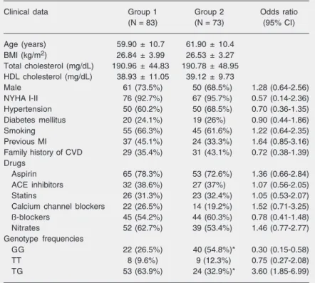
with unstable angina; group 2: patients with stable angina). In univariate analysis, the two groups were similar regarding age, gen-der, body mass index, NYHA functional class, plasma lipid profile, and risk factors for coronary disease such as hypertension, diabetes mellitus, smoking, and a family history of cardiovascular disease. In addi-tion, the two groups were similar regarding the medication in use (aspirin, angiotensin-converting enzyme inhibitors, statins, cal-cium channel blockers, ß-blockers, and ni-trates).
Angiographic characteristics
Table 2 shows the angiographic charac-teristics of the culprit lesion. The two groups of patients were similar regarding type of lesion, luminal diameter of reference and lesion length, but differed in the minimal luminal diameter (P = 0.006, Student t-test) and degree of stenosis (P = 0.005, Student t -test).
Allele and genotype distribution
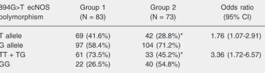
The global genotype (TT = 0.11; TG = 0.49; GG = 0.40) and allele (T = 0.36; G = 0.64) frequencies in the sample studied did not differ from the values predicted by the Hardy-Weinberg model (P = 0.61, chi-square). The distribution of the alleles and genotypes in patients of each group is com-pared in Table 3. The frequency of the T allele was significantly higher in patients with unstable angina compared to patients with stable angina (41.6 vs 28.8%; P = 0.025, Pearson chi-squaretest). Also, assuming a dominant heredity model (combined TT + TG genotypes versus GG genotype), the fre-quency of TT + TG genotypes was signifi-cantly higher in patients with unstable an-gina compared to patients with stable anan-gina (73.5 vs 45.2%; P = 0.001, Pearson chi-squaretest).
Multivariate logistic regression showed
Table 2. Angiographic characteristics of patients with unstable angina (group 1) and patients with stable angina (group 2).
Angiographic characteristics Group 1 (N = 83) Group 2 (N = 73)
Minimal luminal diameter (mm) 0.68 ± 0.47 0.89 ± 0.48*
Reference luminal diameter (mm) 2.84 ± 0.53 2.87 ± 0.53
Degree of stenosis (%) 76.50 ± 15.22 69.54 ± 14.94*
Lesion length (mm) 12.40 ± 5.17 12.93 ± 4.90
Data are reported as mean ± SD. *P < 0.05 (Student t-test).
that the presence of the T allele was the only variable with a predictive value for unstable angina, when controlled for the other vari-ables (previous MI, minimal luminal
diam-Table 1. Clinical, demographic and genetic characteristics of patients with unstable angina (group 1) and patients with stable angina (group 2).
Clinical data Group 1 Group 2 Odds ratio
(N = 83) (N = 73) (95% CI)
Age (years) 59.90 ± 10.7 61.90 ± 10.4
BMI (kg/m2) 26.84 ± 3.99 26.53 ± 3.27
Total cholesterol (mg/dL) 190.96 ± 44.83 190.78 ± 48.95
HDL cholesterol (mg/dL) 38.93 ± 11.05 39.12 ± 9.73
Male 61 (73.5%) 50 (68.5%) 1.28 (0.64-2.56)
NYHA I-II 76 (92.7%) 67 (95.7%) 0.57 (0.14-2.36)
Hypertension 50 (60.2%) 50 (68.5%) 0.70 (0.36-1.35)
Diabetes mellitus 20 (24.1%) 19 (26%) 0.90 (0.44-1.86)
Smoking 55 (66.3%) 45 (61.6%) 1.22 (0.64-2.35)
Previous MI 37 (45.1%) 24 (33.3%) 1.64 (0.85-3.16)
Family history of CVD 29 (35.4%) 31 (43.1%) 0.72 (0.38-1.39)
Drugs
Aspirin 65 (78.3%) 53 (72.6%) 1.36 (0.66-2.84)
ACE inhibitors 32 (38.6%) 27 (37%) 1.07 (0.56-2.05)
Statins 26 (31.3%) 23 (32.4%) 1.05 (0.53-2.07)
Calcium channel blockers 22 (26.5%) 14 (19.2%) 1.52 (0.71-3.25)
ß-blockers 45 (54.2%) 44 (60.3%) 0.78 (0.41-1.48)
Nitrates 52 (62.7%) 39 (53.4%) 1.46 (0.77-2.77)
Genotype frequencies
GG 22 (26.5%) 40 (54.8%)* 0.30 (0.15-0.58)
TT 8 (9.6%) 9 (12.3%) 0.75 (0.27-2.08)
TG 53 (63.9%) 24 (32.9%)* 3.60 (1.85-6.99)
Data are reported as mean ± SD or as the number of patients with percent in parentheses. CI = confidence interval; BMI = body mass index; HDL = high density lipoproteins; NYHA = New York Heart Association functional classification; CVD = cardiovascular disease; MI = myocardial infarction; ACE = angiotensin-converting enzyme.
eter and degree of stenosis of the culprit lesion, age, gender, plasma lipid profile, smoking habit, and the use of statins) (data not shown). The risk of presenting unstable angina for the patients who carried the T allele was 6.1 (95% CI = 2.55-14.43); P < 0.001).
Discussion
We investigated the possible association between 894G>T polymorphism of the ecNOS gene and unstable angina in a South-ern Brazilian population mostly of European ancestry (97.44%). The genotype and allele frequencies of this sample were similar to those reported by Rios et al. (20), who also analyzed subjects of European ancestry from South Brazil, and to those detected for a white Brazilian population (27; data not shown). A significantly higher frequency of the T allele and of the TT + TG genotypes was found among patients with unstable an-gina compared to a group of patients with stable angina (Table 3). Therefore, in the present study, the T allele was significantly associated with unstable angina. Multivari-ate logistic regression analysis demonstrMultivari-ated the presence of the T allele (combined TT and TG genotypes) as the only variable with a predictive value for unstable angina, when controlled for the other variables. It should be emphasized that the two groups of pa-tients were similar in terms of clinical
char-acteristics, risk factors for coronary disease and medication in use (Table 1) and that only patients with angina were included in group 2, excluding patients with coronary atherosclerosis but without symptoms. Thus, the fact that the frequency of genotypes was significantly different in a homogenous popu-lation in repopu-lation to the clinical characteris-tics suggests an association between the 894G>T polymorphism and unstable angina in this group of patients with coronary dis-ease diagnosed by angiography.
EDRF, synthesized from L-arginine by the action of ecNOS, has a fundamental role in the control of vascular homeostasis. There-fore, modifications in the nucleotide se-quence of the gene that codes for ecNOS could result in alterations in phenotypic ex-pression, consequently affecting the clinical status of patients with coronary atheroscle-rosis. In fact, previous studies have demon-strated that this polymorphism is associated with MI in Japanese and English popula-tions (15-17), although the mechanism by which this occurs is unknown. Initial studies have shown that polymorphism of the ecNOS gene may have a functional effect on the enzyme. T allele carriers without coronary disease were shown to present a vasomotor coronary dysfunction due to an increase in microvascular resistance at rest, that was not seen in G allele carriers. It seems that this effect might be allele dose dependent, since TT homozygotes present greater dysfunc-tion (28). In another study, also on healthy people, no effect of the T allele was seen on the vascular response to acetylcholine (ACh), whereas TT homozygotes demonstrated a significantly lower endogenous NO synthe-sis (29). Also, an attenuated response to ACh was found in coronary patient carriers of the T allele, showing an endothelial-de-pendent dysfunction in resistance vessels (30). Cattaruzza et al. (31) demonstrated that there is no difference in the endothelium-dependent NO-mediatedrelaxant response to ACh in the saphenous vein segments
de-Table 3. Allele and genotype distribution of 894G>T ecNOS polymorphism in patients with unstable angina (group 1) and patients with stable angina (group 2).
894G>T ecNOS Group 1 Group 2 Odds ratio
polymorphism (N = 83) (N = 73) (95% CI)
T allele 69 (41.6%) 42 (28.8%)* 1.76 (1.07-2.91)
G allele 97 (58.4%) 104 (71.2%)
TT + TG 61 (73.5%) 33 (45.2%)* 3.36 (1.72-6.57)
GG 22 (26.5%) 40 (54.8%)
Data are reported as the number of patients and percent in parentheses. CI = confidence interval.
rivedfrom patients undergoing aortocoronary bypass with either the TT, TG, or GG geno-type. On the other hand, these investigators found that this response was significantly attenuated in segments derived from the CC genotype of the -786T>C polymorphism as compared with CT or TT genotype donors. Also, no apparent differences were observed between the coronary heart disease-positive and -negative patients regarding the 894G>T polymorphism (TT 7.6%, TG 36.9%, GG 55.6% vs TT 7.3%, TG 38.4%, GG 54.3%, respectively). Therefore, in this population 894G>T polymorphism does not seem to play a major role in the development of coronary heart disease. Moreover, in this regard, the lack of effect of the T-allele on NO-dependent relaxation ex vivo argues against a potential linkage between the -786T>C and 894G>T polymorphism (31). At the molecular level, the 894G>T poly-morphism has been shown to result in en-hanced proteolytic cleavage of the mature ecNOS, suggesting that this polymorphism hasa functional effect on the ecNOSprotein (32). In a study on cultured human umbilical vein endothelial cells from normal deliver-ies treated in vitro with or without cigarette smoking extracts, Senthil et al. (33) observed low-ecNOS protein levels and enzyme ac-tivities in carriers of the TT genotype, but relatively high mRNA levels in both control and cigarette smoking extract-treated endo-thelial cells. The reduced ecNOS protein levels and enzyme activities are in agree-ment with the T allele being associated with increased vascular risk. The relatively higher ecNOS mRNA could be a compensatory up-regulation in transcription, since the muta-tion at exon 7 could result in accelerated protein degradation (32). Thus, the 894G>T polymorphism may indeed affect eNOS pro-tein stability since the TT genotype had a low protein level (33).
In the present study, we used a dominant model pooling TT + TG carriers. It seems that the functional effect of the
polymor-phism is present in both homozygous and heterozygous subjects. In fact, in the study by Tesauro et al. (32), a 100-kDa band, product of the proteolytic cleavage of the mature ecNOS, was observed in cell lysates from endothelial cell lines with the TT and TG genotypes, but not with the GG geno-type. In another study (34) on patients with autosomal dominant polycystic kidney dis-ease, Ca2+-dependent NOS activity was
sys-tematically decreased in renal arteries of patients with the TT genotype or the TG genotype compared to patients with the GG genotype.
Two reports have demonstrated an asso-ciation of 894G>T ecNOS polymorphism with ACS, but only when in combination with other polymorphisms of this gene. In the Korean study (18), the 4a allele for the ecNOS 4a4b polymorphism had a protective effect against the development of ACS and the GG genotype for the ecNOS 894G>T polymorphism exerted an additive benefi-cial effect in 4a allele carriers. In the Italian study (19), an increased predisposition to ACS was observed in subjects carrying the -786CC/894TT genotypes. Some differences between the present study and those cited above should be pointed out. First, in our study the control group consisted of sympto-matic patients with stable angina, while in the Korean and Italian studies the control groups were apparently healthy individuals; thus, our population was more homogeneous. Second, we included patients with indica-tion for coronary intervenindica-tion, i.e., a popula-tion differing from those studied in the Ko-rean and Italian investigations, implying a possible selection bias. Third, the Korean and Italian results were obtained in popula-tions of ethnic backgrounds differing from that of our population. All of these aspects may account for the differences between our results and those obtained in the Korean and Italian studies.
poly-morphic variation in intron 4, and the 894G>T variant in exon 7) in the susceptibility to vascular diseases is still controversial. Senthil et al. (33) argued that while there is little doubt that dysfunctional ecNOS is involved in the pathogenesis of vascular diseases, the reasons for the apparently inconsistent find-ings are: I) false-positive statistical results; II) none of the studied polymorphisms would be functional in regulating ecNOS expres-sion and their associations with vascular diseases are mediated through linkage with other functional ecNOS variant site(s), which may only be polymorphic in some popula-tions but not in others; III) the putative func-tion of these polymorphisms or possibly linked variants at one or more other loci may be conditional on specific environmental factor(s) (33).
To explain such inconsistencies, at least four extensive additional studies would be necessary: i) meta-analyses including stud-ies with a large number of individuals from populations with different clinically relevant phenotypic characteristics and diverse ge-netic backgrounds; ii) molecular in vitro investigations to identify how functional each eNOS polymorphism could be regarding its capacity to quantitatively or qualitatively modify the ecNOS enzyme; iii) sequencing studies of the ecNOS gene to uncover new candidate mutations explaining putative functional ecNOS enzyme alterations, and iv) genomic analyses to identify additional loci that may modulate the expression of the ecNOS gene.
Since there are few studies demonstrat-ing that the 894G>T polymorphism is func-tional, an alternative explanation for our
results is that the association found might be due to linkage disequilibrium with other poly-morphisms in the same or other genes, or to interaction with other factors such as smok-ing habit, physical activity, age, gender, and plasma lipid profile, among others, taking into account that coronary disease is multi-factorial. Thus, the probability of a patient to develop unstable angina might depend on all those factors, besides the 894G>T polymor-phism.
A limitation of our study is the small sample (83 cases and 73 controls). However, since we excluded asymptomatic patients, a fact that permitted us to study a homoge-neous population in terms of clinical charac-teristics (Table 1), and multivariate logistic regression analysis demonstrated the pres-ence of the T allele as the only variable with a predictive value for unstable angina, when controlled for the other variables, we sug-gest that the 894G>T ecNOS polymorphism is associated with unstable angina in patients from the South of Brazil and that this poly-morphism may be considered to be a genetic risk factor for unstable angina.
Studies on other populations are neces-sary to confirm this hypothesis.
Acknowledgments
We thank the statistician Vânia Hirakata (Grupo de Pesquisa e Pós-Graduação - Hos-pital de Clínicas de Porto Alegre, Porto Ale-gre, RS, Brazil)and Mrs. Daniela Benzano (Grupo de Pesquisa e Pós-Graduação - Hos-pital de Clínicas de Porto Alegre, Porto Ale-gre, RS, Brazil)for excellent assistance with the statistical analysis.
References
1. Worthley SG, Osende JI, Helft G, Badimon JJ, Fuster V. Coronary artery disease: pathogenesis and acute coronary syndromes. Mt Sinai J Med 2001; 68: 167-181.
2. Furchgott RF, Zawadzki JV. The obligatory role of endothelial cells in the relaxation of arterial smooth muscle by acetylcholine. Nature 1980; 288: 373-376.
3. Ignarro LJ, Byrns RE, Buga GM, Wood KS. Endothelium-derived relaxing factor from pulmonary artery and vein possesses pharma-cologic and chemical properties identical to those of nitric oxide radical. Circ Res 1987; 61: 866-879.
Nature 1987; 327: 524-526.
5. Azuma H, Ishikawa M, Sekizaki S. Endothelium-dependent inhibi-tion of platelet aggregainhibi-tion. Br J Pharmacol 1986; 88: 411-415. 6. Radomski MW, Palmer RM, Moncada S. Comparative
pharmacol-ogy of endothelium-derived relaxing factor, nitric oxide and prosta-cyclin in platelets. Br J Pharmacol 1987; 92: 181-187.
7. Radomski MW, Palmer RM, Moncada S. Endogenous nitric oxide inhibits human platelet adhesion to vascular endothelium. Lancet 1987; 2: 1057-1058.
8. Radomski MW, Palmer RM, Moncada S. The anti-aggregating prop-erties of vascular endothelium: interactions between prostacyclin and nitric oxide. Br J Pharmacol 1987; 92: 639-646.
9. Irokawa M, Nishinaga M, Funayama H, Shimada K. The effect of endothelium-derived relaxing factor (EDRF) on anticoagulant hepa-ran sulfate on cultured vascular endothelial cell (EC). Circulation 1994; 90: I-396.
10. Moilanen E, Vuorinen P, Kankaanranta H, Metsa-Ketela T, Vapaatalo H. Inhibition by nitric oxide-donors of human polymorphonuclear leucocyte functions. Br J Pharmacol 1993; 109: 852-858.
11. Clancy RM, Leszczynska-Piziak J, Abramson SB. Nitric oxide, an endothelial cell relaxation factor, inhibits neutrophil superoxide an-ion productan-ion via a direct actan-ion on the NADPH oxidase. J Clin Invest 1992; 90: 1116-1121.
12. Gaboury J, Woodman RC, Granger DN, Reinhardt P, Kubes P. Nitric oxide prevents leukocyte adherence: role of superoxide. Am J Physiol 1993; 265: H862-H867.
13. Marsden PA, Heng HH, Scherer SW, Stewart RJ, Hall AV, Shi XM, et al. Structure and chromosomal localization of the human constitu-tive endothelial nitric oxide synthase gene. J Biol Chem 1993; 268: 17478-17488.
14. Yoshimura M, Yasue H, Nakayama M, Shimasaki Y, Sumida H, Sugiyama S, et al. A missense Glu298Asp variant in the endothelial nitric oxide synthase gene is associated with coronary spasm in the Japanese. Hum Genet 1998; 103: 65-69.
15. Hibi K, Ishigami T, Tamura K, Mizushima S, Nyui N, Fujita T, et al. Endothelial nitric oxide synthase gene polymorphism and acute myocardial infarction. Hypertension 1998; 32: 521-526.
16. Shimasaki Y, Yasue H, Yoshimura M, Nakayama M, Kugiyama K, Ogawa H, et al. Association of the missense Glu298Asp variant of the endothelial nitric oxide synthase gene with myocardial infarction. J Am Coll Cardiol 1998; 31: 1506-1510.
17. Hingorani AD, Liang CF, Fatibene J, Lyon A, Monteith S, Parsons A, et al. A common variant of the endothelial nitric oxide synthase
(Glu298 → Asp) is a major risk factor for coronary artery disease in
the UK. Circulation 1999; 100: 1515-1520.
18. Park KW, You KH, Oh S, Chae IH, Kim HS, Oh BH, et al. Association of endothelial constitutive nitric oxide synthase gene polymorphism with acute coronary syndrome in Koreans. Heart 2004; 90: 282-285. 19. Fatini C, Sofi F, Sticchi E, Gensini F, Gori AM, Fedi S, et al. Influence of endothelial nitric oxide synthase gene polymorphisms (G894T, 4a4b, T-786C) and hyperhomocysteinemia on the predisposition to acute coronary syndromes. Am Heart J 2004; 147: 516-521. 20. Rios DL, Callegari-Jacques SM, Hutz MH. Endothelial nitric oxide
synthase and fractalkine chemokine receptor polymorphisms on
angiographically assessed coronary atherosclerosis. Clin Chim Acta 2005; 362: 138-146.
21. Tanus-Santos JE, Desai M, Flockhart DA. Effects of ethnicity on the distribution of clinically relevant endothelial nitric oxide variants. Pharmacogenetics 2001; 11: 719-725.
22. Casas JP, Bautista LE, Humphries SE, Hingorani AD. Endothelial nitric oxide synthase genotype and ischemic heart disease: meta-analysis of 26 studies involving 23028 subjects. Circulation 2004; 109: 1359-1365.
23. Braunwald E. Unstable angina. A classification. Circulation 1989; 80: 410-414.
24. The Criteria Committee of the New York Heart Association. Nomen-clature and criteria for diagnosis of diseases of the heart and great vessels. 9th edn. Boston: Little Brown and Co.; 1994. p 253-256. 25. Ellis SG, Vandormael MG, Cowley MJ, DiSciascio G, Deligonul U,
Topol EJ, et al. Coronary morphologic and clinical determinants of procedural outcome with angioplasty for multivessel coronary dis-ease. Implications for patient selection. Multivessel Angioplasty Prognosis Study Group. Circulation 1990; 82: 1193-1202. 26. Lahiri KD, Nurnberger JI Jr. A rapid non-enzymatic method for the
preparation of HMW DNA from blood for RFLP studies. Nucleic Acids Res 1991; 19: 5444.
27. Marroni AS, Metzger IF, Souza-Costa DC, Nagassaki S, Sandrim VC, Correa RX, et al. Consistent interethnic differences in the distri-bution of clinically relevant endothelial nitric oxide synthase genetic
polymorphisms. Nitric Oxide 2005; 12: 177-182.
28. Naber CK, Baumgart D, Altmann C, Siffert W, Erbel R, Heusch G. eNOS 894T allele and coronary blood flow at rest and during adeno-sine-induced hyperemia. Am J Physiol Heart Circ Physiol 2001; 281: H1908-H1912.
29. Sofowora G, Dishy V, Xie HG, Imamura H, Nishimi Y, Morales CR, et al. In-vivo effects of Glu298Asp endothelial nitric oxide synthase
polymorphism. Pharmacogenetics 2001; 11: 809-814.
30. Hambrecht R, Erbs S, Adams V, Linke A, Gielen S, Schuler G. Attenuated endothelium-dependent vasodilation in patients with cor-onary artery disease: Impact of G894T polymorphism of the endo-thelial nitric oxide synthase. J Am Coll Cardiol 2001; 37 (Suppl A): 286A.
31. Cattaruzza M, Guzik TJ, Slodowski W, Pelvan A, Becker J, Halle M, et al. Shear stress insensitivity of endothelial nitric oxide synthase expression as a genetic risk factor for coronary heart disease. Circ Res 2004; 95: 841-847.
32. Tesauro M, Thompson WC, Rogliani P, Qi L, Chaudhary PP, Moss J. Intracellular processing of endothelial nitric oxide synthase iso-forms associated with differences in severity of cardiopulmonary diseases: cleavage of proteins with aspartate vs. glutamate at posi-tion 298. Proc Natl Acad Sci U S A 2000; 97: 2832-2835.
33. Senthil D, Raveendran M, Shen YH, Utama B, Dudley D, Wang J, et al. Genotype-dependent expression of endothelial nitric oxide syn-thase (eNOS) and its regulatory proteins in cultured endothelial cells. DNA Cell Biol 2005; 24: 218-224.