Noise-induced tinnitus: auditory evoked potential in
symptomatic and asymptomatic patients
Valdete Alves Valentins dos Santos-Filha, Alessandra Giannella Samelli, Carla Gentile Matas
Faculdade de Medicina da Universidade de Sa˜o Paulo (FMUSP), Department of Physical Therapy, Speech-language Pathology and Audiology, Occupational Therapy, Sa˜o Paulo/SP, Brazil.
OBJECTIVES:We evaluated the central auditory pathways in workers with noise-induced tinnitus with normal hearing thresholds, compared the auditory brainstem response results in groups with and without tinnitus and correlated the tinnitus location to the auditory brainstem response findings in individuals with a history of occupational noise exposure.
METHOD:Sixty individuals participated in the study and the following procedures were performed: anamnesis, immittance measures, pure-tone air conduction thresholds at all frequencies between 0.25–8 kHz and auditory brainstem response.
RESULTS:The mean auditory brainstem response latencies were lower in the Control group than in the Tinnitus group, but no significant differences between the groups were observed. Qualitative analysis showed more alterations in the lower brainstem in the Tinnitus group. The strongest relationship between tinnitus location and auditory brainstem response alterations was detected in individuals with bilateral tinnitus and bilateral auditory brainstem response alterations compared with patients with unilateral alterations.
CONCLUSION:Our findings suggest the occurrence of a possible dysfunction in the central auditory nervous system (brainstem) in individuals with noise-induced tinnitus and a normal hearing threshold.
KEYWORDS: Noise-Induced Tinnitus; Auditory Evoked Potentials; Auditory Pathways.
Santos-Filha VA, Samelli AG, Matas CG. Noise-induced tinnitus: auditory evoked potential in symptomatic and asymptomatic patients. Clinics. 2014;69(7):487-490.
Received for publication onDecember 9, 2013;First review completed onFebruary 5, 2014;Accepted for publication onFebruary 14, 2014
E-mail: [email protected]
Tel.: 55 11 3091-8411
& INTRODUCTION
Excessive noise exposure is a major cause of hearing loss and/or tinnitus in adults (1-3). However, the mechanisms associated with tinnitus generation are poorly understood and, although the condition is often associated with hearing loss or ear disease (4), many individuals with tinnitus have no or only mild hearing loss (5,6).
Tinnitus may be generated as a pathological signal in the auditory system, triggering a sequence of events that increases neuronal activity at different levels of the auditory pathway. Additionally, other systems are also involved in the generation and maintenance of tinnitus, particularly the limbic and autonomic nervous systems (the ‘‘neurophysio-logical concept of tinnitus generation’’)(7).
Auditory brainstem response (ABR) is an electrophysio-logical measure that has been used for the assessment of
central auditory pathways in individuals with tinnitus. Although the role of the brainstem in the generation and maintenance of tinnitus is still controversial, ABR electro-physiological abnormalities have been reported in indivi-duals with tinnitus (8-11). However, this procedure has received little study in individuals with tinnitus who have a history of occupational noise exposure and normal hearing. Studying the ABR results of these individuals may further our understanding of the mechanisms involved in tinnitus generation by identifying mechanisms other than those directly associated with known cochlear lesions (6).
Tinnitus can be caused by various diseases that affect different structures along the auditory pathway. Thus, a more homogeneous study population (e.g., individuals exposed to occupational noise) should be investigated to eliminate the variables associated with different etiologies of tinnitus (12).
This study aimed to evaluate the central auditory path-ways in workers with noise-induced tinnitus and normal hearing, to compare the ABR results with those obtained for workers exposed to occupational noise without tinnitus and to correlate tinnitus location to ABR findings. Herein, we investigated the hypothesis that individuals with tinnitus would show a dysfunction in the ABR compared with subjects without tinnitus. Our findings may help improve Copyrightß2014CLINICS– This is an Open Access article distributed under
the terms of the Creative Commons Attribution Non-Commercial License (http:// creativecommons.org/licenses/by-nc/3.0/) which permits unrestricted non-commercial use, distribution, and reproduction in any medium, provided the original work is properly cited.
No potential conflict of interest was reported.
DOI:10.6061/clinics/2014(07)08
CLINICAL SCIENCE
our understanding of the mechanisms involved in tinnitus generation.
& MATERIALS AND METHODS
This was a cross-sectional study conducted in the Speech and Hearing Investigation Laboratory in Auditory Evoked Potentials, Faculty of Medicine, University of Sa˜o Paulo (FMUSP), Brazil, and was approved by the Ethics Com-mittee of the Hospital das Clı´nicas da Faculdade de Medicina da Universidade de Sa˜o Paulo (HCFMUSP) and the Ethics Committee for Analysis of Research Projects of the Hospital das Clı´nicas da Faculdade de Medicina da Universidade de Sa˜o Paulo (HCFMUSP), (CAPPesq), under protocols No. 712/06 and No. 1278/06, respectively. All subjects signed an informed consent form before participat-ing in the study.
The study included 60 subjects exposed to occupational noise (above 85 dBA), including 30 with tinnitus (hereafter ‘‘Tinnitus’’) and 30 without tinnitus (hereafter ‘‘Control’’), with four females (13.3%) and 26 males (86.7%) in each group.
The age of the subjects in the two groups ranged between 27 and 50 years, with a mean of 41 years in the Tinnitus group and 41.6 years in the Control group. There was no significant difference in the mean age between the groups (p= 0.563).
The inclusion criteria were: uni- or bilateral constant or intermittent tinnitus (Tinnitus group); occupational noise exposure; hearing threshold within normal limits (less than or equal to 25 dBHL at all frequencies: 0.25–8 kHz) in both ears; type ‘‘A’’ tympanogram (pressure $-100 daPa and volume between 0.3–1.6 cc); and the presence of a contral-ateral acoustic reflex at 0.5, 1, and 2 kHz (13). Based on medical records, subjects with neurological, psychiatric and behavioral dysfunctions were excluded.
The following procedures were performed: anamnesis; application of a questionnaire on the characteristics of tinnitus (14); inspection of the external auditory canal using a Heine otoscope; pure-tone air audiometry over 0.25–8 kHz and pure-tone bone conduction audiometry over 0.5–4 kHz at frequencies with thresholds higher than 20 dBHL, conducted bilaterally, using Grason-Stadler GSI 68 audiometer; acoustic immittance measurements (226 Hz probe-tone tympanometry and ipsilateral and con-tralateral acoustic reflex of the stapedius muscle at 0.5, 1, and 2 kHz) using a GSI 33 middle ear analyzer.
After the hearing evaluation, the selected individuals underwent an electrophysiological assessment of hearing (ABR) to determine the absolute latencies of waves I, III and V and interpeaks I-III, III-V and I-V.
The subjects were seated in a reclining chair in a dimly lit room and instructed to keep their eyes closed throughout the examination. A Bio-Logic Traveler Express evoked potentials unit with EP317 software was used during the examination after cleaning the patient’s skin with abrasive paste and attaching the electrodes at predeter-mined positions with electrolytic paste and adhesive tape (MicroporeTM).
The electrode impedance was maintained at less than 5 kOhms. The acoustic stimulus was presented monaurally via supra-aural earphones (TDH39) that elicited responses in both the right and left ears. The electrodes were placed in the right (A2) and left (A1) ears and at the vertex (Cz)
and forehead (Fpz), according to the IES 1020 standard (International Electrode System).
We used the acoustic click stimulus with rarefied polarity presented at 80 dBHL and 19 clicks per second with a 0.1-millisecond duration and a total of 2000 stimuli. Two measurements were recorded on each side to confirm the reproduction of traces and the existence of responses. The absolute latencies of waves I, III and V and interpeaks I-III, III-V and I-V were then analyzed.
ABR was initially classified as normal or altered and the types of alterations found were subsequently described. ABR was considered altered when at least one ear, or one side, had an alteration. The results followed the normality criteria of latency and interpeak values for individuals older than 24 months proposed by the Evoked Potential User Manual (15).
Altered ABR results were divided according to alteration site: Lower brainstem (LB): increased wave III and V latencies and, consequently, interpeak latencies I-III and I-V;
Higher brainstem (HB): increased latencies of wave V and interpeaks I-V and III-V in the presence of normal absolute latencies of waves I and III;Both: simultaneous LB and HB alterations, one in each ear, in the same individual. We then compared the tinnitus-affected side (reported by subjects) with the ABR alteration side when it was detected.
Tinnitus severity was assessed by a visual analog scale (1 to 10), with 1 representing mild tinnitus and 10 the worst imaginable tinnitus. The scores were classified as: 1 to 3 (mild tinnitus), 4 to 6 (moderate tinnitus) and 7 to 10 (severe tinnitus), and individuals were grouped according to tin-nitus severity: mild tintin-nitus (group 1); moderate tintin-nitus (group 2); and severe tinnitus (group 3).
The Wilcoxon, Mann-Whitney, Equality of Two Pro-portions, and Chi-square tests were used. The significance level was set atp#0.05, and 95% confidence intervals were constructed.
& RESULTS
Tinnitus location was assessed for laterality of the right ear (RE), left ear (LE), or both ears (BE) in individuals in the Tinnitus group (n = 30). In the Tinnitus group, tinnitus was bilateral in 67% of cases, left sided in 13% and right sided in 20%.
Tinnitus severity was classified as mild, moderate, or severe in individuals in the Tinnitus group (n = 30). Moderate tinnitus was reported by 57% of the individuals with tinnitus, which was significantly higher than the proportions reporting mild (13%;p,0.001) or severe (30%; p= 0.037) tinnitus.
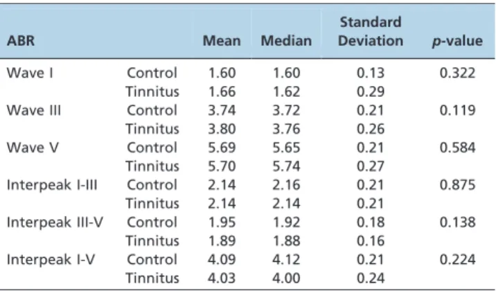
The results of the comparison of the mean absolute latency values of waves I, III and V, and interpeaks I-III, III-V and I-III-V (in ms) between the right and left ears in the Tinnitus and Control groups are shown in Table 1. Because no significant differences were found between the right and left ears in any group, the results were pooled and the mean values were compared between the Tinnitus and Control groups (Table 2). Similarly, there were no significant differences in ABR absolute latencies of waves I, III and V, and interpeaks I-III, III-V and I-V between the Tinnitus and Control groups.
There were significant differences in the proportions of normal and altered ABR results between the Tinnitus and Control groups and between the normal and altered results Tinnitus: auditory evoked potential
Santos-Filha VA et al. CLINICS 2014;69(7):487-490
within the Control group (Table 3). A higher percentage of normal results was observed in the Control group and a higher percentage of altered results was observed in the Tinnitus group. Conversely, there were no significant differences in the type of alteration (lower brainstem, higher brainstem, or both) between the groups. In addition, there were no higher brainstem alterations in either group. Finally, the most frequent ABR alteration was observed in the lower brainstem, as it was observed in 86% and 100% of the subjects in the Control and Tinnitus groups, respectively (Table 4).
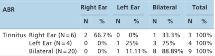
There was a significant positive relationship between the ABR alteration side and the tinnitus-affected side, particu-larly when tinnitus was bilateral or right sided (Table 5).
& DISCUSSION
Quantitative analysis of ABR showed no significant within- or between-group differences for right and left ears. Nevertheless, the mean and standard deviation of the absolute latencies were slightly higher in the Tinnitus group than in the Control group.
Similar results have been reported in other studies, with increased wave I latency (16), significantly increased wave III latency (17,18) and increased absolute latencies of waves I, III and V, with interpeak latencies I-III, I-V and III-V within normal limits (11). These results may be related to differences in the neural transmission of acoustic stimuli (19), as these latencies were slightly increased in the Tinnitus group in our study.
In studies of noise-induced tinnitus, some authors (18,20,21) have reported a possible dysfunction in neural activity at the level of the cochlear nucleus, indicating a possible etiology for this symptom. Other dysfunctions have been observed in different structures along the brainstem, suggesting the participation of the superior olivary complex, inferior colliculus and lateral lemniscus in noise-induced tinnitus, as proposed in some studies (18,22-25).
Although a quantitative analysis did not show any significant differences in ABR between the groups, a qua-litative analysis showed significant differences in the normal and altered results between the groups. Similar findings have been reported in a different study population (11).
ABR alterations were most frequently observed in the lower brainstem, as they were found in 100% of the subjects in the Tinnitus group and 86% of the subjects in the Control group. Although there were no significant differences bet-ween groups, the number of LB alterations was higher in the Tinnitus group. There are no studies describing the types of ABR alterations in individuals with tinnitus exposed to high
Table 1 -Comparison of the mean values of the auditory brainstem response (ABR) absolute latencies of waves I, III and V, and interpeaks I-III, III-V and I-V (in ms) between the right and left ears in the Control and Tinnitus groups (N = 60).
ABR Mean Median
Standard
Deviation p-value
Wave I Control RE 1.61 1.60 0.11 0.191 LE 1.59 1.60 0.14
Tinnitus RE 1.64 1.64 0.28 0.391 LE 1.67 1.60 0.29
Wave III Control RE 3.73 3.72 0.20 0.615 LE 3.75 3.73 0.23
Tinnitus RE 3.80 3.76 0.26 0.431 LE 3.81 3.78 0.26
Wave V Control RE 5.71 5.65 0.21 0.367 LE 5.68 5.66 0.21
Tinnitus RE 5.70 5.74 0.26 0.579 LE 5.70 5.74 0.28
Interpeak I-III Control RE 2.12 2.14 0.19 0.077 LE 2.17 2.18 0.22
Tinnitus RE 2.17 2.14 0.22 0.169 LE 2.12 2.12 0.20
Interpeak III-V Control RE 1.98 1.96 0.20 0.139 LE 1.92 1.90 0.15
Tinnitus RE 1.89 1.88 0.18 0.858 LE 1.88 1.90 0.15
Interpeak I-V Control RE 4.10 4.12 0.18 0.731 LE 4.09 4.04 0.24
Tinnitus RE 4.06 4.02 0.26 0.244 LE 4.00 4.00 0.22
RE: Right ear; LE: Left ear; N: number of individuals; ms: milliseconds.
Table 2 -Comparison of the mean values of the auditory brainstem response (ABR) absolute latencies of waves I, III and V, and interpeaks I-III, III-V and I-V (in ms) between the Control and Tinnitus groups (N = 60).
ABR Mean Median
Standard
Deviation p-value
Wave I Control 1.60 1.60 0.13 0.322 Tinnitus 1.66 1.62 0.29
Wave III Control 3.74 3.72 0.21 0.119 Tinnitus 3.80 3.76 0.26
Wave V Control 5.69 5.65 0.21 0.584 Tinnitus 5.70 5.74 0.27
Interpeak I-III Control 2.14 2.16 0.21 0.875 Tinnitus 2.14 2.14 0.21
Interpeak III-V Control 1.95 1.92 0.18 0.138 Tinnitus 1.89 1.88 0.16
Interpeak I-V Control 4.09 4.12 0.21 0.224 Tinnitus 4.03 4.00 0.24
Note: ms: milliseconds.
Table 3 -Distribution of normal and altered auditory brainstem response (ABR) results in the Control and Tinnitus groups (N = 60).
ABR Control Tinnitus p-value
N % N %
Normal 23 76.7% 14 46.7% 0.017*
Altered 7 23.3% 16 53.3%
p-value ,0.001* 0.606
*statistically significant
p-value; N: number of individuals.
Table 4 -Distribution of auditory brainstem response (ABR) alteration type in the Control and Tinnitus groups.
ABR LB HB Both
N % N % N %
Control 6 86% 0 0% 1 14%
Tinnitus 16 100% 0 0% 0 0%
p-value 0.122 - x - 0.122
Note: LB: lower brainstem; HB: higher brainstem; N: number of individuals; - x -: not available.
CLINICS 2014;69(7):487-490 Tinnitus: auditory evoked potential
Santos-Filha VA et al.
sound pressure levels, although some studies have reported the occurrence of ABR alterations (18,26). Similarly, there are no studies correlating the tinnitus-affected side and the ABR alteration side in individuals with normal hearing with tinnitus complaints who are exposed to high sound pres-sure levels. Nevertheless, our findings suggest a positive relationship between tinnitus location and ABR alteration, and they provide evidence for central nervous system involvement in tinnitus generation and/or maintenance.
Quantitative analysis of ABR latencies between groups showed no significant differences between the Control and Tinnitus groups. However, the number of altered ABRs was higher in individuals with tinnitus complaints than in individuals without tinnitus complaints. Alterations of the auditory pathways in the lower brainstem were the most frequent in tinnitus group, possibly due to changes in synchrony between the generation and transmission of neuroelectrical stimuli in the brainstem. Finally, the rela-tionship between the ABR alteration side and the tinnitus-affected side was stronger in individuals with bilateral tinnitus.
& AUTHOR CONTRIBUTIONS
Santos-Filha VA was in charge of data collection and tabulation and the preparation of the manuscript. Samelli AG collaborated on the data analysis and the preparation of the manuscript. Matas CG was responsible for the data collection, as well as, the general orientation of the stages of execution and the preparation of the manuscript.
& REFERENCES
1. Axelsson A, Prasher D. Tinnitus induced by occupational and leisure noise. Noise Health. 2000;2(8):47-54.
2. Kujawa SG, Liberman MC. Acceleration of age-related hearing loss by early noise exposure: evidence of a misspent youth. J Neurosci. 2006 15;26(7):2115-23, http://dx.doi.org/10.1523/JNEUROSCI.4985-05.2006. 3. Sliwinska-Kowalska M, Davis A. Noise-induced hearing loss. Noise
Health. 2012;14(61):274-80, http://dx.doi.org/10.4103/1463-1741.104893. 4. Prasher D, Ceranic B, Sulkowski W, Guzek W. Objective evidence for tinnitus from spontaneous emission variability. Noise Health. 2001; 3(12):61-73.
5. Henry JA, Dennis KC. General review of tinnitus: prevalence, mechan-isms, effects, and management. J Speech Lang Hear Res. 2005;48(5):1204-35, http://dx.doi.org/10.1044/1092-4388(2005/084).
6. Lindblad A, Rosenhall U, Olofsson A˚ , Hagerman B. The efficacy of N-acetylcysteine to protect the human cochlea from subclinical hearing loss
caused by impulse noise: A controlled trial. Noise Health. 2011; 13(55):392-401, http://dx.doi.org/10.4103/1463-1741.90293.
7. Jastreboff PJ, Hazell WP. A neurophysiological approach to tinnitus: clinical implications. Br J Audiol. 1993;27(1):7-17.
8. Shulman A, Seitz MR. Central tinnitus-diagnosis and treatment. Observations simultaneous binaural auditory brain responses with monoaural stimulation in the tinnitus patient. Laryngoscope. 1981; 91(12):2025-35.
9. Rosenhall U, Axelsson A. Auditory brainstem response latencies in patients with tinnitus. Scand Audiol. 1995;24(2):97-100, http://dx.doi. org/10.3109/01050399509047521.
10. Gerken GM, Hesses PS, Wiorkowski JJ. Auditory evoked responses in control subjects and in patients with problem-tinnitus. Hear Res. 2001;157(1-2):52-64, http://dx.doi.org/10.1016/S0378-5955(01)00277-5. 11. Kehrle HM, Granjeiro RC, Sampaio ALL, Bezerra R, Almeida VF, Oliveira
CA. Comparison of auditory brainstem response results in normal-hearing patients with and without tinnitus. Arch Otolaryngol Head Neck Surg. 2008;134(6):647-51, http://dx.doi.org/10.1001/archotol.134.6.647. 12. Attias J, Urbach D, Gold S, Shemesh Z. Auditory event related potentials
in chronic tinnitus patients with noise induced hearing loss. Hear Res. 1993;71(1-2):106-13, http://dx.doi.org/10.1016/0378-5955(93)90026-W. 13. Gelfand SA. The contralateral acoustic-reflex threshold. In: Silman S.
editor. The acoustic reflex: Basic principles and clinical applications. New York: Academic Press; 1984. p.137-83.
14. Newman CW, Jacobson GP, Spitzer JB. The development of the Tinnitus Handicap Inventory. Arch Otolaryngol Head Neck Surg. 1996;122(2):143-8, http://dx.doi.org/10.1001/archotol.1996.01890140029007.
15. Evoked Potential User Manual. Evoked Potential: Program Version 5.00. User Manual. Bio-logic Systems Corp. Part n˚590-BLSUEP rev.1; 1993. 16. Kadlec E, Mendel LL, editors. Auditory brainstem and middle latency
responses in tinnitus sufferers. Proceedings of the 9th Annual
Convention AAA American Academy of Audiology; 1997 April 12-16; Ft Lauderdall. Miami: AAA;1997. p.116.
17. Sanchez TG. Zumbido: Estudo da Correlac¸a˜o entre Limiar Tonal e Eletrofisiolo´gico e das Respostas Ele´tricas do Tronco encefa´lico [Tese de Doutorado]. Sa˜o Paulo: Universidade de Sa˜o Paulo. Faculdade de Medicina. Departamento de Otorrinolaringologia, 1997.
18. Samelli AG. Estudo das emisso˜es otoacu´sticas e dos potenciais auditivos evocados de tronco encefa´lico em pacientes com zumbido [Dissertac¸a˜o de Mestrado]. Sa˜o Paulo: Universidade de Sa˜o Paulo. Faculdade de Medicina. Curso de Fonoaudiologia. Departamento de Fisioterapia, Fonoaudiologia e Terapia Ocupacional, 2000.
19. Fukuda Y. Zumbido e suas correlac¸o˜es otoneurolo´gicas [Tinnitus and its correlations otoneurologic]. In: Gananc¸a MM, editor. Vertigem tem cura? Sa˜o Paulo: Lemos Editorial; 1998. p.171-6.
20. Attias J, Bresloff I, Furman V. The influence of the efferent auditory system on otoacustic emissions in noise induced tinnitus: clinical relevance. Acta Otolaryngol. 1996;116(4):534-9, http://dx.doi.org/10. 3109/00016489609137885.
21. Kaltenbach JA. Summary of evidence pointing to a role of the dorsal cochlear nucleus in the etiology of tinnitus. Acta Otolaryngol Suppl. 2006;(556):20-6, http://dx.doi.org/10.1080/03655230600895309. 22. Melcher JR, Sigalovsky IS, Guinan JJ, Levine RA. Lateralized tinnitus
studied with functional magnetic resonance imaging: abnormal inferior colliculus activation. J Neurophysiol. 2000;83(2):1058-72.
23. Caspary DM, Salvi RJ, Helfert RH, Brozoski TJ, Bauer C.A. Neuropharmacology of noise induced hearing loss in brainstem auditory structures. In: Henderson D, Prasher D, Kopke R, Salvi RJ, Hamernik R, editors. Noise induced hearing loss: Mechanisms of damage and means of prevention. London: NRN; 2001. p.169-86.
24. Samelli AG. (2004). Hipo´teses atuais sobre a gerac¸a˜o do zumbido [Current hypotheses about the generation of tinnitus]. In: Samelli AG, editor. Zumbido: Avaliac¸a˜o, diagno´stico e reabilitac¸a˜o – Abordagens atuais. Sa˜o Paulo: Lovise; 2004. p.23-35.
25. Santos Filha VAV. Avaliac¸a˜o de indivı´duos com queixa de zumbido por meio de procedimentos psicoacu´sticos e eletroacu´sticos [Dissertac¸a˜o de Mestrado]. Sa˜o Paulo: Pontifı´cia Universidade Cato´lica de Sa˜o Paulo, 2005.
26. Attias J, Pratt H, Reshef I, Bresloff I, Horowitz G, Polyakov A, et al. Detailed analysis of auditory brainstem responses in patients with noise-induced tinnitus. Audiology. 1996b;35(5):259-70, http://dx.doi.org/10. 3109/00206099609071946.
Table 5 -Relationship between the auditory brainstem response (ABR) altered side and the tinnitus-affected side in the Tinnitus group (N = 30).
ABR Right Ear Left Ear Bilateral Total
N % N % N % N %
Tinnitus Right Ear (N = 6) 2 66.7% 0 0% 1 33.3% 3 100% Left Ear (N = 4) 0 0% 1 25% 3 75% 4 100% Bilateral (N = 20) 0 0% 1 11.11% 8 88.89% 9 100%
Note:p-value = 0.033*; *statistically significant p-value; N: number of individuals.
Tinnitus: auditory evoked potential
Santos-Filha VA et al. CLINICS 2014;69(7):487-490