Departamento de Fisioterapia, Fonoaudiologia e Terapia Ocupacional da Faculdade de Medicina da Universidade de São Paulo, São Paulo SP, Brasil (FMUSP): 1Professor Associado Doutor do Curso de Fonoaudiologia da FMUSP; 2Professor Doutor do Curso de Fonoaudiologia da FMUSP; 3Mestre em Ciências, Fonoaudióloga do Curso de Fonoaudiologia da FMUSP; 4Doutor em Medicina (Neurologia) pela Escola Paulista de Medicina, Docente do Curso de Pós-Graduação em Ciências da Reabilitação Neuromotora da Universidade Bandeirante.
Received 24 January 2006, received in final form 12 June 2006. Accepted 4 July 2006.
Dra. Eliane Schochat - Rua Baronesa de Itu 788 / 61 - 01231-001 São Paulo SP - Brasil. E-mail: [email protected]
CENTRAL AUDITORY EVALUATION
IN MULTIPLE SCLEROSIS
Case report
Eliane Schochat
1, Carla G. Matas
2, Seisse Gabriela G. Sanches
3,
Renata M.M. Carvallo
1, Sandro Matas
4ABSTRACT - Herein, we report a case of multiple sclerosis in which peripheral and central hearing, were evaluated through early (brainstem), middle and late auditory evoked potentials before and after corti-costeroid therapy. Auditory evaluation revealed better performance on all post-treatment tests. In this case, central auditory function tests (behavioral and electrophysiological) identified the location of the impairment (brainstem), which was in agreement with the patient complaint. The speech in noise test and brainstem auditory evoked potentials are definitely appropriate in confirming brainstem lesions.
KEY WORDS: multiple sclerosis, electrophysiology, auditory perceptual disorders.
Avaliação auditiva central na esclerose múltipla: relato de caso
RESUMO - Relatamos caso de esclerose múltipla em que foi feita avaliação da audição periférica e central utilizando os potenciais evocados auditivos de curta, média e longa latência antes e depois da terapia com corticosteróides. A avaliação auditiva revelou melhor desempenho em todos os testes após o tratamento. Neste caso, os testes que avaliam a função central da audição (comportamental e eletrofisiológico) foram capazes de identificar o local da lesão (tronco encefálico), o que estava de acordo com as queixas do paciente. Os testes de fala com ruído e os potenciais evocados auditivos de curta latência são apropriados para revelar lesões de tronco encefálico.
PALAVRAS-CHAVE: esclerose múltipla, eletrofisiologia, distúrbios perceptuais auditivos.
The advent of magnetic resonance imaging (MRI) techniques represents a major advance in the diag-nosis of multiple sclerosis (MS). Although neurophys-iological tests, that of evoked potentials in particu-lar, have great value in the diagnosis of MS, they have not been widely used for the diagnosis of MS, despite the fact that MRI is costly and is available at only a few health care facilities. Technical advances, togeth-er with new methods of investigating afftogeth-erent and efferent nervous pathways, seem to have increased the sensitivity of neural dysfunction detection, but the clinical gains have been modest at best. More promising is the use of neurophysiological tests to
quantify the extent of white matter involvement1.
The demyelination or sclerosis (scarring) induces a
slowing of nerve impulse propagation. Impaired con-ductance is reflected in an increase in latency of evok-ed potentials. Abnormal evokevok-ed responses to differ-ent types of stimuli provide clues for the location of plaques or lesions, confirm clinically ambiguous le-sions and confirm the organic basis of symptoms. A large proportion of patients with established MS also show lesions of the central auditory pathways, which can be identified by brainstem auditory evoked tentials (BAEPs), middle-latency auditory evoked po-tentials (MLAEPs) and late auditory evoked poten-tials (LAEPs), as well as by using neuroimaging pro-cedures2-4. Unfortunately, many professionals are not
often go undetected, especially if assessment is lim-ited to a peripheral test battery. Musiek et al.5found
that 18% of their MS subjects had significant hear-ing losses, although more than 40% of their subjects with normal peripheral hearing presented auditory complaints. They also reported that 80% of their sub-jects presented an abnormality on at least one audi-tory test when central as well as peripheral hearing
tests were administered. Celebisoy et al.6studied
MLAEPs and BAEPs in 30 patients with MS. They found BAEP abnormalities in 18 of the patients and MLAEP abnormalities in 22. In 15 of the 30 patients studied, BAEPs and MLAEPs were both abnormal. In 7 of the 12 patients with normal BAEPs, MLAEPs were found to be abnormal. Of the 18 patients with abnor-mal BAEPs, only 3 presented norabnor-mal MLAEPs.
The purpose of this paper is to report behavioral and electrophysiological findings, before and after corticosteroid therapy, in an MS patient presenting hearing complaints.
CASE
The patient gave an informed consented for this case report.
A 27-year-old right-handed man, diagnosed with MS 11 years prior and using interferon beta-1a, presented in December 2002 an acute crisis during which he experienced blurred vision and, for the first time, hearing difficulties in the right ear. The hearing complaints included difficulty hearing in noisy environments and a lack of tolerance for loud sounds. One week after the onset of the symptoms,
the patient visited the Speech and Hearing Department at the University of São Paulo School of Medicine for audio-logic evaluation. After the first evaluation, the neurologist prescribed corticosteroid therapy. After seven weeks, the patient returned for new evaluation involving the same procedures.
Pre- and post-treatment audiologic evaluation includ-ed conventional pure tone audiometry (250-800 Hz), per-formance-intensity functions, immittance measures, the speech in noise test and the staggered spondaic word (SSW) test, as well as measurement of BAEPs, MLAEPs and the P300 component of the LAEPs. A two-channel audiometer (GSI 61; Grason Stadler) was used for pure tone audiome-try, speech audiometry and behavioral tests of central audi-tory processing. A GSI 33 middle ear analyzer (Grason Sta-dler) was used for immittance measures.
Electrophysiological procedures were carried out using Biologic Traveler Express equipment. For BAEP we used rar-efaction clicks at 80 dB HL, presented at 19 clicks/sec. The presence and absolute latencies of waves I, III and V, as well as the interwave latencies I-III, III-V and I-V, were analyzed. For MLAEP 70-dB HL rarefaction clicks were presented at 10 clicks/sec using the following electrode sites: forehead (as ground), left and right ears (A1 and A2) and both tem-poral lobes (C3 and C4). The MLAEP wave Pa latency and amplitude measures were registered ipsilaterally (C3/A1 and C4/A2) and contralaterally (C3/A2 and C4/A1). For P300 we used clicks at 75 dB HL, presented at 1.1 clicks/sec, to analyze the presence and latency of P300.
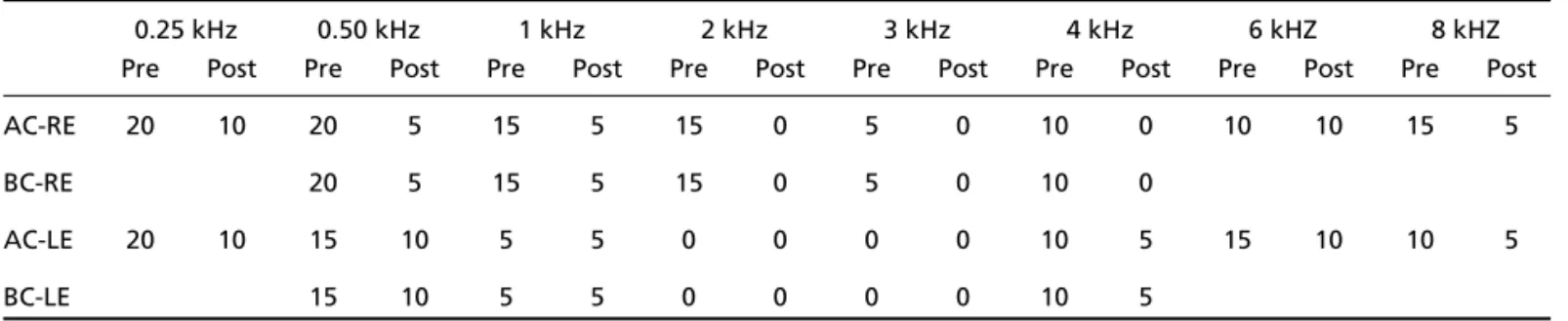
Results – After corticosteroid therapy, pure tone thresh-olds in the right ear improved by 10-15 dB at all frequen-cies (Table 1). As can be seen in Table 2, the acoustic reflex-es in the right ear also improved by 10-15 dB after
treat-Table 1. Pre- and post-treatment pure tone audiometry.
0.25 kHz 0.50 kHz 1 kHz 2 kHz 3 kHz 4 kHz 6 kHZ 8 kHZ
Pre Post Pre Post Pre Post Pre Post Pre Post Pre Post Pre Post Pre Post
AC-RE 20 10 20 5 15 5 15 0 5 0 10 0 10 10 15 5
BC-RE 20 5 15 5 15 0 5 0 10 0
AC-LE 20 10 15 10 5 5 0 0 0 0 10 5 15 10 10 5
BC-LE 15 10 5 5 0 0 0 0 10 5
AC, air conduction; RE, right ear; BC, bone conduction; LE, left ear.
Table 2. Pre- and post-treatment acoustic reflexes.
0.50 kHZ 1 kHz 2 kHz 4 kHZ
Pre Post Pre Post Pre Post Pre Post
IPSI RE 100 85 95 85 95 90 95 90
CONTRA RE 115 105 100 95 105 95 95 85
IPSI LE 95 90 85 85 80 85 80 90
CONTRA LE 95 100 90 95 90 95 85 85
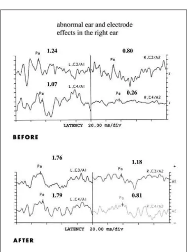
ment. In addition, the speech in noise performance of the right ear improved after treatment (Table 3). Figure 1 shows all BAEP waves for both ears before and after treatment. The Pa amplitudes for all of the electrode sites (C3A1, C3A2, C4A1 and C4A2) increased after treatment (Fig 2). In Figure 3, it can be seen that there was no significant difference between pre- and post-treatment values for P300 latency in either ear.
Table 3. Pre- and post-treatment behavioral evaluation of the auditory pathway.
Speech in noise test SSW test
Pre Post Pre Post
RE 64% 84% 90% 90%
LE 80% 84% 90% 90%
SSW, staggered spondaic word; RE, right ear; LE, left ear.
Fig 1. BEAPs before and after treatment. LE, left ear; RE, right ear.
Fig 2. MLAEPs before and after treatment.
DISCUSSION
The patient presented noticeable improvement in auditory symptoms after corticosteroid therapy. Although post-treatment improvement was observed in the pure tone audiometry results, as well as in tho-se of the acoustic reflex test, it is of note that periph-eral hearing was found to be within normal limits. Therefore, had a more thorough hearing examina-tion not been performed in order to evaluate cen-tral auditory processing, it would not have been pos-sible to identify any alterations.
The post-treatment results of the speech in noise test were normal for the right ear, and the SSW test results were essentially normal before and after treat-ment (Table 3). These results confirm the fact that the patient presented abnormalities at the level of the brainstem but not at the more central level.
Impairment of binaural processing can occur in MS due to the demyelination of many pons structu-res, such as the superior olives and a region between the two inferior colliculi, that are responsible for the function involved in processing speech in noise.
In the electrophysiological evaluation of the right ear, the first BAEP evaluation demonstrated no wave III and a 2-ms delay (7.84 ms) in absolute latency of wave V in comparison to the left ear, evidencing brainstem impairment on the right side. In the sec-ond evaluation, we noticed a decrease in absolute latency of wave V (6.84 ms), 1 ms sooner than in the first evaluation. Matathias et al.2also reported BAEP
abnormalities in 50% of the MS patients studied. Walsh, Kane and Butler7reported that BAEPs are
mo-re likely to be abnormal when demyelination affects the brainstem clinically, but they can also detect “si-lent” lesions in approximately 40% of patients who do not have symptoms or signs of brainstem involve-ment. Delay is probably indicative of the MS-related demyelination of the auditory pathways. It is impor-tant to remember that vascular, inflammatory, neu-rodegenerative, metabolic and infectious conditions may mimic MS lesions on MRIs. Therefore, a diagno-sis of MS may not be made based on the presence of MRI lesions alone and requires corroboration through appropriate clinical or other tests. When MS presents as a clinically isolated syndrome, the criteria for disse-mination over time must be met in order for a defin-itive clinical diagnosis of MS to be made. Dissemi-nation in space may often be demonstrated by evok-ed potential testing.
Bergamaschi et al.3found that BAEP
abnormali-ties decreased progressively to normalization that
coincided with clinical recovery. Although, for our patient, we had no access to BAEP test results obtain-ed prior to symptom onset, recovery was coincident with the decreased wave V latency. Therefore, we strongly recommend that all patients diagnosed with MS (even those presenting no auditory symptoms) be submitted to BAEP testing as a baseline evalua-tion for later comparison.
The first MLAEP test revealed ear and electrode effects for the right ear. The ear effect was not found in the second evaluation. Myelin not only protects nerve fibers but enables their function. When myelin or nerve fiber is damaged or destroyed, the ability of the nerves to conduct electrical impulses to and from the brain is disrupted, and this produces the various symptoms of MS.
Celebisoy et al.6found that 60% of the patients
with confirmed brainstem involvement presented BAEP abnormalities. In addition, the MLAEP results were abnormal in 73.3% of the patients.They con-cluded that the use of BAEP measures in combina-tion with MLAEP measures is a more comprehensive means of evaluating brain function in such patients.
Another study8showed that up to 79% of MS
pati-ents with clinical evidence of brain stem involvement presented abnormal BAEPs.
The P300 and SSW test results were within nor-mal limits before and after treatment, demonstrat-ing that the disease had not affected the higher lev-els (cortical areas) of the auditory pathway. However some researchers have found abnormalities in the LAEPs of patients with MS4,9,10. In such cases, one
might infer that the disease had not affected the au-ditory areas related to the generation of such poten-tial. Therefore, the behavioral and electrophysiolog-ical results were in agreement, contributing to the determination of the site of lesion2,3,4,6,10.
Therefore, we must prioritize the allocation of exist-ing resources. It is feasible to use those resources with precision, thereby achieving two main objectives: the follow-up treatment of pre-established profiles; and the diagnosis of new lesions that might develop.
Using central auditory tests (behavioral and elec-trophysiological), we were able to identify the exact location of the impairment (brainstem), which was in agreement with the patient complaint. We found the speech in noise test and the BAEP test both to be definitely appropriate in confirming brainstem lesions.
REFERENCES
1. Leocani L, Comi G. Neurophysiological investigations in multiple scle-rosis. Curr Opin Neurol 2000;13:255-261.
2. Matathias O, Sohmer H, Biton V. Central auditory tests and auditory
nerve-brainstem evoked responses in multiple sclerosis. Acta Otola-ryngol (Stockholm) 1985;99:369-376.
3. Bergamaschi R, Romani A, Zappoli F, Versino M, Cosi V. MRI and brain-stem auditory evoked potential evidence of eighth cranial nerve involve-ment in multiple sclerosis. Neurology 1997;48:270-272.
4. Jones SJ, Sprague L, Vaz Pato J. Electrophysiological evidence for a defect in the processing of temporal sound patterns in multiple scle-rosis. J Neurol Neurosurg Psychiatry 2002;73:561-567.
5. Musiek FE, Gollegly KM, Kibbe KS, Reeves AG. Electrophysiologic and behavioral auditory findings in multiple sclerosis. Am J Otol 1989;10:343-350.
6. Celebisoy N, Aydogdu I, Ekmekci O, Akurekli O. Middle latency audi-tory evoked potentials (MLAEPs) in MS. Acta Neurol Scand 1996; 93:318-321.
7. Walsh P, Kane N, Butler S. The clinical role of evoked potentials. J Neurol Neurosurg Psychiatry 2005;76(Suppl 2):S16-S22.
8. Robinson K, Rudge P. Abnormalities of the auditory evoked potentials in patients with multiple sclerosis. Brain 1997;100:19-40.
9. Joy JE, Johnson RB. Multiple sclerosis: current status and strategies for the future. Neurology 1997;48:270-272.