REVIEW
aDivision of Physiology, School of Medicine, University of California - San
Diego/CA.
bDepartment of Kinesiology, California State University - Fullerton/CA.
Received for publication on 26/11/07 Accepted for publication on 29/12/07
STRATEGIES FOR CARDIOPULMONARY EXERCISE
TESTING OF PECTUS EXCAVATUM PATIENTS
Moh H. Malek,a Jared W. Coburnb
Malek MH, Coburn JW. Strategies for cardiopulmonary exercise testing of pectus excavatum patients. Clinics. 2008;63:245-54.
The purpose of this paper is to provide strategies for cardiopulmonary exercise testing of pectus excavatum patients. Currently, there are no standardized methods for assessing cardiovascular and pulmonary responses in this population; therefore, making comparisons across studies is difficult if not impossible. These strategies are intended for physicians, pulmonary technicians, exer-cise physiologists, and other healthcare professionals who conduct cardiopulmonary exerexer-cise testing on pectus excavatum patients. By using the strategies outlined in this report, comparisons across studies can be made, and the effects of pectus excavatum on cardiopulmonary function can be assessed with greater detail.
KEYWORDS: Funnel Chest/diagnosis. Circulatory and Respiratory Physiology. Thoracic Wall/abnormalities.
BACKGROUND
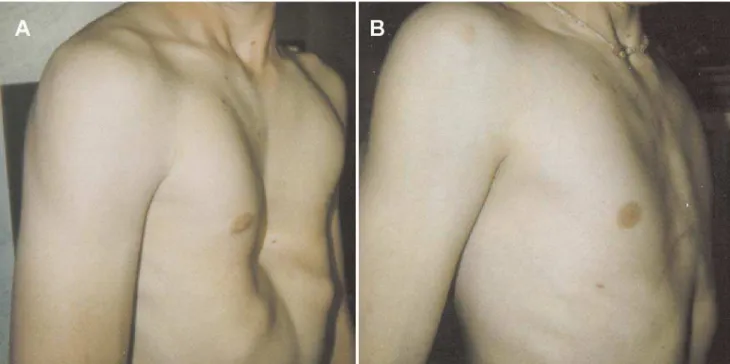
Pectus excavatum (Figure 1) is a relatively common con-genital deformity of the chest wall, with an incidence of ap-proximately one in every 300 to 400 Caucasian male births.1
This condition is more common than Down syndrome, which occurs one in every 600 to 1,000 births.2 Although
the pathogenesis of pectus excavatum remains unclear, investigators have hypothesized that the deformity results from unbalanced overgrowth in the costochondral regions. As a result, the chest appears concave, and a displaced heart is often palpable on the left mid-axillary line slightly below the armpit. Pectus excavatum occurs more often in males than females (6:1) and accounts for 90% of congenital chest wall deformities.3,4 Approximately 40% of pectus excavatum
patients are aware of one or more members of their family who have pectus deformities; however, a genetic link has not been established.4
The severity of pectus excavatum can be calculated by di-viding the inner width of the chest at the widest point (a) by the distance between the posterior surface of the sternum and anterior surface of the spine (b) as determined by computed tomography (CT) scans or chest radiographs (Figure 2).5 The
normal chest has an index of 2.5, however, in our experience we have observed symptomatic pectus excavatum patients with severity indices ranging from 3.2 to 8.0.6,7 It should be
noted that older patients often experience more severe symp-toms with a lower index than do adolescents.4
Recently, Malek and colleagues8-10 conducted two
meta-analyses examining the effects of surgical repair on cardiovascular and pulmonary function in pectus excava-tum patients. The investigators reported that cardiovascular function significantly and clinically improved after surgical repair (ES = 0.59; P < 0.05),9 whereas pulmonary function
did not significantly improve after surgical repair (ES = 0.08;P > 0.05).10 A salient finding in the meta-analyses of
Malek et al.9,10 was the fact that many studies did not
uti-lize a standardized method of determining cardiovascular function, pectus severity index, or control for potentially confounding variables, such as the subject’s habitual physi-cal activity level preoperatively and postoperatively. Based on the results of Malek et al.9,10 and our experience6,7 with
strategies related to assessing cardiopulmonary function in pectus excavatum patients is warranted in order to more effectively compare results between studies without em-ploying a meta-analytic approach. Although there are two textbooks11,12 and one paper13 on exercise testing and
inter-pretation, they are not specific to pectus excavatum patients and do not provide step-by-step strategies for performing cardiopulmonary exercise testing (CPET). Therefore, the purpose of this paper is to provide specific strategies for examining cardiopulmonary function in pectus excavatum patients.
Introduction to Cardiopulmonary Testing (CPET)
There are various invasive and noninvasive methods of assessing cardiovascular and ventilatory function, however, the use of CPET allows examination of the integrative re-sponses of the cardiovascular and ventilatory response to maximal incremental exercise.11-13 CPET is an invaluable
assessment tool used to 1) classify individuals for health risk, 2) quantify training intensity for aerobic exercise prescription, and 3) monitor the effects of aerobic training programs in healthy and clinical populations13-15. Although
other assessment techniques such as electrocardiograph, questionnaires, and/or submaximal exercise protocols have been used to estimate exercise tolerance, Meyers16 stated, “…
measurements of ventilation and gas exchange responses… [are] the only modality that provides an accurate and objec-tive expression of exercise capacity.” (p.S49). CPET can be performed using different modes of exercise that include the treadmill,17 arm ergometry,18 single-leg knee extension
ergometry,19,20 or cycle ergometry.7 Although each mode has
associated advantages and disadvantages, it is our position that studies examining cardiopulmonary function in pec-tus excavatum patients should use cycle ergometry. Cycle ergometry provides a number of advantages over the other modes of exercise testing: 1) subject comfort, 2) reduction of potential joint injuries associated with weight-bearing exercise, 3) control of cadence, and 4) researcher control of the external work rate.12,13 This last feature is particularly
important since small increments in work rate over the dura-tion of the test will allow for detecdura-tion of subtle changes in
Figure 2 - Measurement of the pectus severity index using a CT scan. This is calculated by dividing the inner width of the chest at the widest point (a) by the distance between the posterior surface of the sternum and anterior surface of the spine (b)
cardiovascular and ventilatory function, which may provide insight into the patient’s exercise tolerance.
Exercise Physiology of CPET
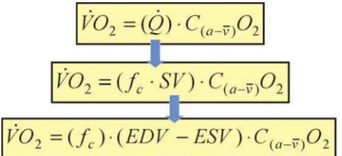
As shown in Figure 3, one of the primary components used to describe cardiopulmonary function is oxygen uptake (VsO2) which is represented by the Fick equation where Qs
is cardiac output and C(a-v–
)O2is the difference between the
oxygen content of the arterial ( 20 ml of O2 ml O2·100 ml-1
of blood) and venous sides (varies with exercise intensity). Cardiac output, however, is a product of fc (cardiac frequen-cy) and stroke volume (SV). Stroke volume can be further decomposed to end-diastolic volume (EDV) and end-systolic volume (ESV). During incremental cycle ergometry, SV in-creases to a percentage of VsO2max and plateaus,21 whereas f
c
continues to increase and may eventually plateau at Vs
O2max. The C(a-v–
)O2 gradient during rest is approximately 4 ml
O2·100 ml-1of blood, whereas at near-maximal exercise
ca-pacity this gradient increases to approximately 16 ml O2·100 ml-1. Thus, the maximal volume of oxygen uptake (Vs
O2max) represents the maximal amount of oxygen transported and utilized during aerobic metabolism.11,13 Related to pectus
excavatum patients, the impairment of one or more compo-nents in this equation results in decreased exercise tolerance. It should be noted, however, that VsO2max can be influenced by various other factors, such as age, gender, and habitual exercise history.7,22-25 Also, direct and indirect measurements
ofSV, EDV, ESV, and C(a-v–
)O2are possible; however, these
procedures often require specialized equipment, are imprac-tical, and do not offer the same advantages as CPET. There-fore, the examination of gas exchange indices measured at the mouth via a two-way breathing valve and metabolic cart provides a more practical alternative that is easily tolerable with healthy and clinical populations.11
The following sections are guidelines for assessing pectus excavatum patients during CPET. The preparation
phase is described, followed by the testing phase and then an interpretation of various physiological indices.
Cardiopulmonary Test (Preparation Phase)
Documenting Patient’s Exercise History
In order to assess the severity of pectus excavatum, in-vestigators need to document the patient’s habitual exercise history. Studies have found that in untrained individuals,
VsO2max can increase as much as 20% following 12 weeks of endurance training.11,26 The adaptations to endurance
train-ing, however, will not be retained (i.e., deconditioning) if a minimal level of exercise is not maintained. Studies27-29 have
shown that seven to eight weeks of deconditioning, in mod-erately training individuals, resulted in a complete reversal ofVsO2max values. Related to components of the Fick equa-tion, Perhonen et al.30 found that after two weeks of bed rest,
Qs and SV decreased by 17% and 25%, respectively, from baseline values. Furthermore, the investigators30 reported
an 8.0% decrease in left ventricular volume after six weeks of bed rest, whereas the right ventricular volume decreased
by 10% during the same amount of time. Coyle et al.31
hypothesized that the decrease in SV may be linked to the reduction in blood volume and not necessarily decreases in cardiac dimensions. Perhonen et al.32 reported, however, that
reductions in SV measured in an upright position were “… greater after bed rest than after acute hypovolemia alone…” (p.1856). With regard to C(a-v–
)O2, studies have reported a
re-duction in the oxygen extraction capabilities of the skeletal muscle, which may be mediated by factors such as capillary density, myoglobin concentration, mitochondria size and density, and oxidative enzymes.27,33
When evaluating the pectus excavatum patient preopera-tively and postoperapreopera-tively, investigators need to document the patient’s habitual exercise history in order to minimize the confounding effects of deconditioning. This is a critical component of the evaluation process, because in the months following the surgical repair, patients may either reduce their level of physical activity or adopt a sedentary lifestyle in order to prevent displacement of the Adkins strut1 or
Lorenz metal bar.34 Therefore, it is recommended that the
mode (i.e., type of exercise performed), frequency (i.e., ses-sions per week), duration (hours per week), length of time the exercise regimen has been consistently maintained, and intensity of exercise be documented for each patient. Malek and colleagues24,25 used the following series of questions to
document the habitual exercise history of individuals tested in their laboratory: “What type of exercise do you perform?”; “How many sessions per week do you exercise?”; “How many hours per week do you exercise?; “How long have you consistently, no more than one month without exercise, been
Figure 3 - The Fick equation for calculating oxygen uptake (9s
2). Refer
to text for detailed discussion. 4s
is cardiac output; C(a-v–
)O2 is the difference
between the oxygen content of the arterial ( 20 ml of O2 mL O2·100 mL-1
of blood) and venous sides; fc is cardiac frequency; SV is stroke volume;
exercising?; and “Indicate [using the Borg Rating of Per-ceived Exertion (RPE) 6-20 scale], in general, the intensity at which you perform your exercise regimen.
Subject Preparation
Before the CPET can begin, a number of procedures must be performed. In their meta-analyses, Malek and colleagues9,10 reported that studies varied in their approach
to measuring pectus severity. Therefore, in order to com-pare findings across studies in the future, pectus severity should be assessed using the procedures of Haller et al.5
This approach uses the CT scan and provides a more objective method of estimating pectus severity than do other approaches such as the Welch index,35 which utilizes
lateral chest x-ray.5 It should be noted, however, that CT
scans are not part of standard assessment procedures for evaluating pectus severity; they may be cost-prohibitive. Nevertheless, investigators should justify this cost in their grant or department budget. In addition, if a complete pulmonary function test has not been conducted, then an abbreviated version should be performed to determine forced expiratory volume in the first second (FEV1), forced vital capacity (FVC), and maximum voluntary ventilation (MVV) values (see ventilatory response variables section for details). The 12-lead ECG electrodes should be placed in accordance with standard guidelines.15 Although there
is little information in the literature regarding the patterns of heart rhythm in pectus excavatum patients, we recom-mend placement of ECG electrodes such that any abnormal rhythms may be documented. Depending on the severity of the pectus excavatum, however, electrode placement may have to be modified (particularly leads v1, v2 and v3) at the discretion of the attending physician and/or exercise physiologist.
Cardiopulmonary Test (Exercise Phase)
Prior to beginning the CPET, the seat height of the cycle ergometer should be adjusted so that the patient has a slight bend ( 5°) in their knees. The handle bars should be ad-justed so the patient is not leaning forward but rather has an erect posture. Furthermore, the mouthpiece and breathing valve should be adjusted so that the patient is not strug-gling to maintain the apparatus in their mouth. The nose clip should be properly placed over the nostrils so that no air is escaping as the patient exhales. Also, the metabolic cart should be calibrated prior to each testing session. The CPET protocol that is recommended includes three minutes of rest (i.e., baseline), followed by three minutes of warm-up at unloaded pedaling (i.e., 0 W), and then an incremental (i.e., ramp) increase in power output every minute (15
to 20 W min-1) thereafter.6,7,11,12 The determination of the
ramp rate often depends on the patient’s level of physical conditioning and, therefore, should be individualized. For example, a faster ramp rate (i.e., 20 or 25 W min-1) is
appro-priate for those individuals who engage in regular endurance exercise, whereas a slower ramp rate (i.e., 10 or 15 W min-1)
may be more appropriate for those individuals who are disabled or sedentary.13 The ramp protocol is recommended
over a step protocol (i.e., increase in power output every two or three minutes), because the physiological responses are more uniform.36 The preferred cycling cadence, in a
clini-cal setting, may range between 60 and 70 rev min-1. In our
experience with pectus excavatum patients, we have found that 60 rev min-1is well tolerated across different degrees of
severity.6,7
The criteria for terminating the CPET may range from absolute (i.e., acute myocardial infarction, sustained ven-tricular tachycardia, or request to stop by the subject) to relative (i.e., hypertensive response, changes in ST segment depression or elevation, or exercise-induced bundle branch block not distinguishable from ventricular tachycardia) indicators.12,13 If, however, none of the above indicators are
present, patients should receive strong verbal encouragement in order to achieve the upper limits of their physiological capacity. In our laboratory,7,17,23,24,37,38Vs
O2max is considered achieved if two of the following three criteria during the test are met:13,39,40 a) 90% of age-predicted maximum heart rate
(i.e., 220–age), b) respiratory exchange ratio > 1.20, and c) a plateauing of oxygen uptake ( 150 mL min-1 in Vs
O2 over the last 30 seconds of the test). In addition, Vs
O
2max is
deter-mined by taking the highest Vs
O2 value in the last 30 seconds of the CPET. It is important to note that the patient be al-lowed to cool-down following the CPET for as long as they want, with f
c, blood pressure, and ECG being continually
monitored until these indices return to those values observed during warm-up.
Physiological Response Variables and Interpretation
Due to the lack of consistent physiological variables that are reported in the pectus excavatum literature resulting from CPETs,9,10 the following section will focus on aerobic
the same information in their manuscripts without incurring additional cost for specialized equipment and/or personnel. In addition, data from the metabolic cart should be reported using moving averages. Most metabolic carts have a range of sampling options from breath-by-breath to 2-minute aver-ages. We recommend that researchers use a moving average (5- or 8-breath). Myers et al.41 reported that 5- and 10-second
averages of breath-by-breath data resulted in high variability ( 4.5 and 3.5 mL kg-1min-1, respectively), whereas moving
averages (i.e., 5- or 8-breath) resulted in lower variability ( 1.7 and 1.6 mL kg-1min-1). The investigators41 concluded that
the “…gain in accuracy is attained by using larger samples or averaging breaths. Although 60-s sampling offers the least variability (SD = 0.08 mL kg-1min-1), it could be argued
that samples this large may be too imprecise for evaluating certain interventions.” (p.409).
Aerobic Capacity Response Variables
The response to exercise is a function of numerous physiological mechanisms. The ability to sustain high-intensity exercise is contingent on four aerobic parameters: 1) VsO2max; 2) the gas exchange threshold (GET), above which there is a sustained increase in blood lactic acid con-centration; and 3) work efficiency represented as the slope of Vs
O2/ Ws
42. It should be noted that the fourth aerobic
parameter, the time constant for oxygen uptake kinetics ( Vs
O2max), requires multiple constant-power output exer-cises, which are performed over a number of visits to the laboratory;43 therefore, it is not a practical assessment tool
for this population. Thus, we focus our discussion on the first three aerobic parameters.
As discussed earlier, Vs
O
2max is a measure of aerobic
power and should be reported in both absolute (L min-1)
and relative (mL kg-1min-1) terms. The normalization of
Vs
O2max to body weight in kilograms allows the researcher to compare the patient’s Vs
O
2max to normative data as well as
other pectus excavatum patients within a sample and across studies. In addition, the patient’s Vs
O
2max value should be
re-ported as a percentage of their predicted Vs
O2max. Although there are numerous Vs
O
2max prediction equations in the
lit-erature, we recommend that investigators use the equations by Cooper and Storer,12 because these were derived from a
composite of five to six prediction equations that included a wide range of clinical populations as well as sedentary to mildly active individuals:
Vs
O2max L min-1 = ((0.0716*ht)–0.0518)*(44.22 –
(0.394*age))+(0.0058*ABW) [men]
Vs
O
2max L min
-1 = ((0.0626*ht)–0.0455)*(37.03–
(0.371*age))+(0.0058*ABW) [women]
where ht is the patient’s height in meters, age is measured in years, and ABW is the actual body weight of the patient in kilograms. The equations of Cooper and Storer,12 however,
are not appropriate to predict VsO2max for aerobically trained individuals/patients, which Malek et al.24,37 operationally
defined as “…[individuals] who had participated in continu-ous aerobic exercise three or more sessions per week for a minimum of one hour per session, for at least the past 18 months.” Therefore, in such cases, the following equations by Malek et al.24,37 should be used to predict Vs
O2max in aerobically trained pectus excavatum patients:
Vs
O2max mL min-1 = 27.387(wt) + 26.634(ht) - 27.572(age) +
26.161(HPW) + 114.904(INT) + 506.752(YRT) - 4609.791 [men]
Vs
O2max mL min-1 = 18.528(wt) + 11.993(ht) - 17.197(age) +
23.522(HPW) + 62.118(INT) + 278.262(YRT)-1375.878 [women]
where wt is the patient’s weight in kilograms, ht is the patient’s height in meters, age is measured in years, HPW is the hours per week of training, INT is the overall intensity of each training session as measured by the Borg RPE (6-20) scale, and YRT is the natural log of years of training. Regardless of which prediction equation is used, the researcher should also report the pectus excavatum patient’s
VsO2max relative to the percentage of the predicted value (i.e., %predict. Vs
O2max = [observed Vs
O2max / predicted
Vs
O
2max] x 100).
The GET is also known as the lactate acidosis threshold, or ventilatory threshold, but these terms are essentially used to describe the disproportionate increase in CO2 output relative to oxygen uptake resulting from an accelerated reliance on glycolysis for energy production during incre-mental exercise11,12. The GET has been used to identify
an individual’s level of aerobic fitness in clinical44,45 and
sports46,47 related settings, as well as for monitoring training
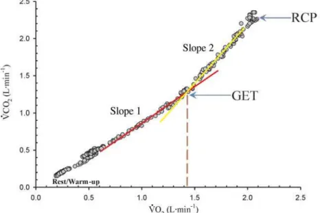
adaptations.22 The GET is determined through the V-slope
method48 by using either regression analysis or visual
in-spection by trained personnel. Briefly, as shown in Figure 4,Vs
O 2 and V
s
CO
2 increase proportionately at the beginning
of incremental exercise, thus yielding a linear slope. As the exercise bout continues, a second slope develops due to the disproportionate increase in CO2 output relative to oxygen uptake.11,13 However, because of the potential of acute
Gaskill et al.49 recently reported that determining the GET
using a combination of the V-slope, ventilatory equivalents, and end-tidal gas tensions methods was more reliable than using any of these methods separately. The GET values should be reported in terms of oxygen uptake similar to the
VsO2max values (i.e., L min-1 and mL kg-1min-1) as well as
a percentage of the predicted Vs
O2max (i.e., %predVs
O2max =
[measured GET / predicted VsO2max] x 100).12 It is
recom-mended that the predicted Vs
O2max value be used, and not the measured Vs
O
2max value, because the “…measured
Vs
O2max can produce serious errors of interpretation in the case of a suboptimal effort…” (p.103),12 which is more
likely in clinical settings. Typically, in healthy individuals, the %predVs
O2max for GET occurs at 50% to 60%, whereas
in diseased populations, this value is often equal to or less than 40%.12 For example, Malek et al.7 reported that
physi-cally active pectus excavatum patients with a severity index greater than 5.0 had GET values 39%. Thus, investigators can use %predVs
O2max to characterize the level of conditioning
in their sample, which may provide insight into the patient’s muscle energetics.
The measure of work efficiency, Vs
O
2/ W s
, is inde-pendent of gender, age, or height and has a consistency for apparently healthy subjects, being 10.3 mL min-1W-1 (SD =
1.0 mL min-1W-1) with a 95% confidence interval of 8.3 to
12.3 mL min-1W-1.12,13 The Vs
O
2/ W s
examines the oxygen utilization by the skeletal muscles in their performance of work. Poole et al.50 found that the Vs
O
2/ W s
calculated from pulmonary gas exchange measurements and that measured directly from the leg (i.e., constant-infusion thermodilution) had similar values (9.9 vs. 9.2 mL·min-1·W-1). Therefore,
the investigators50 concluded that Vs
O2/ Ws
represents the muscular efficiency of the exercising limb. A reduction in
Vs
O2/ Ws
may indicate inadequacies with factors related to oxygen transport [for a detailed review, refer to citation 33].
Cardiovascular Response Variables
Heart Rate (fc) and Cardiac Reserve
The cardiovascular response to incremental exercise is one potential limitation to exercise tolerance.11 In
pec-tus excavatum patients, the assessment of cardiovascular variables can provide valuable information related to the efficiency of surgical repair as well as the patient’s response to maximal exercise. Some researchers have used the pre-dictedf
cmax as a marker for achieving the upper-limits of
exercise tolerance, and thus terminate the CPET on this basis. It is well-agreed in the exercise physiology literature that this approach is inappropriate because of the vari-ances associated with the available prediction equations for
fcmax.16 Despite this drawback, however, the patient’s f
cmax
relative to their predicted value should be reported. Typi-cally, one of two prediction equations are used to estimate af
cmax: 1) 220–age or 2) 210-(age x 0.65).
11,12 Regardless
of which equation is used to estimate fcmax, investigators should report the mean and standard deviation of the actual
fcmax and the percentage of predicted (i.e., [actual fcmax / predictedf
cmax] x 100) for their sample. In addition to
re-porting fcmax, the resting fc value, which is recorded during the baseline phase of the CPET, should also be reported by the investigator.
Figure 4 - Determination of GET by V-slope method (9s
&29
s
2 following completion of a CPET. Note: Rest and warm-up data, as well as data points
Related to f
cmax is cardiac reserve, defined as the
dif-ference between the measured and predicted fcmax values. This information can be used to determine whether the sub-ject achieved cardiovascular limitation. As a general rule, a small cardiac reserve value and a high ventilatory reserve value (discussed below) indicates cardiovascular limitation to incremental exercise.11,12
Oxygen Pulse (Vs
O2max / fcmax)
Due to the close relationship between fc and VsO2 (Fig-ure 3), the slope of the fc / Vs
O2 relationship can provide information related to the patient’s level of cardiovascular conditioning. fc/ Vs
O2 is the slope of the relationship between heart rate and VsO2 during incremental exercise. This slope is related both to stroke volume and to the differ-ence in oxygen content between arterial and mixed venous blood11-13. f
c/ V
s
O2is also the reciprocal of the asymptotic oxygen pulse (VsO2/fc), which is a measure of cardiovascular efficiency with units of milliliters per beat.11-13 Thus, Vs
O2/fc
is closely related to SV and may be used to estimate SV at various stages of incremental exercise testing.11-13
Ventilatory Response Variables
The examination of ventilatory indices during incremental CPET provides information related to the pectus excavatum patient’s pulmonary function. Previous studies have found that values for pulmonary function indices, such as FEV1, FVC, and MVV, are in the low normal range and that pectus excavatum does not influence the patient’s overall pulmonary function.6,7,10,51-53 (For a detailed review of the effects of
pec-tus excavatum on pulmonary function, refer to citation 10). Because pulmonary function tests are effort-dependent, it is recommended that the patient perform several trials with strong verbal encouragement from the pulmonary technician. For detailed guidelines regarding pulmonary function testing, refer to citation.54 Consistent with these findings, Malek et
al.10 found a small mean-weighted effect size (ES = 0.07, P
>0.05) for pulmonary function following surgical repair, but noted that many of the indices were measured at rest and not during exercise. It is well-established in the clinical exercise physiology literature that examination of ventilatory function during CPET is equally as important in patient assessment as cardiovascular function.11-13,55 Therefore, future studies should
examine and report the following ventilatory responses to CPET in order to more accurately assess the potential impair-ment of pectus excavatum on pulmonary function.
Minute Ventilation (Vs
E)
At rest, breathing frequency (f
R) in healthy individuals
is approximately 12 to 15 beats·min-1, whereas tidal volume
Table 1 - Step-by-step approach for CPET of pectus exca-vatum patients
Preparation Phase
– Document patients’ exercise history using questions outlined in the text. Subject Preparation
– Use of CT scan to estimate Haller pectus severity index.
– At a minimum, measure FEV1, FVC, and MVV at part of pulmonary function test.
–Place ECG electrodes. Exercise Phase
– Ensure cycle ergometer, subject, breathing valve, and metabolic cart are appropriately set up.
Aerobic capacity response variables
– Report 9s
2max (L min-1 and mL kg-1min-1) and as a percentage of
predicted9s
2max.
– Estimate GET using the V-slope method and report value in terms of
9s
2 (L min-1 and mL kg-1min-1), as well as a percentage of
predicted 9s
2max.
– Report work efficiency ( 9s
2 :
s ). Cardiovascular response variables
– Report resting heart rate (fc) as well as fc at 9
s
2max (i.e., fcmax).
– Report cardiac reserve (i.e., fcmax - predicted fcmax).
Ventilatory response variables
– Report minute ventilation at 9s
2max (i.e., 9
s
(max, L min-1) as a
per-centage of MVV.
– Report ventilatory reserve (i.e., measured MVV - 9s (max). Note: Refer to text for discussion of each variable and procedure.
(V
T) is 0.500 liters. Minute ventilation (V
s
E) is thus the
prod-uct of fR and VT and is typically 6.0 L·min-1 at rest. However,
during vigorous exercise fR and VT increase to 50 beats·min-1
and 3.0 L·min-1, respectively, resulting in a Vs
E of 150 L·min
-1.
It should be noted, however, that during low-intensity exer-cise, VT primarily increases, whereas during higher-intensity exercise (i.e., 80% of Vs
O2max), both VT and fR increase, with onlyfR increasing as the individual approaches VsO2max.11-13
Vs
E is a critical component of regulating acid-base balance
during increased metabolic demands by the exercising muscles.11-13 During the CPET, the relationship between CO
2
output and VsE is mostly linear, up to approximately 85 to 90% of Vs
O2max.11-13 Thereafter, Vs
E increases
disproportion-ately to CO2 output. This breakpoint is called the respira-tory compensation point (RCP). The underlying mechanism of the RCP is related to the stimulation of the peripheral chemoreceptors of the carotid bodies.
Ventilatory Reserve
Vs
Emaxvalue attained at V
s
O
2max as a percentage of the
pa-tient’s MVV value (i.e., %MVV = [VsEmax/MVV] x 100). In healthy individuals, the %MVV at Vs
O2max is approximately 50-70%.,11-13 whereas in patients with respiratory disease
(e.g., COPD), this value may be greater than 80%.11,56,57
Johnson et al.58 reported that, in healthy individuals, the
Vs
Emax achieved at maximal exercise capacity was highly
correlated with the measured MVV. In order to better under-stand the physiological limitation of pectus excavatum to exercise, future studies should report the %MVV value for their sample in addition to the Vs
EmaxandMVV values.
CONCLUSION
In summary, Table 1 provides a step-by-step approach that should be used when assessing cardiopulmonary re-sponses in a pectus excavatum patient before and after surgical repair. Although investigators are encouraged to use other laboratory techniques to assess the effects of pectus excavatum on cardiovascular and pulmonary function, it is imperative that, at a minimum, they conduct a CPET as out-lined above and report the corresponding indices.
REFERENCES
1. Fonkalsrud EW, Dunn JCY, Atkinson JB. Repair of pectus excavatum deformities: 30 years experience with 375 patients. Ann Surg. 2000; 231:443-448.
2. Hook EB. Epidemiology of Down Syndrome. In: Pueschel SM, Rynders JE, eds. Down syndrome: Advances in biomedicine and the behavioral sciences. Cambridge: Ware Press. 1982;11-88.
3. Fonkalsrud EW. Chest wall abnormalities. In: Baue AE, Geha AS, Hammond GL, et al., eds. Glenn’s thoracic and cardiovascular surgery. Stamford: Appleton and Lange. 1995;581-592.
4. Fonkalsrud EW. Current management of pectus excavatum. World J Surg. 2003;27:502-508.
5. Haller JA, Jr., Kramer SS, Lietman SA. Use of CT scans in selection of patients for pectus excavatum surgery: a preliminary report. J Pediatr Surg. 1987;22:904-906.
6. Malek MH, Fonkalsrud EW. Cardiorespiratory outcome after corrective surgery for pectus excavatum: A case study. Med Sci Sports Exerc. 2004; 36:183-190.
7. Malek MH, Fonkalsrud EW, Cooper CB. Ventilatory and cardiovascular responses to exercise in patients with pectus excavatum. Chest. 2003; 124:870-882.
8. Malek MH, Berger DE, Marelich WD, Coburn JW. On the application of meta-analysis in pectus excavatum research. Am J Cardiol. in press.
9. Malek MH, Berger DE, Housh TJ, Marelich WD, Coburn JW, Beck TW. Cardiovascular function following surgical repair of pectus excavatum: a meta-analysis. Chest. 2006;130:506-16.
10. Malek MH, Berger DE, Marelich WD, Coburn JW, Beck TW, Housh TJ. Pulmonary function following surgical repair of pectus excavatum: a meta-analysis. Eur J Cardiothorac Surg. 2006;30:637-43.
11. Wasserman K, Hansen JE, Sue DY, Stringer WW, Whipp BJ. Principles of exercise testing and interpretation : including pathophysiology and clinical applications. 4th ed. Philadelphia: Lippincott Williams & Wilkins, 2005.
12. Cooper CB, Storer TW. Exercise testing and interpretation: A practical approach. London: Cambridge University Press, 2001.
13. ATS/ACCP. ATS/ACCP Statement on cardiopulmonary exercise testing. Am J Respir Crit Care Med. 2003;167:211-77.
14. American College of Sports Medicine., Pollock ML, Gaesser GA, Butcher JD, Després J, Dishman RK, Franklin BA, Garber CW. American College of Sports Medicine Position Stand. The recommended quantity and quality of exercise for developing and maintaining cardiorespiratory and muscular fitness, and flexibility in healthy adults. Med Sci Sports Exerc.1998;30:975-91.
15. Fletcher GF, Balady GJ, Amsterdam EA, Chaitman B, Eckel R, Fleg J, Froelicher VF, Leon AS, Pina IL, Rodney R, Simons-Morton DA, Williams MA, Bazzarre T. Exercise standards for testing and training: a statement for healthcare professionals from the American Heart Association. Circulation. 2001;104:1694-740.
16. Myers J. Applications of cardiopulmonary exercise testing in the management of cardiovascular and pulmonary disease. Int J Sports Med. 2005;26:S49-S55.
17. Malek MH, Housh TJ, Schmidt RJ, Coburn JW, Beck TW. Proposed tests for measuring the running velocity at the oxygen consumption (RVVO2)
and heart rate (RVHRT) thresholds for treadmill exercise. J Strength Cond Res. 2005;19:847-852.
19. Richardson RS, Knight DR, Poole DC, Kurdak SS, Hogan MC, Grassi B, Wagner PD. Determinants of maximal exercise Vo2 during single leg knee-extensor exercise in humans. Am J Physiol. 1995;268:H1453-1461.
20. Knight DR, Poole DC, Schaffartzik W, Guy HJ, Prediletto R, Hogan MC, Wagner PD. Relationship between body and leg Vo2 during maximal cycle ergometry. J Appl Physiol. 1992;73:1114-21.
21. Wilmore JH, Costill DL. Physiology of sport and exercise. 3rd ed. Champaign, IL: Human Kinetics, 2004.
22. Jones AM, Carter H. The effect of endurance training on parameters of aerobic fitness. Sports Med. 2000;29:373-86.
23. Malek MH, Berger DE, Housh TJ, Coburn JW, Beck TW. Validity of Vo2max equations for aerobically trained males and females. Med Sci Sports Exerc. 2004;36:1427-32.
24. Malek MH, Housh TJ, Berger DE, Coburn JW, Beck TW. A new non-exercise based Vo2max prediction equation for aerobically trained
females. Med Sci Sports Exerc. 2004;36:1804-10.
25. Malek MH, Housh TJ, Berger DE, Coburn JW, Beck TW. A new non-exercise-based Vo2max prediction equation for aerobically trained men. J Strength Cond Res. 2005;19:559-65.
26. Wilmore JH, Stanforth PR, Gagnon J, Leon AS, Rao DC, Skinner JS, Bouchard C. Endurance exercise training has a minimal effect on resting heart rate: the HERITAGE Study. Med Sci Sports Exerc. 1996;28:829-35.
27. Wilber RL, Moffatt RJ. Physiological and biochemical consequences of detaining in aerobically trained individuals. J Strength Cond Res. 1994;8:110-24.
28. Fringer MN, Stull GA. Changes in cardiorespiratory parameters during periods of training and detraining in young adult females. Med Sci Sports. 1974;6:20-5.
29. Klausen K, Andersen LB, Pelle I. Adaptive changes in work capacity, skeletal muscle capillization and enzyme levels during training and detraining. Acta Physiol Scand. 1981;113:9-16.
30. Perhonen MA, Franco F, Lane LD, Buckey JC, Blomqvist CG, Zerwekh JE, et al. Cardiac atrophy after bed rest and spaceflight. J Appl Physiol. 2001;91:645-53.
31. Coyle EF, Hemmert MK, Coggan AR. Effects of detraining on cardiovascular response to exercise: Role of blood volume. J Appl Physiol. 1986;60:95-99.
32. Perhonen MA, Zuckerman JH, Levine BD. Deterioration of left ventricular chamber performance after bed rest : “cardiovascular deconditioning” or hypovolemia? Circulation. 2001;103:1851-7. 33. Wagner PD. Determinants of maximal oxygen transport and utilization.
Annu Rev Physiol.1996;58:21-50.
34. Park HJ, Lee SY, Lee CS, Youm W, Lee KR. The Nuss procedure for pectus excavatum: evolution of techniques and early results on 322 patients. Ann Thorac Surg. 2004;77:289-95.
35. Welch KJ. Satisfactory surgical correction of pectus excavatum deformity in childhood: A limited opportunity. J Thorac Surg.1958;36:697-713. 36. Myers J, Bellin D. Ramp exercise protocols for clinical and
cardiopulmonary exercise testing. Sports Med. 2000;30:23-9.
37. Malek MH, Housh TJ, Coburn JW, Weir JP, Schmidt RJ, Beck TW. The effects of interelectrode distance on electromyographic amplitude and mean power frequency during incremental cycle ergometry. J Neurosci Methods. 2006;151:139-47.
38. Malek MH, Coburn JW, Weir JP, Beck TW, Housh TJ. The effects of innervation zone on electromyographic amplitude and mean power frequency during incremental cycle ergometry. J Neurosci Methods. 2006;155:126-33.
39. Baumgartner TA, Jackson AS. Measurement for evaluation in physical education and exercise science. 7th ed. Boston: WCB/McGraw-Hill, 2003.
40. Day JR, Rossiter HB, Coats EM, Skasick A, Whipp BJ. The maximally attainable VO2 during exercise in humans: The peak vs. maximum issue. J Appl Physiol. 2003;95:1901-7.
41. Myers J, Walsh D, Sullivan M, Froelicher V. Effect of sampling on variability and plateau in oxygen uptake. J Appl Physiol. 1990;68:404-10.
42. Whipp BJ, Davis JA, Torres F, Wasserman K. A test to determine parameters of aerobic function during exercise. J Appl Physiol.1981;50:217-21. 43. Jones AM, Poole DC. Oxygen uptake kinetics in sport, exercise and
medicine : a practical handbook. New York: Routledge, 2005. 44. Tesiorowski AM, Harris M, Chan KJ, Thompson CR, Montaner JS.
Anaerobic threshold and random venous lactate levels among HIV-positive patients on antiretroviral therapy. J Acquir Immune Defic Syndr. 2002;31:250-1.
45. Thin AG, Linnane SJ, McKone EF, Freaney R, FitzGerald MX, Gallagher CG, et al. Use of the gas exchange threshold to noninvasively determine the lactate threshold in patients with cystic fibrosis. Chest. 2002;121:1761-70.
46. Maffulli N, Testa V, Capasso G. Anaerobic threshold determination in master endurance runners. J Sports Med Phys Fitness. 1994;34:242-9. 47. Tanaka K, Matsuura Y. Marathon performance, anaerobic threshold, and onset of blood lactate accumulation. J Appl Physiol.1984;57:640-3. 48. Beaver WL, Wasserman K, Whipp BJ. A new method for
detecting anaerobic threshold by gas exchange. J Appl Physiol. 1986;60:2020-7.
49. Gaskill SE, Ruby BC, Walker AJ, Sanchez OA, Serfass RC, Leon AS. Validity and reliability of combining three methods to determine ventilatory threshold. Med Sci Sports Exerc. 2001;33:1841-8. 50. Poole DC, Gaesser GA, Hogan MC, Knight DR, Wagner PD. Pulmonary
and leg VO2 during submaximal exercise: implications for muscular
efficiency. J Appl Physiol. 1992;72:805-10.
51. Kaguraoka H, Ohnuki T, Itaoka T, Kei J, Yokoyama M, Nitta S. Degree of severity of pectus excavatum and pulmonary function in preoperative and postoperative periods. J Thorac Cardiovasc Surg. 1992;104:1483-8. 52. Kowalewski J, Barcikowski S, Brocki M. Cardiorespiratory function
before and after operation for pectus excavatum: Medium-term results. Eur J Thorac Cardiovasc Surg. 1998;13:275-9.
54. Miller MR, Hankinson J, Brusasco V, Burgos F, Casaburi R, Coates A, et al. Standardisation of spirometry. Eur Respir J. 2005;26:319-338. 55. Poole DC, Richardson RS. Determinants of oxygen uptake: Implications
for exercise testing. Sports Med. 1997;24:308-320
56. Montes de Oca M, Rassulo J, Celli BR. Respiratory muscle and cardiopulmonary function during exercise in very severe COPD. Am J Respir Crit Care Med, 1996;154:1284-1289
57. Bauerle O, Younes M. Role of ventilatory response to exercise in determining exercise capacity in COPD. J Appl Physiol. 1995; 79:1870-7.
58. Johnson BD, Scanton PD, Beck KC. Regulation of ventilatory capacity during exercise in .asthmatics. J Appl Physiol.1995;79:892-901. 59 Fonkalsrud EW, Beanes S, Hebra A, Adamson W, Tagge E. Comparison