Clinical and laboratorial features of spontaneous bacterial
peritonitis in southern Brazil
Características clínicas e laboratoriais da peritonite bacteriana espontânea
no sul do Brasil
Gabriela Bicca Thiele
I, Otávio Marcos da Silva
I, Leonardo Fayad
II, César Lazzarotto
III, Mariana do Amaral Ferreira
I,
Maíra Luciana Marconcini
I, Esther Buzaglo Dantas-Corrêa
IV, Leonardo de Lucca Schiavon
IV, Janaína Luz Narciso-Schiavon
IVNúcleo de Estudos em Gastroenterologia e Hepatologia (NEGH), Universidade Federal de Santa Catarina (UFSC), Santa Catarina, Brazil
ABSTRACT
CONTEXT AND OBJECTIVE: Spontaneous bacterial peritonitis (SBP) is a severe complication that occurs in 8-27% of hospitalized patients with liver cirrhosis and ascites, with high mortality rates. This study aimed to identify the clinical characteristics associated with SBP.
DESIGN AND SETTING: Cross-sectional study, conducted in a public university.
METHODS: The study consecutively included individuals with liver cirrhosis and ascites between Sep-tember 2009 and March 2012. Forty-ive patients were included: mean age 53.2 ± 12.3 years, 82.2% male, 73.8% Caucasian, mean Model of End-stage Liver Disease (MELD) score of 19.5 ± 7.2, and 33.3% with SBP. The subjects were divided into two groups: SBP and controls.
RESULTS: Comparison between individuals with SBP and controls showed that those with SBP had lower mean prothrombin activity time (36.1 ± 16.0% versus 47.1 ± 17.2%; P = 0.044) and lower median serum-asci-tes albumin gradient (SAAG) (1.2 versus 1.7, P = 0.045). There was a tendency towards higher mean MELD in the SBP group, not signiicant (22.2 ± 7.6 versus 17.9 ± 6.7; P = 0.067). There was a strong positive correlation between the neutrophil count in ascitic luid and serum leukocyte count (r = 0.501; P = 0.001) and a negative correlation between the neutrophil count in ascitic luid with prothrombin activity time (r = -0.385; P = 0.011).
CONCLUSION: A few characteristics are associated with the presence of SBP, especially liver dysfunction, SAAG and peripheral leukocytosis.
RESUMO
CONTEXTO E OBJETIVO: Peritonite bacteriana espontânea (PBE) é uma complicação grave que ocorre em 8-27% dos pacientes hospitalizados com cirrose hepática e ascite, e apresenta altas taxas de mortali-dade. O objetivo deste estudo é identiicar as características clínicas associadas à PBE.
TIPO DE ESTUDO E LOCAL: Estudo transversal, conduzido em uma universidade pública.
MÉTODOS: O estudo incluiu, consecutivamente, indivíduos com cirrose hepática e ascite entre setembro 2009 e março 2012. Foram incluídos 45 indivíduos com média de idade de 53,2 ± 12,3 anos, sendo 82,2% homens, 73,8% brancos, com MELD (Modelo para Doença Hepática Terminal) de 19,5 ± 7,2, e 33,3% com PBE. Os indivíduos foram divididos em dois grupos: PBE e controles.
RESULTADOS: Quando se compararam os indivíduos com PBE aos controles, observou-se menor média de tempo de atividade da protrombina (TAP; 36,1 ± 16,0% versus 47,1 ± 17,2%; P = 0,044) e menor mediana de gradiente albumina soro-ascite (GASA; 1,2 versus 1,7; P = 0,045). Houve tendência do grupo com PBE de apre-sentar maior média de MELD, sem signiicância estatística (22,2 ± 7,6 versus 17,9 ± 6,7; P = 0,067). Foi obser-vada forte correlação positiva entre neutróilos do líquido ascítico e contagem sérica de leucócitos (r = 0,501; P = 0,001) e correlação negativa de neutróilos do líquido ascítico com TAP (r = -0,385; P = 0,011).
CONCLUSÃO: Poucas características estão associadas à presença de PBE, em especial a disfunção hepáti-ca, o GASA e a leucocitose periférica.
IMedical Student. Universidade Federal de Santa Catarina (UFSC), Florianópolis, Santa Catarina, Brazil.
IIMD. Resident in Gastroenterology, Núcleo de Estudos em Gastroenterologia e Hepatologia (NEGH), Universidade Federal de Santa Catarina (UFSC), Florianópolis, Santa Catarina, Brazil. IIIMD, MSc. Resident in Gastroenterology, Núcleo de Estudos em Gastroenterologia e Hepatologia (NEGH), Universidade Federal de Santa Catarina (UFSC), Florianópolis, Santa Catarina, Brazil. IVMD, PhD. Adjunct Professor in Gastroenterology, Núcleo de Estudos em Gastroenterologia e Hepatologia (NEGH), Universidade Federal de Santa Catarina (UFSC), Florianópolis, Santa Catarina, Brazil.
KEY WORDS: Liver cirrhosis. Ascites. Peritonitis. Ascitic luid. Paracentesis.
INTRODUCTION
Spontaneous bacterial peritonitis (SBP) is found in 8% to 27% of the patients hospitalized with liver cirrhosis and ascites, and presents high rates of intra-hospital mortality, of between 20 and 40%.1-3 Studies have suggested that the recurrence rates are high: more than 70% within one year.4.5
In the great majority of the cases, the bacteria that cause SBP come from the digestive tract. Extra-intestinal bacteria, such as those from the respiratory and urogenital tracts or the skin, are much less frequent. Catheters and other equipment used during invasive procedures represent another possible source of infec-tion. Currently, the most accepted hypothesis regarding the pathogenesis of SBP consists of a bacteremia episode during luid exchange between the peritoneal and intravascular cavities, with consequent infection of the ascitic luid.6 Aerobic Gram-negative bacteria (most frequently Escherichia coli) are considered respon-sible for the majority of SBP cases, through translocation of the intestinal lumen.5,7
In fact, only a few patients with SBP present typical symptoms suggestive of peritoneal infection, such as fever, abdominal pain and peripheral leukocytosis. SBP is most frequently suspected when the patient develops signs of hepatic encephalopathy, increased abdominal volume or renal dysfunction, without any apparent precipitating factor. In addition to this, in a signiicant proportion of the cases, SBP may be completely asymptomatic and the diagnosis can only be made by analyzing the paracente-sis results.8 If ascitic luid infection is suspected (fever, abdominal pain, unexplained encephalopathy, azotemia, acidosis, hypoten-sion or hypothermia), total and diferential cellularity tests and ascitic luid culturing need to be requested, with inoculation of the material into blood culturing bottles at the bedside.9,10
he diagnosis for SBP consists of polymorphonuclear (PMN) counts ≥ 250 cells/mm³ and a positive ascitic luid culture, with-out any evidence of external or intra-abdominal infectious source. Neutrophilic ascites is deined by negative cultures and also by PMN counts in the ascitic luid higher than 250 cells/mm³. Presence of a positive ascitic luid culture with neutrophil count less than 250 cells/mm³ is diagnosed as bacterascites, and manage-ment of this condition varies from patient to patient.11
Given the scarcity of studies assessing characteristics of patients with SBP in Brazil, and also the regional diferences that exist, there is a need to evaluate this infection in our setting. hrough observing its behavior, new strategies aiming towards diagnostic improvement can be sought.
OBJECTIVE
he main objective of this study was to identify the characteris-tics associated with presence of SBP in individuals with decom-pensated liver cirrhosis with ascites. We also aimed to describe the clinical proile of individuals with SBP.
METHODS
his cross-sectional analytical study was conducted by review-ing the medical records of individuals with decompensated liver cirrhosis with ascites who were admitted to the gastroenterology ward of the “Polydoro Ernani de São hiago” university hospital of the Federal University of Santa Catarina (Universidade Federal de Santa Catarina, UFSC), between September 2009 and March 2012. Over the same period, we evaluated the results from ascitic luid cultures made in the university hospital laboratory of UFSC, for inclusion in the study. Among these, ater analysis of the medical iles, we excluded the patients who did not have cirrhosis (patients with ascites from other causes) and also those with insuicient clinical and laboratory data registered in their medical records.
he study protocol conformed to the ethical guidelines of the 1975 Helsinki Declaration and had been approved by our institu-tional review board under the number 885/10.
Patients were interviewed regarding their demographic and clin-ical characteristics as stated below. Additional clinclin-ical and laboratory variables relating to all the individuals from medical charts. he fol-lowing clinical variables were studied: age; gender; skin color; SBP (which was deined as a neutrophil count in ascitic luid higher than 250 cells/mm³ and/or a positive culture); jaundice; hepatic encepha-lopathy; upper gastrointestinal bleeding (UGIB) during hospitaliza-tion; ascitic luid cultures; maximum axillary temperature; abdomi-nal pain; diarrhea; comorbidities: diabetes mellitus (DM), systemic arterial hypertension (SAH), dyslipidemia and HIV; etiology of the cirrhosis: alcohol, hepatitis C or hepatitis B; and duration of prophy-lactic antibiotics for paracentesis. Among the laboratory variables, the following were evaluated: neutrophil count in ascitic luid; pro-thrombin activity (PA); serum-ascites albumin gradient (SAAG); hemoglobin; leukocytes; platelet count; aspartate aminotransferase (AST), alanine aminotransferase (ALT), alkaline phosphatase (ALP) and gamma glutamyl transferase (GGT); albumin; total and direct bilirubin; creatinine; sodium; glucose; total protein in the ascitic luid; and albumin in the ascitic luid. he results from the liver biochemical tests (AST, ALT, ALP and GGT) were expressed as the number of times the upper limit of normal (xULN). he other laboratory variables were expressed as absolute values. he biliru-bin tests, international normalized ratio (INR) and creatinine were used for calculating MELD (Model of End-stage Liver Disease).12 For analysis purposes, the patients were divided into two chronolog-ical groups: SBP and controls (cirrhotic individuals with ascites but no evidence of infection).
Statistical analysis
luid and laboratory variables was assessed using Pearson’s cor-relation coeicient. All tests were performed using the Statistical Package for the Social Science (SPSS, Chicago, Illinois, United States), version 17.0.
RESULTS
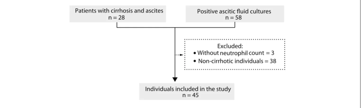
From September 2009 to March 2012, 86 patients were evaluated for inclusion in the study because they presented decompen-sated cirrhosis with ascites and/or positive results from ascitic luid cultures in the laboratory. hree individuals were excluded from the study because no neutrophil count in the ascitic luid was available, along with another 38 patients who did not present cirrhosis (Figure 1).
We included 45 patients with decompensated cirrhosis with ascites in the study, and 15 of these (33.3%) presented SBP (Table 1). he mean age was 53.2 ± 12.3 years; 82.2% of the patients were men and 73.8% of them were Caucasian. About 60% of the individu-als were classiied as presenting Child-Pugh C and a mean MELD score of 19.5 ± 7.2. No individual was classiied as Child-Pugh A, and only one presented MELD score ≤ 10. Besides SBP, we observed that 63.6% had jaundice, 35% hepatic encephalopathy and 37.1% UGIB. Only two individuals (6.5%) were using prophylactic antibi-otics (norloxacin), and none of them presented SBP.
With regard to the etiology of the cirrhosis, it was observed that 46.7% showed alcoholism alone (from 73.8% patients with alcohol-ism), 20% HCV and alcoholism, 20% hepatitis C alone, 6.7% hepa-titis B alone and 2.2% HBV and alcoholism. Table 2 describes the biochemical characteristics of the individuals with decompensated cirrhosis, and shows that 37.9% had DM, 42.9% SAH and 4.3% dys-lipidemia. Acquired immunodeiciency syndrome was found in 9.4% of the individuals, in co-infection with HCV.
Evaluation of the individuals included in the study, according to the presence of SBP
In the SBP group, the mean neutrophil count in ascitic luid was 1,393.5 ± 1,115.1 cells/mm3, and three patients (21.4%) presented
positive ascitic luid cultures: two with Klebsiella pneumoniae and one with Streptococcus sp. he diagnostic paracentesis was per-formed, on average, ater 3.6 ± 3.5 days of hospitalization.
In comparing the patient with SBP and the controls (Tables 1
and 2), it was observed that the patients with SBP presented lower mean PA (36.1 ± 16.0% versus47.1 ± 17.2%; P = 0.044) and lower median SAAG (1.2 versus1.7; P = 0.045). here was a tendency for the SBP group to present higher mean MELD scores (22.2 ± 7.6 ver-sus 17.9 ± 6.7; P = 0.067). No diference was observed in relation to analyses on the following clinical and laboratory variables: age, gen-der, race, maximum axillary temperature, abdominal pain, diarrhea, Child-Pugh, jaundice, presence of encephalopathy, UGIB, death during hospitalization, hemoglobin, leukocytes, platelets, AST, ALT, GGT, ALP, albumin, total bilirubin, creatinine, sodium, glucose, proteins in the ascitic luid and albumin in the ascitic luid.
We observed a strong positive correlation between the neutro-phil count in ascitic luid and the serum leukocyte count (r = 0.501; P = 0.001). A negative correlation was also noted between the neutrophils count in ascitic luid and PA (r = -0.385; P = 0.011). Signiicant correlations with hemoglobin, platelets, AST, ALT, ALP, GGT, serum albumin, INR, total bilirubin, creatinine, sodium, glu-cose, total proteins in the ascitic luid, albumin in the ascitic luid, SAAG and maximum axillary temperature were observed.
DISCUSSION
SBP has previously been described more frequently among males, at percentages ranging from 72.8% to 83.7%, which was similar to what was found in this study.13-16 Although the mean age among the individuals with SBP was somewhat lower than previously described by other authors (52.8 to 58.4 years),13-16 most patients were classi-ied as Child-Pugh C (72.7%) with a high MELD score (22.2). Other authors have noted prevalences of Child-Pugh Class C ranging from 50.9% to 77.7%14,16-18 and mean MELD scores between 16.6 and 23.2, in line with what was found in the present study.14,16,19
With regard to the etiology of cirrhosis, Heo et al. reported that the greatest prevalence of cirrhosis was due to HBV (71.3%),
Figure 1. Flowchart of potential candidates for inclusion in the study, application of exclusion criteria and individuals included. Positive ascitic fluid cultures
n = 58
Individuals included in the study n = 45
Excluded:
•
•Without Non-cirrhotic individuals = 38 count = 3 neutrophil
followed by alcoholic cirrhosis (19.7%) and cirrhosis by HCV (6.4%),16 and these data are consistent with the high prevalence of HBV in Asia.20-22 In North America, Heidelbaugh et al. reported that alcohol was the main cause of cirrhosis (60-70%), fol-lowed by viral hepatitis (10%) and non-alcoholic fatty liver dis-ease (10%).23 hese indings were similar to those of a Brazilian study, conducted in Rio de Janeiro, which showed that in 39.9% of the cases, the etiology related to alcohol, 28.7% viruses, 11.9% mixed causes (alcohol and virus) and 14.7% a variety of causes.18
he prevalence of alcoholism in Brazil has been reported to range from 7.6% to 9.2%, which emphasizes the importance of this eti-ology as a cause of cirrhosis in our setting.24
Even though SBP is diagnosed as an ascitic liquid neutrophil count ≥ 250 cells/mm3, cellularities of up to 8,000 neutrophils per mm³ have been described.25,26 Despite the high cellularity found in this study, only 21.4% of the cases presented positive ascitic luid cultures. In the literature, the prevalence has ranged from 12.6% and 68.4% of the cases.13,15,18,25,27,28
Total With SBP
n = 15
Without SBP
n = 30 P
Age (years)* 53.2 ± 12.3 (52.0) 49.7 ± 13.0 (46.0) 55.0 ± 11.8 (55.0) 0.175†
Male gender, n (%) 37 (82.2) 12 (80.0) 25 (83.3) 1.000‡
Caucasian, n (%) 31 (73.8) 10 (71.4) 21 (75.0) 1.000‡
Child-Pugh C, n (%) 16 (59.3) 8 (72.7) 8 (50.0) 0.427‡
MELD* 19.5 ± 7.2 (19) 22.2 ± 7.6 (20.0) 17.9 ± 6.7 0.067†
Complications
SBP, n (%) 15 (33.3) 15 (100.0) 0 (0.0)
-Jaundice, n (%) 28 (63.6) 11 (73.3) 17 (58.6) 0.336‡
Encephalopathy, n (%) 14 (35.0) 5 (41.7) 9 (32.1) 0.720‡
Upper gastrointestinal bleeding, n (%) 13 (37.1) 5 (41.7) 8 (34.8) 0.726‡
Etiology of the cirrhosis
Hepatitis B, n (%) 4 (12.1) 1 (10.0) 3 (13.0) 1.000‡
Hepatitis C, n (%) 18 (47.4) 5 (38.5) 13 (52.0) 0.428§
Alcohol, n (%) 31 (73.8) 12 (85.7) 19 (67.9) 0.283‡
Comorbidities:
Diabetes mellitus, n (%) 11 (37.9) 2 (20.0) 9 (47.4) 0.234‡
Hypertension, n (%) 12 (42.9) 5 (50.0) 7 (38.9) 0.698‡
Dyslipidemia, n (%) 1 (4.3) 0 (0.0) 1 (6.3) 1.000‡
AIDS, n (%) 3 (9.4) 11 (73.3) 17 (58.6) 0.336§
Table 1. Comparative analysis of the clinical characteristics of 45 individuals with decompensated cirrhosis with ascites, in accordance with the presence of spontaneous bacterial peritonitis (SBP)
MELD = Model of End Stage Liver Disease; AIDS = acquired immunodeiciency syndrome. *Mean ± standard deviation (median); †Student’s t test, ‡Fisher’s exact
test; §chi-square test.
Total With SBP
n = 15
Without SBP
n = 30 P
Hemoglobin (g/dl)* 9.5 ± 2.4 (9.6) 9.1 ± 2.7 (9.1) 9.7 ± 2.3 (9.7) 0.385†
Leukocytes (/mm³)* 9.298.9 ± 6.768.7 (8.550.0) 11.921.3 ± 9.546.1 (8880.0) 7.987.7 ± 4.492.7 (8.000.0) 0.647†
Platelets (/mm³)* 134.266.7 ± 104.811.4 (109.000.0)
142.600.0 ± 102.083.2 (111.000.0)
130.100.0 ± 107.623.7
(76.500.0) 0.289
‡
AST (U/l xULN)* 4.7 ± 7.7 (2.1) 7.8 ± 12.7 (2.2) 3.1 ± 2.2 (2.0) 0.804‡
ALT (U/l xULN)* 1.4 ± 1.6 (0.9) 1.7 ± 2.5 (0.6) 1.2 ± 0.8 (0.9) 0.144‡
ALP (U/l xULN)* 1.0 ± 0.8 (0.9) 1.1 ± 0.6 (1.0) 1.0 ± 0.9 (0.6) 0.126‡
GGT (U/l xULN)* 3.6 ± 3.5 (2.3) 3.2 ± 2.7 (2.4) 3.8 ± 3.8 (2.2) 0.559†
Albumin (g/dl)* 2.0 ± 0.6 (2.0) 1.9 ± 0.7 (1.9) 2.1 ± 0.6 (2.1) 0.466†
PA (%)* 43.5 ± 17.4 (42.8) 36.1 ± 16.0 (35.4) 47.1 ± 17.2 (44.5) 0.044†
INR* 1.7 ± 0.5 (1.6) 1.7 ± 0.6 (1.6) 1.6 ± 0.4 (1.6) 0.639‡
BRBT (mg/dl)* 5.5 ± 6.0 (2.8) 6.7 ± 5.9 (4.9) 4.8 ± 6.0 (2.0) 0.109‡
Creatinine (mg/dl)* 1.4 ± 0.7 (1.2) 1.6 ± 0.9 (1.4) 1.3 ± 0.5 (1.2) 0.303‡
Sodium (mEq/l)* 134.3 ± 5.9 (136.0) 133.1 ± 8.2 (133.0) 134.9 ± 4.3 (136.0) 0.245‡
Glucose (mg/dl)* 94.3 ± 23.4 (92.0) 91.4 ± 20.8 (91.0) 95.5 ± 24.8 (100.0) 0.524‡
Ascitic fluid analysis
T proteins (g/dl)* 1.1 ± 0.7 (0.9) 1.2 ± 0.5 (1.2) 1.0 ± 0.8 (0.9) 0.153‡
Albumin (g/dl)* 0.3 ± 0.3 (0.3) 0.3 ± 0.2 (0.3) 0.4 ± 0.3 (0.3) 0.674‡
SAAG (g/dl)* 1.6 ± 0.6 (1.6) 1.3 ± 0.4 (1.2) 1.7 ± 0.6 (1.7) 0.045‡ Table 2. Comparative analysis on the laboratory characteristics of 45 individuals with decompensated cirrhosis with ascites, in accordance with the presence of spontaneous bacterial peritonitis (SBP)
From reviewing the clinical and laboratory characteristics in relation to the presence of SBP, it was seen that there is controversy regarding the indings in the literature. he heterogeneity of the indings that have been correlated with the presence of SBP justi-ies indication of diagnostic paracentesis for all patients with decom-pensated cirrhosis with ascites who are admitted to hospital.13 Evans et al.29 assessed 427 patients with ascites and observed that 3.5% had SBP, but there were no signiicant diferences in relation to serum albumin, serum bilirubin or INR between patients with and without SBP. Similarly to what was found in this study, it was reported that individuals with moderate to high MELD scores presented a sub-stantially greater risk of development of SBP.19,28 At the same time, leukocytosis in peripheral blood can help to predict the appearance of SBP in asymptomatic patients with ascites.14 Other variables that have been described as predictors of SBP include C-reactive protein, erythrocyte sedimentation rate,14 UGIB and hypoalbuminemia.18
Figueiredo et al.18 evaluated 143 individuals with decompensated cirrhosis with ascites, among whom 20.3% presented a diagnosis of SBP. Among the variables analyzed, serum albumin (P < 0.001), C4 of ascitic luid (P < 0.001) and UGIB in the previous week (P = 0.03) were identiied as independent predictors for diagnosing SBP, and it was found that these combined variables could predict almost 97% of episodes of ascitic luid infection. Kim et al.30 assessed 188 patients with cirrhosis and showed that, in comparison with patients with serum sodium ≥ 136 mmol/l, cirrhotic individuals with serum sodium concentration of ≤ 130 mmol/l presented signiicantly higher risk of development of SBP (33.3% versus 16.3%; P = 0.037) Guarner et al.31 evaluated 109 patients with ascites and cirrhosis, and discov-ered that 25.6% had developed SBP. Of the 20 variables evaluated in their study, 5 presented positive values that predicted the emergence of SBP: Child-Pugh score (P = 0.08); presence of encephalopathy (P = 0.06); serum bilirubin concentration (P = 0.007); total platelet count (P = 0.02); and total proteins in the ascitic luid (P = 0.05). Only serum bilirubin count and platelet count presented independent cor-relations with the risk of SBP development.
Such et al.32 evaluated 33 patients who had been hospitalized due to cirrhosis, of whom 21.2% had been diagnosed with SBP. Among the SBP patients, the complement C3 concentration in the ascitic luid was signiicantly lower than in patients who had not developed ascitic luid infection (9.0 ± 2.67 versus 18.26 ± 8.11; P < 0.01). he complement C4 concentration did not show any signiicant diference. Some serum markers were also indi-cated as predictors of SBP, namely: albumin (P < 0.05), bilirubin (P < 0.05) and PA (P < 0.05). Girón-González et al.33 studied 32 patients with cirrhosis and found that 20 of them had ascitic luid infection. heir study showed that SBP was signiicantly associ-ated with high serum levels of ICAM-I (P < 0.05), IL-8 (P < 0.01) and Gro-alpha (P < 0.01) and also with high levels of ICAM-I in the ascitic luid (P < 0.05). A positive correlation was detected between PMN count in the ascitic luid and IL-8 concentration
(r = 0.65; P < 0.01). Coşkun et al.34 studied 50 individuals with cirrhosis, of whom 20% presented SBP. In their analysis, they demonstrated that nitrate levels were signiicantly higher in the patients with SBP than in the patients without SBP (282.4 ± 111.3 versus 186.4 ± 87.6; P < 0.05). In the same way, they showed that there were higher nitrate levels in the ascitic luid of patients with SBP (302.4 ± 66 versus 135.4 ± 65.8; P < 0.001).
From comparing the factors associated with SBP in the present study with what has already been described in the literature, we per-ceived that these studies are discordant. hus, each study found difer-ent variables to predict the appearance of SBP. Nonetheless, the pres-ent study, as well as the others, idpres-entiied some variables as factors for the existence of SBP. hese characteristics conirm that there is higher prevalence of SBP among individuals with advanced liver disease.
he present study reinforces the recommendation that diag-nostic paracentesis should be performed on hospital admission for all cirrhotic patients with ascites, in order to investigate the presence of SBP, even for patients admitted for reasons other than ascites, since no clinical characteristics other than the severity of liver disease can predict ascitic luid infection.
CONCLUSION
A few characteristics are associated with the presence of SBP, especially liver dysfunction (prothrombin activity), SAAG and peripheral blood leukocytes.
REFERENCES
1. Arroyo V, Jiménez W. Complications of cirrhosis. II. Renal and circulatory dysfunction. Lights and shadows in an important clinical problem. J Hepatol. 2000;32(1 Suppl):157-70.
2. Sort P, Navasa M, Arroyo V, et al. Efect of intravenous albumin on renal impairment and mortality in patients with cirrhosis and spontaneous bacterial peritonitis. N Engl J Med. 1999;341(6):403-9.
3. Toledo C, Salmerón JM, Rimola A, et al. Spontaneous bacterial peritonitis in cirrhosis: predictive factors of infection resolution and survival in patients treated with cefotaxime. Hepatology. 1993;17(2):251-7. 4. Runyon BA. Management of adult patients with ascites caused by
cirrhosis. Hepatology. 1998;27(1):264-72.
5. Guarner C, Soriano G. Spontaneous bacterial peritonitis. Semin Liver Dis. 1997;17(3):203-17.
6. Fernández J, Bauer TM, Navasa M, Rodés J. Diagnosis, treatment and prevention of spontaneous bacterial peritonitis. Baillieres Best Pract Res Clin Gastroenterol. 2000;14(6):975-90.
7. Guarner C, Runyon BA. Macrophage function in cirrhosis and the risk of bacterial infection. Hepatology. 1995;22(1):367-9.
8. Angeloni S, Lebofe C, Parente A, et al. Eicacy of current guidelines for the treatment of spontaneous bacterial peritonitis in the clinical practice. World J Gastroenterol. 2008;14(17):2757-62.
10. Runyon BA. Strips and tubes: improving the diagnosis of spontaneous bacterial peritonitis. Hepatology. 2003;37(4):745-7.
11. Kamani L, Mumtaz K, Ahmed U, Ali AW, Jafri W. Outcomes in culture positive and culture negative ascitic luid infection in patients with viral cirrhosis: cohort study. BMC Gastroenterol. 2008;8:59.
12. Kamath PS, Wiesner RH, Malinchoc M, et al. A model to predict survival in patients with end-stage liver disease. Hepatology. 2001;33(2):464-70. 13. Thanopoulou AC, Koskinas JS, Hadziyannis SJ. Spontaneous bacterial peritonitis (SBP): clinical, laboratory, and prognostic features. A single-center experience. Eur J Intern Med. 2002;13(3):194-8. 14. Kasztelan-Szczerbinska B, Slomka M, Celinski K, et al. Prevalence of
spontaneous bacterial peritonitis in asymptomatic inpatients with decompensated liver cirrhosis – a pilot study. Adv Med Sci. 2011;56(1):13-7. 15. Mostafa MS, El-Seidi EA, Kassem AM, et al. Detection of ascitic
luid infections in patients with liver cirrhosis and ascites. Arab J Gastroenterol. 2011;12(1):20-4.
16. Heo J, Seo YS, Yim HJ, et al. Clinical features and prognosis of spontaneous bacterial peritonitis in korean patients with liver cirrhosis: a multicenter retrospective study. Gut Liver. 2009;3(3): 197-204. 17. Coral G, Mattos AA, Damo DF, Viégas AC. Prevalência e prognóstico
da peritonite bacteriana espontânea. Experiência em pacientes internados em um hospital geral de Porto Alegre, RS, Brasil (1991-2000) [Prevalence and prognosis of spontaneous bacterial peritonitis. Experience in patients from a general hospital in Porto Alegre, RS, Brazil (1991-2000)]. Arq Gastroenterol. 2002; 39(3):158-62.
18. Figueiredo FAF, Coelho HSM, Soares JAS. Peritonite bacteriana espontânea na cirrose hepática: prevalência, fatores preditivos e prognóstico [Spontaneous bacterial peritonitis in hepatic cirrhosis: prevalence, predictive factors and prognosis]. Rev Assoc Med Bras (1992). 1999;45(2):128-36. 19. Kraja B, Sina M, Mone I, et al. Predictive Value of the Model of End-Stage
Liver Disease in Cirrhotic Patients with and without Spontaneous Bacterial Peritonitis. Gastroenterol Res Pract. 2012;2012:539059. 20. Liu Y, Pan S, Liu L, et al. A genetic variant in long non-coding RNA
HULC contributes to risk of HBV-related hepatocellular carcinoma in a Chinese population. PLoS One. 2012;7(4):e35145.
21. Zhang S, Ristau JT, Trinh HN, et al. Undertreatment of Asian chronic hepatitis B patients on the basis of standard guidelines: a community-based study. Dig Dis Sci. 2012;57(5):1373-83.
22. Hur K, Wong M, Lee J, Lee J, Juon HS. Hepatitis B infection in the Asian and Latino communities of Alameda County, California. J Community Health. 2012;37(5):1119-26.
23. Heidelbaugh JJ, Bruderly M. Cirrhosis and chronic liver failure: part I. Diagnosis and evaluation. Am Fam Physician. 2006;74(5):756-62. 24. Costa JS, Silveira MF, Gazalle FK, et al. Consumo abusivo de álcool
e fatores associados: estudo de base populacional [Heavy alcohol consumption and associated factors: a population-based study]. Rev Saude Publica. 2004;38(2):284-91.
25. Reginato TJ, Oliveira MJ, Moreira LC, et al. Characteristics of ascitic luid from patients with suspected spontaneous bacterial peritonitis in emergency units at a tertiary hospital. Sao Paulo Med J. 2011;129(5):315-9.
26. Siersema PD, de Marie S, van Zeijl JH, Bac DJ, Wilson JH. Blood culture bottles are superior to lysis-centrifugation tubes for bacteriological diagnosis of spontaneous bacterial peritonitis. J Clin Microbiol. 1992;30(3):667-9.
27. Shizuma T, Fukuyama N. Investigation into bacteremia and spontaneous bacterial peritonitis in patients with liver cirrhosis in Japan. Turk J Gastroenterol. 2012;23(2):122-6.
28. Gayatri AA, Suryadharma IG, Purwadi N, Wibawa ID. The relationship between a model of end stage liver disease score (MELD score) and the occurrence of spontaneous bacterial peritonitis in liver cirrhotic patients. Acta Med Indones. 2007;39(2):75-8.
29. Evans LT, Kim WR, Poterucha JJ, Kamath PS. Spontaneous bacterial peritonitis in asymptomatic outpatients with cirrhotic ascites. Hepatology. 2003;37(4):897-901.
30. Kim JH, Lee JS, Lee SH, et al. The association between the serum sodium level and the severity of complications in liver cirrhosis. Korean J Intern Med. 2009;24(2):106-12.
31. Guarner C, Solà R, Soriano G, et al. Risk of a irst community-acquired spontaneous bacterial peritonitis in cirrhotics with low ascitic luid protein levels. Gastroenterology. 1999;117(2):414-9.
32. Such J, Guarner C, Enriquez J, et al. Low C3 in cirrhotic ascites predisposes to spontaneous bacterial peritonitis. J Hepatol. 1988;6(1):80-4. 33. Girón-González JA, Rodríguez-Ramos C, Elvira J, et al. Serial analysis
of serum and ascitic luid levels of soluble adhesion molecules and chemokines in patients with spontaneous bacterial peritonitis. Clin Exp Immunol. 2001;123(1):56-61.
34. Coşkun U, Ozenirler S, Sancak B, Bukan N. Serum and ascitic luid nitrate levels in patients with cirrhosis. Clin Chim Acta. 2001; 306(1-2):127-32.
Acknowledgments: This paper was presented as a partial fulillment of the requirements for the Medical Doctor (MD) degree from the Universidade Federal de Santa Catarina (UFSC)
Sources of funding: None
Conflict of interest: None
Date of irst submission: April 3, 2013
Last received: August 7, 2013
Accepted: August 15, 2013
Address for correspondence: Janaína Luz Narciso-Schiavon Departamento de Clínica Médica
Hospital Universitário Polydoro Ernani de São Thiago, 3o andar
Universidade Federal de Santa Catarina (UFSC) Rua Professora Maria Flora Pausewang, s/no
Trindade — Florianópolis (SC) — Brasil CEP 88040-900
Tel. (+55 48) 3721-9149