Efficiency of different protocols for enamel clean-up
after bracket debonding: an
in vitro
study
Lara Carvalho Freitas Sigilião1, Mariana Marquezan2, Carlos Nelson Elias3, Antônio Carlos Ruellas4,Eduardo Franzotti Sant’Anna4
How to cite this article: Sigilião LCF, Marquezan M, Elias CN, Ruellas AC, Sant’Anna EF. Efficiency of different protocols for enamel clean-up after bracket debonding: an in vitro study. Dental Press J Orthod. 2015 Sept-Oct;20(5):78-85. DOI: http://dx.doi.org/10.1590/2177-6709.20.5.078-085.oar
Submitted: December 08, 2014 - Revised and accepted: February 05, 2015
Contact address: Eduardo Franzotti Sant’Anna
Av. Professor Rodolpho Paulo Rocco, 325, Ilha do Fundão, Rio de Janeiro/RJ E-mail: [email protected]
1 Dentist, Brazilian Navy, Rio de Janeiro, Rio de Janeiro, Brazil.
2 Postdoc resident in Orthodontics, Universidade Federal do Rio de Janeiro (UFRJ), Rio de Janeiro, Rio de Janeiro, Brazil.
3 Professor, Instituto Militar de Engenharia (IME), Rio de Janeiro, Rio de Janeiro, Brazil.
4 Professor, Universidade Federal do Rio de Janeiro (UFRJ), Rio de Janeiro, Rio de Janeiro, Brazil.
» The authors report no commercial, proprietary or financial interest in the products or companies described in this article.
Objective: This study aimed to assess the efficiency of six protocols for cleaning-up tooth enamel after bracket debond-ing. Methods: A total of60 premolars were divided into six groups, according to the tools used for clean-up: 12-blade bur at low speed (G12L), 12-blade bur at high speed (G12H), 30-blade bur at low speed (G30L), DU10CO ORTHO polisher (GDU), Renew System (GR) and Diagloss polisher (GD). Mean roughness (Ra) and mean roughness depth (Rz) of enamel surface were analyzed with a profilometer. Paired t-test was used to assess Ra and Rz before and after enamel clean-up. ANOVA/Tukey tests were used for intergroup comparison. The duration of removal procedures was recorded. The association between time and variation in enamel roughness (∆Ra, ∆Rz) were evaluated by Pearson’s correlation test. Enamel topography was assessed by scanning electron microscopy (SEM). Results: In Groups G12L and G12H, original enamel roughness did not change significantly. In Groups G30L, GDU, GR and GD, a smoother surface (p < 0.05) was found after clean-up. In Groups G30L and GD, the protocols used were more time-consuming than those used in the other groups. Negative and moderate correlation was observed between time and (∆Ra, ∆Rz); Ra and (∆Ra, ∆Rz); Rz (r = - 0.445, r = - 0.475, p < 0.01). Conclu-sion: All enamel clean-up protocols were efficient because they did not result in increased surface roughness. The longer the time spent performing the protocol, the lower the surface roughness.
Keywords:Orthodontic brackets. Dental enamel. Dental debonding.
DOI: http://dx.doi.org/10.1590/2177-6709.20.5.078-085.oar
Objetivo: esse estudo objetivou avaliar a eficiência de seis protocolos de remoção de resina do esmalte após a descolagem de braquetes. Métodos: sessenta (60) pré-molares foram divididos em seis grupos conforme as ferramentas utilizadas: broca de 12 lâminas em baixa rotação (G12L), broca de 12 lâminas em alta rotação (G12H), broca de 30 lâminas em bai-xa rotação (G30L), polidor DU10CO-ORTHO (GDU), Renew System (GR) e polidor Diagloss (GD). As médias de rugosidade (Ra) e profundidade média de rugosidade (Rz) da superfície do esmalte foram analisadas com perfilômetro. Teste t pareado foi utilizado para avaliar Ra e Rz antes e depois da limpeza do esmalte; testes de ANOVA/Tukey foram utilizados para avaliar a diferença intergrupos. A duração dos procedimentos de remoção da resina foi registrada. Ainda, a associação entre o tempo e a variação da rugosidade do esmalte (∆Ra, ∆Rz) foi avaliada pelo teste de correlação de Pearson. A topografia do esmalte também foi avaliada, por microscopia eletrônica de varredura (MEV). Resultados: nos grupos G12L e G12H, a rugosidade do esmalte original não se alterou significativamente. Nos Grupos G30L, GDU, GR e GD, foi verificada superfície mais lisa após a limpeza (p < 0,05). Nos Grupos G30L e GD, os protocolos utilizados foram mais demorados que nos demais grupos. Foi observada correlação negativa e moderada entre tempo, ∆Ra e ∆Rz (r = -0.445, r = -0.475, p < 0,01). Conclusão: todos os protocolos de limpeza do esmalte foram eficientes, pois não resul-taram no aumento da rugosidade superficial. Quanto maior o tempo gasto, menor a rugosidade da superfície.
INTRODUCTION
Direct bracket bonding to tooth surface became possible with the advent of acid etching which
revo-lutionized the orthodontic practice.1 On
comple-tion of orthodontic treatment, the residual resin left behind after bracket debonding must be cleaned ef-ficiently and rapidly while preserving enamel sur-face; in addition, enamel surface must be smoothed and polished to prevent plaque accumulation. Several factors are involved in these procedures, in-cluding the tools used for debonding, protocols for
residual resin removal, the type of adhesive used2
and the operator’s skill.
Although there is no consensus in the literature regarding this matter, one of the most common methods of removing residual adhesive from the enamel surface is using a tungsten carbide bur at low
speed.3-6 Several new and more conservative multiple
and one-step systems for enamel clean-up, such as
fiber-reinforced composites,7 polishers with diamond
particles, aluminum oxide rubber and sandblasting,6
have been developed and gained popularity among orthodontists. However, many of these tools have not been tested as a method of providing characteristics similar to those of the original enamel.
The aims of this study were to compare in vitro
enamel surface roughness by using six protocols for removal of adhesive remnant and enamel polishing after bracket debonding; assess the time spent to re-move residual resin in each one of them; and assess the correlation between roughness and removal time.
MATERIAL AND METHODS
This study was approved by the Research and Ethics Committee of the Institute of Public Health and Research at Universidade Federal do Rio de Janeiro, Brazil (#05/2012).
A total of 60 human caries-free premolars extracted for orthodontic purposes were stored in aqueous solution of thymol (0.1%) to prevent bac-terial growth and dehydration. Teeth were selected based on visual observation of soundness of the buccal surfaces, absence of caries and cracks in the coronal portion, and no previous exposure to adhe-sive agents. The teeth roots were removed and the crowns were embedded in self-polymerizing acryl-ic resin with the buccal surfaces facing upwards.
The bond area was limited by marks made on the base of the specimens to ensure that roughness as-sessments were made in the same area.
Samples were randomly divided into six equal groups (n = 10) to compare different protocols for removal of adhesive remnant and enamel polishing (Table 1). Sample size was calculated at a level of significance set at 5% and test power of 80%, based
on data from a previous study.8
Teeth were cleaned with fine pumice slurry us-ing a rubber cup in a low-speed handpiece for ap-proximately 10 seconds, followed by rinsing and dry-ing with moisture-free air spray. Subsequently, teeth were etched for 20 seconds with 37% phosphoric
acid gel (Magic Acid Vigodent®, Rio de Janeiro, RJ,
Brazil), rinsed for 20 seconds and dried. Premolar
metal brackets (Morelli®, Sorocaba, SP, Brazil) were
bonded to teeth with Transbond XT (3M Unitek, Monrovia, Calif, USA), following the manufacturer’s instructions. Brackets were placed on teeth surfaces and firmly pressed into position for the base to fit perfectly, providing uniform resin layer in all spec-imens. After removing excess resin from the edges of bracket bases with the aid of a dental probe, teeth were light-polymerized for 10 seconds on each side of the bracket by means of a conventional LED curing
unit (Optilight Max - Gnatus®, Ribeirão Preto, SP,
Brazil). Specimens were then stored in artificial saliva
at 37 oC for 24 hours to facilitate maximum
polymer-ization and hydration of the material.
Brackets were then removed by gently squeezing their
mesial and distal wings with How Reto pliers.9 Enamel
surfaces were evaluated under Olympus SZ40
stereomi-croscope (Olympus, Japan) under 15X magniication.10
They were classiied according to the Adhesive Remnant
Index (ARI)11: score 0 = no adhesive on enamel, score 1
The same operator performed debonding and adhesive removal without water cooling, and with a new bur or rubber used after treating every two teeth. The overall extent of resin removal was de-termined by visual inspection under the light of an operative lamp. The time required for completion of each resin removal protocol was recorded in seconds with a digital chronometer.
Quantitative and qualitative enamel evaluations were performed. For quantitative evaluation, roughness was measured at two time points: before bonding, to estab-lish initial roughness; and ater debonding and removal of adhesive remnants with inishing and polishing pro-tocols, to establish inal roughness. A proilometer (Mi-tutoyo Surtest SJ-400, Japan), with a cut-of value of 0.8 mm, was used to measure the roughness proile of each surface. Two measurements were performed on each specimen, parallel to one another, traversing the entire 4-mm bonding surface. The mean value of the two measurements of each specimen was recorded. This process involved recording two roughness parameters: 1) Mean roughness (Ra), in µm, determined as the arith-metic mean of all absolute distances of the roughness proile from the center line within the measuring length; and 2) Mean roughness depth (Rz) which describes the average maximum peak-to-valley height of ive
consec-utive sampling lengths.5,12 Variation in roughness was
calculated by the equations: ∆Ra = inal Ra – initial Ra and ∆Rz = inal Rz – initial Rz.
For qualitative evaluation of enamel surface, scanning electron microscopy (Quanta Feg 250, FEI Company, Oregon, USA) was performed to com-pare enamel surface of experimental groups.
STATISTICAL ANALYSIS
Results were collected and statistically analyzed by means of SPSS version 20.0 software (Statisti-cal Package for Social Sciences, SPSS Inc., Chi-cago, IL, USA). Distribution of variables was as-sessed for normality by Kolmogorov-Smirnov and Shapiro-Wilk tests. Paired t-test was used to assess the mean values of roughness parameters (Ra and Rz) before and after enamel surface clean-up, and ver-ify whether this processes altered enamel surface roughness. Intergroup differences for ∆Ra, ∆Rz and time required for cleaning the residual resin after bracket debonding were assessed by ANOVA/Tukey
tests. Pearson’s correlation test was performed to as-sess the association between ∆Ra and ∆Rz and time spent on each enamel clean-up protocol. A level of significance of 0.05 was used for all analyses.
RESULTS
Results showed that all protocols tested for removal of adhesive remnant from enamel did not lead to increase in the original surface roughness significantly.
Ra results for measurements taken before bracket bonding and ater residual resin removal are summa-rized in Table 2. Groups G12H and G12L, in which a 12-blade tungsten carbide bur was used at low and high speed, respectively, showed no signiicant diferences before bonding and ater debonding. Groups G30L, GDU, GR and GD showed a smoother surface ater 30-blade tungsten carbide bur (low speed), DU10CA ORTHO points, 12-blade tungsten carbide bur (high speed) + Renew™ Finishing System, and Diagloss
polisher were used, respectively (p < 0.05).
Rz results for measurements taken before bracket bonding and after residual resin removal are summa-rized in Table 3. Groups G12H and G12L showed no significant differences before bonding and after debonding, and so did Group GDU. Groups G30L, GR and GD showed a reduction in maximum
peak-to-valley height (p < 0.05).
When ∆Ra was compared by means of ANOVA/ Tukey tests, there was no statistically significant dif-ference among the six groups (Table 4). All values were negative because the final Ra value was lower than the initial Ra value. When the six groups were compared in terms of ∆Rz, some statistical differ-ences were observed (Table 4). Groups G30L and GD presented a decrease in vertical irregularities, while the positive value of ∆Rz for G12H implied an increase in vertical irregularities.
The time spent for resin remnant removal is shown in Table 5. The protocols used in Groups G30L and GD were more time-consuming than
those used in the other groups (p < 0.05). Correlation
between time-∆Ra and time-∆Rz was negative and moderate (Table 6). Scatter plots illustrate these re-sults (Figs 1 and 2).
Table 1 - Distribution of groups according to the protocol applied for removal of adhesive remnant.
a Ref. H23R.21.012 (Brasseler®, Savannah, GA, USA), 20,000 rpm; b Ref. H23R.31.012 (Brasseler®, Savannah, GA, USA);
c Ref. FF9714 ( Jet - Beavers Dental®, Ontario, Canada), 20,000 rpm; d DU10CA ORTHO (DhPro®, Paranaguá, PR, Brazil), 9,000 rpm; e Renew™ Finishing System (Reliance Orthodontics® – Illinois, USA); f Diagloss polisher (Edenta, Switzerland), 10,000 a 12,000 rpm
Groups N Protocols
G12L 10 12-blade tungsten carbide bur (low speed)a
G12H 10 12-blade tungsten carbide bur (high speed)b
G30L 10 30-blade tungsten carbide bur (low speed)c
GDU 10 DU10CA ORTHO Pointsd
GR 10 12-blade tungsten carbide bur (high speed) + Renew™ Finishing System Pointe
GD 10 Diagloss polisherf
Table 2 - Mean and standard deviation (SD) for initial and final Ra and results of paired t-test.
* Indicates statistical significance (p < 0.05).
Groups Initial Ra (µm) Final Ra (µm) p-value Mean (SD) Mean (SD)
G12L 1.60 (0.50) 1.39 (0,15) 0.289
G12H 1.99 (0.34) 1.79 (0.38) 0.187
G30L 1.96 (0.50) 1.45 (0.43) 0.003 *
GDU 1.65 (0.34) 1.45 (0.24) 0.045 *
GR 1.64 (0.32) 1.31 (0.32) 0.025 *
GD 2.04 (0.43) 1.45 (0.22) 0.001 *
Table 3 - Mean and standard deviation (SD) for initial and final Rz and results of paired t-test.
* Indicates statistical significance (p < 0.05).
Groups Initial Rz (µm) Final Rz (µm) p-value Mean (SD) Mean (SD)
G12L 6.03 (3.04) 5.48 (0.59) 0.595
G12H 8.16 (2.16) 8.66 (1.75) 0.634
G30L 7.90 (2.33) 5.16 (1.77) 0.001*
GDU 6.26 (2.31) 5.82 (1.62) 0.404
GR 6.04 (1.50) 4.65 (1.00) 0.023*
GD 8.07 (2.47) 5.35 (1.06) 0.002*
Table 4 - Mean and standard deviation (SD) for ∆Ra and ∆Rz and results of ANOVA/Tukey.
Each column indicates an independent statistical analysis.
Different letters indicate statistically significant difference (p < 0.05) for ANO-VA/Tukey.
Groups ∆Ra ∆Rz
Mean (SD) Mean (SD)
G12L - 0.20 (0.58)a - 0.55 (3.15)AB
G12H - 0.19 (0.43)a 0.49 (3.17)B
G30L - 0.51 (0.39)a - 2.74 (1.82)A
GDU - 0.20 (0.27)a - 0.44 (1.59)AB
GR - 0.32 (0.38)a - 1.39 (1.60)AB
GD - 0.59 (0.38)a - 2.71 (2.00)A
Table 5 - Time required for cleaning residual resin after debracketing (sec-onds) p < 0.05.
SD - Standard deviation.
Different letters indicate statistically significant difference.
G12L G12H G30L GDU GR GD
Mean (SD)
34.0 (5.73)
23.5 (5.01)
57.5 (19.9)
31.8 (4.56)
31.9 (5.85)
63.5 (13.8)
A A B A A B
Table 6 - Pearson’s Linear Correlation Coefficient between the time required and the variations in roughness.
** p ≤ 0.01.
ΔRa ΔRz
(95% confidence interval) (95% confidence interval)
Time - 0.445 ** -0.475 **
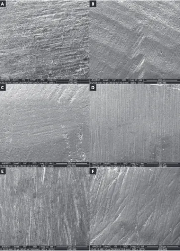
- (-0.685 _ -0.143) (- 0.627 _ -0.214) after debonding and enamel clean-up (Fig 4). Scratch-es produced by the 12-blade burs at low speed are presented in Figure 4A. Deeper scratches were pro-duced by the burs at high speed (Fig 4B). The highest degree of surface smoothness was obtained in Group G30L (Fig 4C) This group presented surface more similar to the original tooth, as shown in Figure 3. In Groups GDU and GR, there was loss of periky-mata with fine scratches caused by polishers of vary-ing abrasiveness (Fig 4D and Fig 4E). Fine scratches, which appeared to be well-marked and deep, caused by the diamond particles embedded in rubber, were also seen in Group GD (Fig 4F).
DISCUSSION
In this study, six protocols for removal of ad-hesive remnant from enamel after bracket debond-ing were assessed. The choice of burs and abrasive points was based on the protocols most used by or-thodontists, in other words, tungsten carbide burs
in low and high-speed handpieces,3-6 and products
launched on the market in recent years.
Figure 1 - Scatter plot of variation in roughness (∆Ra) in relation to time in all groups.
Figure 3 - Scanning electron microscopy (200 X magnification) of original enamel; perikymata (P); prism end openings (arrows).
Figure 4 - Scanning electron microscopy (500 X magnification) showing the effect of enamel clean-up procedures on the surface. A) 12-blade tungsten carbide bur (low speed) (G12L); B) 12-blade tungsten carbide bur (high speed) (G12H); C) 30-blade tungsten carbide bur (low speed) (G30L); D) DU10CA ORTHO polisher; E) Renew Finishing System; F) Diagloss polisher.
Figure 2 - Scatter plot of variation in roughness (∆Rz) in relation to time in all groups.
Group Group
A
C
E
B
D
used alone5,7 because it does not determine the
pro-file of irregularities and makes no distinction be-tween peaks and valleys. The association of other parameters used in this study, such as Rz, enabled us to study the shape of the vertical profile.
In this study, the protocols involving 12-blade tungsten carbide burs at low and high speed pro-duced similar results considering Ra. The Rz pa-rameter, however, was markedly affected when the 12-blade tungsten carbide bur was used at high speed. A ∆Rz value of 0.49 was the only positive value (indicating increase in roughness) and statis-tically different from Groups G30L and GD. This outcome demonstrated the increase in irregularities with sporadic deep scratches, which were not de-tected by ∆Ra, because Ra is an indicator of mean roughness and does not account for the presence of an occasional peak or valley. In SEM evaluation, the 12-blade bur produced deeper scratches at high speed. However, no statistically significant differ-ence was observed for both roughness parameters (∆Ra and ∆Rz) between G12L and G12H.
The literature reports that the use of tungsten carbide burs at high speed to remove resin remnant after debonding leaves the surface similar to that of
intact enamel;2,8,13,14 however, at the cost of a
sub-stantial loss in enamel thickness (19.2 µm).2,15 Other
studies recommend the use of tungsten carbide burs
at low speed3,16-18, which create fine scratches19 with
a lower level of enamel loss (7.9 µm to 11.3 µm)2,10.
In this study, enamel loss was not measured, although this factor should be an important con-sideration when choosing the method for resin
removal. According to Smith et al,21 the average
enamel thickness of a maxillary central incisor is ap-proximately 0.6 mm (600 µm). Considering a single bracket/resin removal, a loss of 10 or 20 µm might seem harmless, but it is necessary to consider the possibility of multiple rebondings due to bracket loss (caused by the patient) or bonding errors (caused by the orthodontist). Therefore, the orthodontist should minimize enamel damage and loss.
The use of high-speed burs without wa-ter cooling has been previously described by
Bicakci et al.22 They observed heating in the pulp
chamber, leading to vascular hyperemia and oc-casional breakage of odontoblasts. However, this
condition is transient, thereby indicating that the damage caused by this protocol is reversible, and pulp repair occurs within about 20 days. The authors recommend removing most of the resin under wa-ter cooling and turning the wawa-ter cooling off when removing the last layer of resin, so that it is pos-sible to successfully distinguish between enamel and resin, thereby preventing further damage to enamel. Considering the results of the present study, low-speed burs without water cooling could be used to remove the last layer of resin, so the risk of enamel scratches might be reduced. In this study, all resin remnant was removed without water cooling. It is suggested that future studies assess enamel rough-ness and loss when following the aforementioned recommendations.
Group GR, which involved the use of 12-blade tungsten carbide burs at high speed followed by
Renew polisher, showed a significant (p < 0.05)
de-crease in the two roughness parameters between the two time points, indicating the importance of gently eliminating the last layer of resin with polishers
af-ter using burs at high speed.1,6,8 The literature shows
that the sequential use of multiple tools for polishing
is more efficient than one-step procedures2,3,17,20,23,24
in terms of reduction in surface roughness. In this study, GR resulted in a low level of surface rough-ness, with negative values for ∆Ra and ∆Rz. However, the final variation in ∆Rz roughness of GR was not statistically different from the majority of the other groups, except for G12L (Table 4).
Roughness values obtained after clean-up in Groups G30L, GDU, GR and GD were lower than the initial roughness values. Similar results were
found in other studies,7,18,20 in which abrasive points
and 30-blade tungsten carbide burs were used to eliminate adhesive remnant. In a previous study, microscopic evaluation showed that the use of abra-sive points (Optimize Discs – TDV – and Onegloss Discs – Shofu) maintained the enamel surface of the study groups in a similar condition to the enamel
surface of the control groups.25
The time required for removing resin differed among the six groups, mainly due to
differenc-es in the cutting power of tools used,7 which was
mainly determined by the speed of rotation,17 type
and pressure applied to the handpiece.2 The latter
variable was minimized because the same operator performed all resin removal procedures. The time required in the groups in which Diagloss polishers (63.5 seconds) and 30-blade tungsten carbide burs at low speed (57.5 seconds) were used, was signifi-cantly longer than that required in the other groups
(p < 0.05). The protocol used in GD involved two
steps: use of rubber with a high gradient of dia-mond particle concentration, which ensured resin reduction, followed by another point for polishing. Hence, the procedure consumed more chair time. In Group G30L, the higher number of blades of the bur used at low speed decreased its cutting power and removed the resin layer by layer, which results in a smoother and scratch-free surface; however, it increased the time required for resin removal.
The fastest protocol was the use of the 12-blade tungsten carbide bur at high speed (23.5 sec-onds), followed by DU10CA ORTHO polisher (31.8 seconds), and the Renew system (31.9 seconds), which also made use of 12-blade tungsten carbide burs at high speed, however, in a two-step procedure.
Ryf et al20 assessed the Renew system, and showed
it required a considerably longer time to remove and polish the enamel (83.6 seconds); however, the burs were used were at low speed. The potential reasons for this difference were the use of lower-speed hand-pieces (under 20,000 rpm) and the use of the same bur every 10 specimens; thus, the bur became worn and had its cutting power diminished.
Our findings corroborate those of other
studies,3,14,19,20,25,26 indicating that all rotary
instru-ments cause varying changes in enamel surface. The association between the time spent and change in roughness (∆Ra, ∆Rz) showed a negative and moderate correlation: the longer the time spent on removing the remaining resin, the lower was the roughness left on the enamel surface, which is in
agreement with a previous study.1 Instruments with
low cutting power perform slower resin removal,
leaving a smoother surface less prone to plaque adhe-sion and pigmentation.
Ater orthodontic treatment, it is impossible to re-store the surface of teeth to their original condition. Prophylaxis with pumice, acid etching, debonding and aggressive resin removal procedures cause
enam-el loss.15 Rotating instruments create some degree
of enamel irregularities, and when rebonding is fre-quently necessary, the surface is modiied and the
peri-kymata pattern of young teeth is probably damaged.3
Therefore, ine scratches, such as those made when us-ing the protocols tested in this study, appear to cause minimum damage and must be placed in an expected clinical perspective. It is up to the orthodontist to
ap-ply methods to minimize damage to tooth enamel.25
Thorough resin removal and polishing ater debonding
is entirely dependent on the operator27 who is
respon-sible for selecting the instruments, using points with particles with a lower degree of hardness than enamel
to minimize iatrogenic abrasions and scratches;2 for the
pressure applied to the handpiece and for eliminating resin from the tooth surface.
CONCLUSIONS
1) All finishing and polishing protocols were considered satisfactory for residual resin removal without increasing enamel roughness.
2) The time spent on enamel clean-up varied from 23.5 (12-blade tungsten carbide bur at high speed) to 63.5 seconds (Diagloss polishers).
3) The longer the time spent on removing the remain-ing resin, the smaller the variation in roughness level.
Author contributions
1. Ulusoy C. Comparison of inishing and polishing systems for residual resin removal after debonding. J Appl Oral Sci. 2009;17(3):209-15.
2. Zarrinnia K, Eid NM, Kehoe MJ. The efect of diferent debonding techniques on the enamel surface: an in vitro qualitative study. Am J Orthod Dentofacial Orthop. 1995;108(3):284-93.
3. Zachrisson BU, Arthun J. Enamel surface appearance after various debonding techniques. Am J Orthod. 1979;75(2):121-7.
4. Hong YH, Lew KK. Quantitative and qualitative assessment of enamel surface following ive composite removal methods after bracket debonding. Eur J Orthod. 1995;17(2):121-8.
5. Eliades T, Gioka C, Eliades G, Makou M. Enamel surface roughness following debonding using two resin grinding methods. Eur J Orthod. 2004;26(3):333-8. 6. Brauchli LM, Baumgartner EM, Ball J, Wichelhaus A. Roughness of enamel
surfaces after diferent bonding and debonding procedures: an in vitro study. J Orofac Orthop. 2011;72(1):61-7.
7. Karan S, Kircelli BH, Tasdelen B. Enamel surface roughness after debonding. Angle Orthod. 2010;80(6):1081-8.
8. Albuquerque GS, Filho MV, Lucato AS, Boeck EM, Degan V, Kuramae M. Evaluation of enamel roughness after ceramic bracket debonding and clean-up with diferent methods. Braz J Oral Sci. 2010;9(2):81-4.
9. Bennett CG, Shen C, Waldron JM. The efects of debonding on the enamel surface. Am J Orthod Dentofacial Orthop. 1995;108(3):284-93.
10. Montasser MA, Drummond JL. Reliability of the adhesive remnant index score system with diferent magniications. Angle Orthod. 2009;79(4):773-6. 11. Artun J, Bergland S. Clinical trials with crystal growth conditioning as an
alternative to acid-etch enamel pretreatment. Am J Orthod. 1984;85(4):333-40. 12. 4287 ANI. Geometrical Product Speciications (GPS) - Surface texture: Proile
method -Terms, deinitions and surface texture parameters; 2002. 13. Rouleau BD Jr, Marshall GW Jr, Cooley RO. Enamel surface evaluations
after clinical treatment and removal of orthodontic brackets. Am J Orthod. 1982;81(5):423-6.
14. Pignatta LMB, Duarte Júnior S, Santos ECA. Evaluation of enamel surface after bracket debonding and polishing. Dental Press J Orthod. 2012;17(4):77-84. 15. Hosein I, Sherrif M, Ireland AJ. Enamel loss during bonding, debonding, and
cleanup with use of a self-etching primer. Am J Orthod Dentofacial Orthop. 2004;126(6):717-24.
REFERENCES
16. Eliades T, Kakaboura A, Eliades G, Bradley TG. Comparison of enamel colour changes associated with orthodontic bonding using two diferent adhesives. Eur J Orthod. 2001;23(1):85-90.
17. Radlanski RJ. A new carbide inishing bur for bracket debonding. J Orofac Orthop. 2001;62(4):296-304.
18. Giampaolo ET, Machado AL, Pavarina AC, Vergani CE. Diferent methods of inishing and polishing enamel. J Prosthet Dent. 2003;89(2):135-40. 19. Macieski K, Rocha R, Locks A, Ribeiro GU. Efects evaluation of remaining resin
removal (three modes) on enamel surface after bracket debonding. Dental Press J Orthod. 2011;16(5):146-54.
20. Ryf S, Flury S, Palaniappan S, Lussi A, van Meerbeek B, Zimmerli B. Enamel loss and adhesive remnants following bracket removal and various clean-up procedures in vitro. Eur J Orthod. 2012;34(1):25-32.
21. Smith TM, Olejniczak AJ, Zermeno JP, Taforeau P, Skinner MM, Hofmann A, et al. Variation in enamel thickness within the genus Homo. J Hum Evol. 2012;62(3):395-411.
22. Bicakci AA, Kocoglu-Altan B, Celik-Ozenci C, Tekcan M, Babacan H, Gungor E. Histopathologic evaluation of pulpal tissue response to various adhesive cleanup techniques. Am J Orthod Dentofacial Orthop. 2010;138(1):12.e1-e7; discussion 12-3.
23. Campbell PM. Enamel surfaces after orthodontic bracket debonding. Angle Orthod. 1995;65(2):103-10.
24. Joo HJ, Lee YK, Lee DY, Kim YJ, Lim YK. Inluence of orthodontic adhesives and clean-up procedures on the stain susceptibility of enamel after debonding. Angle Orthod. 2011;81(2):334-40.
25. De Marchi R, De Marchi LM, Terada RSS, Terada HH. Comparison between two methods for resin removing after bracket debonding. Dental Press J Orthod. 2012;17(6):130-6.
26. Retief DH, Denys FR. Finishing of enamel surfaces after debonding of orthodontic attachments. Angle Orthod. 1979;49(1):1-10.