A Biomechanical Analysis of the Interlock Suture and
a Modified Kessler-Loop Lock Flexor Tendon Suture
Wenfeng Yang, Dan Qiao, Yuanfei Ren, Yvjin Dong, Yaohua Shang, Tiehui Zhang*
Dalian Municipal Central Hospital, Dalian, China.
OBJECTIVE:In this work, we attempted to develop a modified single-knot Kessler-loop lock suture technique and compare the biomechanical properties associated with this single-knot suture technique with those associated with the conventional modified Kessler and interlock suture techniques.
METHODS:In this experiment, a total of 18 porcine flexor digitorum profundus tendons were harvested and randomly divided into three groups. The tendons were transected and then repaired using three different techniques, including modified Kessler suture with peritendinous suture, interlock suture with peritendinous suture, and modified Kessler-loop lock suture with peritendinous suture. Times required for suturing were recorded and compared among groups. The groups were also compared with respect to 2-mm gap load, ultimate failure load, and gap at failure.
RESULTS:For tendon repair, compared with the conventional modified Kessler suture technique, the interlock and modified Kessler-loop lock suture techniques resulted in significantly improved biomechanical properties. However, there were no significant differences between the interlock and modified Kessler-loop lock tech-niques with respect to biomechanical properties, gap at failure, and time required.
CONCLUSIONS:The interlock and modified Kessler-loop lock techniques for flexor tendon sutures produce similar mechanical characteristicsin vitro.
KEYWORDS: Tendon Repair; Animal Experimentation; Flexor Tendon.
Yang W, Qiao D, Ren Y, Dong Y, Shang Y, Zhang T. A Biomechanical Analysis of the Interlock Suture and a Modified Kessler-Loop Lock Flexor Tendon Suture. Clinics. 2017;72(9):582-587
Received for publication onFebruary 23, 2017;First review completed onJune 3, 2017;Accepted for publication onJuly 13, 2017 *Corresponding author. E-mail: [email protected]
’ INTRODUCTION
Flexor tendon injury is a common clinical trauma. Early postoperative active functional exercise can not only effec-tively reduce the formation of local adhesions and edema but also facilitate sliding function of the tendon, thereby pro-moting endogenous tendon healing (1,2). The choice of tendon repair technique plays an important role in tendon healing and postoperative functional exercise. The ideal repair should be easy to perform; provide sufficient strength for healing (over 30 N) (3), which can ensure minimal inter-ference with tendon vascularity; and involve secure sut-ure knots and smooth junction of tendon ends. Currently, various suture techniques (4,5) have been proven to exhi-bit sufficient resistance to gap formation. However, many techniques are demanding with respect to surgical experi-ence and suturing skills. It is essential to explore a rela-tively simple tenorrhaphy with good mechanical strength
to reduce clinical complications associated with flexor tendon suture.
The modified Kessler suture is regarded as the most widely used clinical suture technique for tendon repair. How-ever, the optimal surgical approach for achieving a success-ful outcome remains controversial (6). In the interlock stitch suture technique, locking loops produce improved strength and gap resistance after flexor tendon repair compared with the grasping loops used in the modified Kessler suture tech-nique (7). Additionally, various cross-locked cruciate tendon sutures (8) with diverse biomechanical properties have been developed from interlock stitch sutures and have become common alternatives for flexor tendon repair. However, the interlock stitch suture technique is time-consuming and rela-tively complex, hindering its wide application in clinical practice. Liu et al. (9) modified the Kessler-loop lock suture to enhance resistance strength. However, the use of two knots in the repaired area could lead to a high risk of early failure, suture slippage, and tendon torsion (10).
It is widely accepted that the number of suture passages at the injury site is an extremely important factor in the bio-mechanical outcomes for different types of tendon sutures. Four-strand suture techniques (such as the Strickland and interlock approaches) have been the most widely investi-gated techniques in recent decades. In the present study, we modified the Kessler-loop lock suture technique by changing DOI:10.6061/clinics/2017(09)10
Copyright&2017CLINICS–This is an Open Access article distributed under the terms of the Creative Commons License (http://creativecommons.org/licenses/by/ 4.0/) which permits unrestricted use, distribution, and reproduction in any medium or format, provided the original work is properly cited.
the double-knot suture to a single-knot suture and compa-red our modified technique with the conventional modified Kessler suture and interlock suture techniques with respect to biomechanical properties and suturing time.
’ METHODS
Tendon Harvest and Repair
Porcine flexor tendons are similar in structure and dia-meter to human flexor tendons and are thus appropriate for use in biomechanical studies. Eighteen porcine flexor digitorum profundus tendons (circumference: 8-10 mm; approximate weight: 300-400 g) were harvested from the hind legs of adult pigs (Dalian Chuming Co., Ltd.). This study was approved by the Institutional Animal Care and Use Committee of Dalian Medical University. All operations were performed in accordance with international guidelines regarding the care and treatment of experimental animals. The tendons were randomly divided into three groups (with six tendons in each group). In this experiment, 4-0 nylon (Weigao, China) was used for core sutures, and 6-0 nylon (Weigao, China) was used for peritendinous sutures.
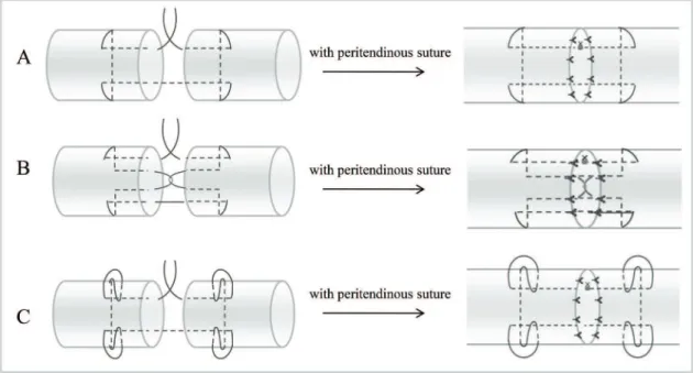
The middle of each tendon was transected with a scalpel and then repaired using one of the following techniques: (A) modified Kessler suture with peritendinous suture (MK); (B) interlock suture with peritendinous suture (IS); and (C) modified Kessler-loop lock suture with peritendinous suture (MKL). Details regarding these techniques are presen-ted in Figure 1. During the Kessler suture technique, sutures were tightened to provide proper tension in the first locking loop. A final range of 10-12 mm was maintained for stitches on both sides. Three surgical knots were used for all suture knots, which consisted of six alternating positive and nega-tive single knots. All tendon repairs were performed by a single experienced surgeon within one day. To minimize tissue injury, the surgeon performed the tendon repairs under an operating microscope with a magnification of 10. Times required for suturing were recorded for each group.
After suturing was completed, the repaired tendons were covered with wet gauze (wet with normal saline), stored at 4o
C overnight, and subjected to biomechanical testing the following morning.
Load-to-failure Tests
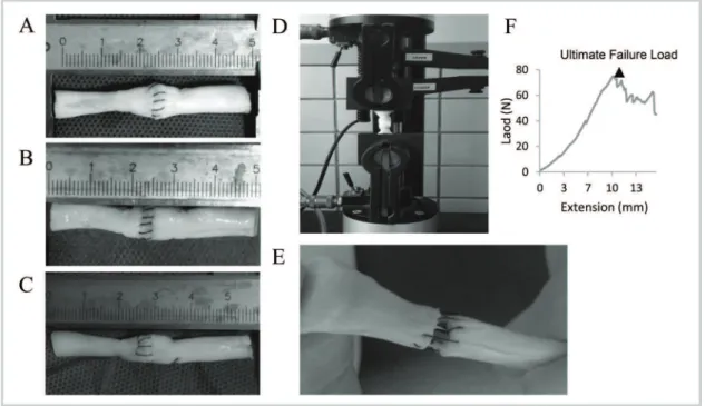
The tendon samples were 50 mm in length with the repair site in the middle. The ends of the tendons were clamped 15 mm from the repair site in a testing machine (Model 5567A, Instron, UK). Bluehill software (Instron, UK) was used to set and record the test parameters (Figure 2). Each tendon was subjected to a preload of 1.0 N and then elongated at a constant velocity of 10 mm/min until the suture site had completely ruptured. The load-to-failure tests were recorded using a camera. During the test, the gap at the repair site was measured using a vernier caliper. In this study, 2-mm gap load was defined as the tensile force needed to produce a 2-mm gap at the repair site. The ultimate load was recorded as the peak force during the stretching process. A computer was used to draw load-displacement curves.
Statistical Analysis
Stata 12.0 (Computer Sciences Corporation, US) was used for statistical analysis. ANOVA followed by post hoc Tukey tests was used to evaluate differences in2-mm gap load, ultimate failure load, gap at failure, and suture time among the three groups.po0.05 was regarded as indicative of sta-tistical significance.
’ RESULTS
Two-millimeter Gap Load
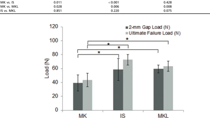
Two-millimeter gap load is an important index for eval-uating resistance strength after tendon repair. During tendon healing, the formation of a gap of 2 mm or more increases the risks of tendon adhesion and impacts tendon healing. As shown in Tables 1 and 3 and Figure 3, compared with the MK technique, both the IS technique (p=0.011) and the
Figure 1 - Various suture techniques used for mechanical testing. A. Modified Kessler suture with peritendinous suture (MK);
B. Interlock suture with peritendinous suture (IS); C. Modified Kessler-loop lock suture with peritendinous suture (MKL).
MKL technique (p=0.028) could produce significantly improved 2-mm gap loads. The IS and MKL techniques did not significantly differ with respect to 2-mm gap load (p=0.851).
Ultimate Failure Load
Ultimate failure load is another essential indicator for describing the biomechanical properties of tendon repair. As shown in Tables 1 and 3 and Figure 3, compared with the MK technique, both the IS technique (po0.001) and the MKL technique (p=0.006) could produce significantly improved ultimate failure loads. However, the IS and MKL techniques did not significantly differ with respect to ultimate failure load (p=0.220).
Failure Profile
As shown in Table 4, suture breakage always occurred in the IS group. The breakage locations were usually at the gap site in the peritendinous suture, although a small portion of ruptures occurred at the root of a knot. Compared with the MK and MKL groups, the IS group had less suture pullout and knot failure but a higher risk of suture breakage.
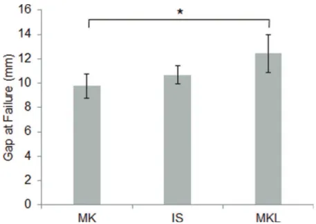
Gap at Failure
As shown in Tables 2 and 3 and Figure 4, the largest gap at failure was observed in the MKL group, whereas stati-stically similar findings were obtained for the MK and IS groups (p=0.428).
Time Required for the 3Suture Techniques
As shown in Tables 2 and 3 and Figure 5, the MK tech-nique was faster to perform than the IS techtech-nique (p=0.011). There was no significant difference in suturing time between the IS and MKL groups (p=0.577).
’ DISCUSSION
The early mobilization of reconstructed flexor tendons is highly significant for tendon recovery. By establishing stress orientation, early active movement can promote the regular arrangement of collagen fibers and break adhesion between the impaired tendon and surrounding tissues. Various suture techniques, such as the MK, double-strand Kessler suture, Silfverskiold (11), Strickland (12), cross-locked cruciate suture (13), Tsuge (14), and combined Kessler-Tsuge (15) techniques, have been used in clinical practice. However, research has demonstrated that these approaches produce different outcomes. Early active mobilization after tendon repair imposes high load at the repair site and requires resistance strength against a 2-mm gap load of more than
Table 2-The gaps at failure and times required for three suture techniques.
Group Gap at Failure (mm) Time Required (min)
Mean SD Mean SD
MK 9.8 1.0 11.2 2.5
IS 10.7 0.7 15.4 1.3
MKL 12.4 1.5 14.2 1.8
Figure 2 -Porcine tendons repaired using various suture techniques. A. Modified Kessler with peritendinous suture (MK); B. Interlock
suture with peritendinous suture (IS); C. Modified Kessler-loop lock suture with peritendinous suture (MKL); D. Repaired porcine tendon in mechanical testing; E. Suture failure in mechanical testing; F. Load-deformation curve.
Table 1-The 2-mm gap loads and ultimate failure loads for three suture techniques.
Group 2-mm Gap Load (N) Ultimate Failure Load (N)
Mean SD Mean SD
MK 39.1 11.6 43.4 9.6
IS 58.8 15.9 72.6 7.7
30 N (16). Certain of the most commonly used suture methods (17) do not allow the repaired tendon to withstand over 30 N of tension. Although other repair techniques (18) can provide adequate strength, these techniques require complex surgical procedures or affect blood supply and tendon healing.
Suture methods for flexor tendons mainly involve grasp-ing loops and lockgrasp-ing loops. Compared with graspgrasp-ing loops, locking loops improve the strength and gap resistance of the tendon repair by tightly holding tendon bands together. It has also been reported that a grasping loop-locking loop combination can maximize the advantages of both suture techniques and dramatically improve the biomechanical properties achieved in tendon repair (19). In the current work, both the IS and MKL suture techniques exhibited the advantages of locking and grasping loops and produced significantly improved 2-mm gap loads and ultimate tensile strengths relative to the MK technique. The IS and MKL approaches did not significantly differ with respect to 2-mm gap load, ultimate failure load, gap at failure, or time requi-red for suturing.
However, different biomechanical properties are obser-ved for the IS and MKL techniques. To withstand greater tensile force with an intra-tendinous locking structure and
stitches running through the tendon fibers, the IS techni-que is based on the grasping loops used in the Kessler suture technique combined with locking loops in the tendon’s core. However, the crossing shear force in the locking suture of the IS pattern increases the risk of suture breakage. If pre-load is insufficient during tendon suturing, the rotational stress caused by loose sutures typically leads to twisting and instability of the injured tendon ends; this phenome-non impacts endogenous tendon healing. Therefore, the IS technique is more suitable for experienced surgeons and requires the use of high-quality sutures to withstand tensile forces.
In the Kessler-loop lock suture technique, the tendon is locked by friction between loop sutures and the tendon, with the objective of withstanding greater tensile force and pre-venting gap formation (20). This approach requires fewer knots than the MK technique. The cross-linked collagen fibers in the tendon generate upward force on the suture, reducing tendon slippage and splitting. The low tension between the injured ends of the tendon may be associated with better tendon healing, although further research is necessary to confirm this possibility.
Therefore, we altered the double-strand Kessler-loop lock suture approach by utilizing single-strand sutures and performed suturing under an operating microscope with a magnification of 10. This approach is easier for less experienced surgeons to understand and accept. Since this approach does not require special sutures, it is relatively readily applied in clinical practice.
Nevertheless, there are certain limitations of our study. This investigation involved the use of devitalized porcine
Table 3-p-values for comparisons between flexor tendon suture techniques.
Comparison 2-mm Gap Load Ultimate Failure Load Gap at Failure Time Required
MK vs. IS 0.011 o0.001 0.428 0.011
MK vs. MKL 0.028 0.006 0.008 0.070
IS vs. MKL 0.851 0.220 0.075 0.577
Figure 3 -Ultimate failure loads and 2-mm gap loads for different suture techniques. Compared with the MK technique, the IS and MKL
techniques resulted in significantly greater 2-mm gap loads and ultimate failure loads. Error bars represent the SD from five porcine flexor tendons. *po0.05vs. MK.
Table 4-Failure modes for three suture techniques.
Group Suture Breakage Suture Pullout Knot Failure
MK (n=6) n=4 n=4 n=3
IS (n=6) n=6 n=2 n=1
MKL (n=6) n=4 n=4 n=2
flexor tendons that may have different biomechanical pro-perties than living tissues. However, in the future, our results could be evaluated in animal models and via clinical studies. Our approach was compared with only the Kessler and inter-lock suture techniques; this comparison could be extended to other representative 4-strand suture techniques, such as the double-strand Kessler and Strickland approaches.
’ ACKNOWLEDGMENTS
The authors thank Jianfeng Li for helping to perform mechanical tests in the School of Chemical Engineering (Dalian University of Technology).
’ AUTHOR CONTRIBUTIONS
Yang W performed biomechanical testing, analyzed data and wrote the manuscript. Qiao D and Ren Y harvested porcineflexor digitorum
pro-fundus tendons. Dong Y and Shang Y contributed to writing the manuscript. Zhang T directed the work. All authors read and approved thefinal version of the manuscript.
’ REFERENCES
1. Gelberman RH, Amifl D, Gonsalves M, Woo S, Akeson WH. The influence of protected passive mobilization on the healing of flexor tendons: a biochemical and microangiographic study. Hand.1981;13(2):120-8, http:// dx.doi.org/10.1016/S0072-968X(81)80051-4.
2. Singh R, Rymer B, Theobald P, Thomas PB. A Review of Current Concepts in Flexor Tendon Repair: Physiology, Biomechanics, Surgical Technique and Rehabilitation. Orthop Rev (Pavia). 2015;7(4):6125, http://dx.doi. org/10.4081/or.2015.6125.
3. Lawrence TM, Davis TR. A biomechanical analysis of suture materials and their influence on a four-strand flexor tendon repair. J Hand Surg Am. 2005;30(4):836-41, http://dx.doi.org/10.1016/j.jhsa.2005.03.011. 4. Diao E, Hariharan JS, Soejima O, Lotz JC. Effect of peripheral suture depth
on strength of tendon repairs. J Hand Surg Am. 1996;21(2):234-9, http:// dx.doi.org/10.1016/S0363-5023(96)80106-7.
Figure 4 -Gaps at failure for different suture techniques. The largest gap at failure was seen in the MKL group (12.4±1.5 mm), whereas
gaps at failure were similar for the MK and IS techniques (9.8±1.0 mmvs. 10.7±0.7 mm, respectively). *po0.05vs. MK.
Figure 5 -Times required for different suture techniques. Less time was required for the MK technique (11.2±2.5 min) than for the
5. Wade PJ, Muir IF, Hutcheon LL. Primary flexor tendon repair: the mechanical limitations of the modified Kessler technique. J Hand Surg Br. 1986;11(1):71-6, http://dx.doi.org/10.1016/0266-7681(86)90018-5. 6. Lee H. Double loop locking suture: a technique of tendon repair for early
active mobilization. Part I: Evolution of technique and experimental study. J Hand Surg Am.1990;15(6): 945-52, http://dx.doi.org/10.1016/ 0363-5023(90)90021-I.
7. Robertson GA, al-Qattan MM. A biomechanical analysis of a new inter-lock suture technique for flexor tendon repair. J Hand Surg Br. 1992;17(1): 92–3, http://dx.doi.org/10.1016/0266-7681(92)90020-3.
8. Tahmassebi R, Peltz TS, Haddad R, Scougall P, Gianoutsos M, Walsh W. The Interlocking Modification of the Cross Locked Cruciate Tendon Repair (Modified Adelaide Repair): A Static and Dynamic Biomechanical Assessment. J Hand Microsurg. 2015;7(1):6–12, http://dx.doi.org/10.1007/
s12593-014-0144-4.
9. Qinghe L, Zuoshan Z, Kai Z. Semi-Knot Locking Suture (SKLS) and It Experimental Study. The Journal of Bone and Joint Injury. 2003;18(10):692-4. 10. Le SV, Chiu S, Meineke RC, Williams P, Wongworawat MD. Number of suture throws and its impact on the biomechanical properties of the four-strand cruciate locked flexor tendon repair with FiberWire. J Hand Surg Eur. 2012;37(9):826–31, http://dx.doi.org/10.1177/1753193412447503.
11. Dwyer CL, Dominy DD, Cooney TE, Englund R, Gordon L, Lubahn JD. Biomechanical comparison of double grasping repair versus cross-locked cruciate flexor tendon repair. Hand (N Y). 2015;10(1):16-22.
12. Iamaguchi RB, Villani W, Rezende MR, Wei TH, Cho AB, Santos GB, et al. Biomechanical comparison of the four-strand cruciate and Strickland techniques in animal tendons. Clinics. 2013;68(12):1543-7, http://dx.doi. org/10.6061/clinics/2013(12)11.
13. Wada A, Kubota H, Hatanaka H, Hotokezaka S, Miura H, Iwamoto Y. The mechanical properties of locking and grasping suture loop configurations in four-strand core suture techniques. J Hand Surg Br. 2000;25(6):548-51, http://dx.doi.org/10.1054/jhsb.2000.0441.
14. Hatanaka H, Manske PR. Effect of suture size on locking and grasp-ing flexor tendon repair techniques. Clin Orthop Relat Res. 2000;(375): 267-74.
15. Renner C, Corella F, Fischer N. Biomechanical evaluation of 4-strand flexor tendon repair techniques, including a combined Kessler-Tsuge approach. J Hand Surg Am. 2015;40(2):229-35, http://dx.doi.org/10.1016/ j.jhsa.2014.10.055.
16. Gan AW, Neo PY, He M, Yam AK, Chong AK, Tay SC. A biomechanical comparison of 3 loop suture materials in a 6-strand flexor tendon repair technique. J Hand Surg Am. 2012;37(9):1830-4, http://dx.doi.org/10.1016/ j.jhsa.2012.06.008.
17. Jiahao L, Haisheng Q, Jianwei W, Chuanpu L. Biomechanical Study on a new method of suturing the tendon. Zhejiang Journal of Traumatic Sur-gery. 2015;20(4):653-5.
18. Lister GD, Kleinert HE, Kutz JE, Atasoy E. Primary flexor tendon repair followed by immediate controlled mobilization. J Hand Surg Am. 1977; 2(6):441-51, http://dx.doi.org/10.1016/S0363-5023(77)80025-7. 19. Nelson GN, Potter R, Ntouvali E, Silva MJ, Boyer MI, Gelberman RH,
et al. Intrasynovial flexor tendon repair: a biomechanical study of varia-tions in suture application in human cadavera. J Orthop Res. 2012;30(10): 1652-9, http://dx.doi.org/10.1002/jor.22108.
20. Lotz JC, Hariharan JS, Diao E. Analytic model to predict the strength of tendon repairs. J Orthop Res. 1998;16(4):399-405, http://dx.doi.org/10.1002/ jor.1100160402.