Malignant transformation of abdominal wall endometriosis
to clear cell carcinoma: case report
João Kleber de Almeida Gentile
I, Renato Migliore
I, Fábio Jorge Neubaner Kistenmacker
I, Marcio Menezes de Oliveira
IIRodrigo Biscuola Garcia
III, Fang Chia Bin
IV, Pedro Marcos Santinho Bueno de Souza
III, José César Assef
IVHospital do Servidor Público Municipal (HSPM-SP), São Paulo (SP), Brazil
ABSTRACT
BACKGROUND: Malignant transformation of endometriosis in the abdominal wall is a rare and still poorly understood event. Less than 30 cases have been reported in the worldwide literature. Most cases of solid tumors are report in a previous abdominal scar with malignant transformation of a focus of endometriosis. Presence of lymph node metastases in nearby chains is frequent and is associated with poor prognosis.
CASE REPORT: We report a case of a 42-year-old woman with a history of abdominal surgery (Pfannen-stiel) to resect abdominal wall endometriosis. Physical examination revealed a solid mass of approximately 10 cm x 6 cm in the anterior wall of the abdomen. Computed tomography (CT) of the abdomen and pel-vis showed a heterogeneous, predominantly hypoattenuating expansive formation measuring 10.6 cm x 4.7 cm x 8.3 cm. The patient underwent exploratory incisional laparotomy, block resection of the abdomi-nal mass and lymphadenectomy of the exterabdomi-nal and inguiabdomi-nal iliac chains. The abdomiabdomi-nal wall was recon-structed using a semi-absorbable tissue-separating screen to reconstitute the defect caused by resection of the tumor. Histological evaluation revealed iniltration by malignant epithelioid neoplasia, thus conirm-ing the immunohistochemical proile of adenocarcinoma with clear cell components. Lymphadenectomy showed metastatic involvement of an external iliac chain lymph node.
CONCLUSION: Resection of the mass along with the abdominal wall, with wall margins, is the most efec-tive treatment. Reconstruction is a challenge for surgeons. The patient has been followed up postopera-tively for eight months, without any evidence of disease to date.
IMD. Resident Physician, Department of Digestive Surgery, Hospital do Servidor Público Municipal (HSPM-SP), São Paulo (SP), Brazil.
orcid.org/0000-0001-8650-2703
IIMD. Resident of General Surgery, Department of General Surgery, Hospital do Servidor Público Municipal (HSPM), São Paulo (SP), Brazil. IIIMD. Attending Physician, Department of Digestive Surgery, Hospital do Servidor Público Municipal (HSPM-SP), São Paulo (SP), Brazil. IVMD. Department of Digestive System Surgery, Hospital do Servidor Público Municipal (HSPM-SP), São Paulo (SP), Brazil.
KEY WORDS:
Endometriosis. Abdominal wall.
Cell transformation, neoplastic. Adenocarcinoma, clear cell.
INTRODUCTION
Endometriosis is deined as the presence of stroma and endometrial glands outside the uter-ine cavity. It afects approximately 15-40% of women of childbearing age. he most common
site is the abdominal cavity, speciically in the pelvis and occasionally at extra-pelvic sites.1,2
Abdominal wall endometriosis accounts for 0.4-2% of the cases, and is mostly found in the umbilical scar and in the scar of previous abdominal incisions, especially in cesarean scars,
lap-aroscopies and appendectomies.2
In patients with abdominal wall endometrioma, the mean time taken to reach the diagnosis is 6 to 20 years ater the initial surgery, and 14.3-26% of the cases show an association with
pel-vic endometriosis.2 he endometrioma is diagnosed preoperatively only in 20-50% of the cases,
and the typical complaint is most frequently cyclical menstrual pain. he diferential diagnoses for an abdominal mass associated with a previous surgical incision in the abdominal wall include
abscess, hematoma, hernia, desmoid tumors, sarcomas and metastatic disease.1
Malignant transformation of an abdominal wall endometrioma is an extremely rare event. Extensive local excision with surgical margins seems to be the only efective treatment, and it is almost always necessary to correct the defect of the abdominal wall with prosthetic surgical or cutaneous laps for the closure of the abdominal wall.
mass (Figure 2) with margins to the peritoneum, along with lymph-adenectomy of the external and inguinal iliac chains. he abdom-inal wall was reconstructed to reconstitute the defect caused by resection of the tumor, using a semi-absorbable tissue-separating screen composed of a polypropylene parietal face and a visceral face coated with carboxymethyl cellulose. his rectangular sodium hyaluronate mesh measured 20.3 cm x 30.5 cm (Sempramesh IP Composite Bard Davol Inc.).
Histological analysis on the abdominal mass revealed infil-tration by malignant epithelioid neoplasia into soft tissues, thus confirming the immunohistochemical profile of
adeno-carcinoma with clear cell components (Figure 3). The antigens
investigated in the immunohistochemical evaluation are listed
in Table 1. Lymphadenectomy showed metastatic involvement
of an external iliac chain lymph node (1/8), and that other CASE REPORT
he patient was a 42-year-old female, with one previous preg-nancy, with a history of cesarean section seven years previously and resection of endometriosis of the cephalic scar (Pfannenstiel) two years previously at another service, for which a histopatho-logical diagnosis of abdominal wall endometriosis was made.
Her condition evolved with progressive expansion in the region previously resected, for eight months, leading to presence of a bulging mass in the right side of the anterior abdominal wall, with cyclical local pain. During the investigation period, the patient said that she did not have any genitourinary or gastrointestinal symptoms, or any presence of lymph nodes or systemic symptoms.
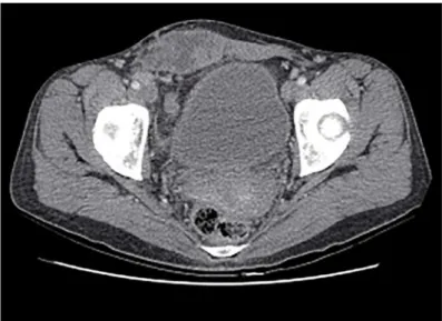
Physical examination revealed a solid mass of approximately 10 cm x 6 cm in the anterior wall of the abdomen bordering the pubis. It extended inferiorly to the umbilical scar and laterally to the upper border of the iliac crest. At the time of the physical exami-nation, there was no lymph node swelling in the inguinal region. Laboratory tests and tumor marker investigations (CA 125, CA 19-9, CEA and alpha-fetoprotein) were requested and these were found to be within normal limits. Computed tomography (CT) of the abdomen and pelvis revealed a heterogeneous expansive formation that was predominantly hypoattenuating, with images suggestive of internal septation. It measured around 10.6 cm x 4.7 cm x 8.3 cm along the major transverse, anteroposterior and longitudinal axes, respectively, and was located in the anterior pel-vic wall, with the largest axis to the right of the midline, involving
the rectus abdominis muscle (Figure 1).
he patient underwent exploratory laparotomy by means of a Pfannenstiel incision, followed by block resection of the abdominal
Figure 1. Computed tomography scan of the abdomen and pelvis
(portal phase) showing an expansive process in the anterior abdominal wall and pelvis and lymph node enlargement in the external and inguinal iliac chains.
Figure 2. Macroscopic appearance demonstrating areas of
cystic and trabecular components.
lymph nodes of the iliac and inguinal chains presented lym-phoid hyperplasia (0/11).
Our patient is in her second postoperative month, without having presented any clinical or surgical intercurrence to date. She is being followed up by the oncology sector and an adjuvant chemotherapy scheme has been indicated.
DISCUSSION
Malignant transformation of endometriosis is quite rare and afects less than 1% of the female population sufering from this condition. In the literature, the most common site of malignant transformation of endometriosis foci is the ovaries, while 20% of the cases occur at extragonadal sites, including the rectovaginal septum, colon and dis-tal organs such as the abdominal wall. Less than 5% of these cases are
carcinomas of clear cell origin like the case reported here.3
his malignant transformation in the abdominal wall is an extremely rare event, with less than 30 case reports in the world-wide literature. hese cases consisted of endometrioid carcinoma
(70%), sarcoma (25%) and clear cell carcinoma (5%).4
Sampson was one of the irst authors to report a case of malig-nancy of an endometriosis outbreak. He proposed three criteria for diagnosing malignant transformation of endometriosis:
1. Demonstration of neoplastic and benign endometrial tissue in the tumor;
2. Histological type compatible with endometrial origin;
3. No other primary site identiied.5
In 1953, Scott added a fourth criterion: histological presence of benign endometriosis and carcinoma with glandular transformation
with atypias. Few reported cases have met all four proposed
crite-ria, and the etiogenesis of such cases remains unknown.6
Malignancy of a focus of endometriosis on a previous scar on the abdominal wall is very rare, with a reported prevalence of 0.03%. It can afect all layers of the abdominal wall and the growth of such
masses is exponential, reaching diameters greater than 10 cm.7
In our case, the mass appeared in the anterior wall of the abdo-men without evidence of abdominal cavity involveabdo-ment from abdominal CT. he abdominal wall itself was limited by the peri-toneum. Our patient underwent preoperative screening for a pri-mary focus of neoplasia, by means of upper gastrointestinal endos-copy, colonoscopy and thyroid ultrasonography. All of these were negative for neoplasms.
We reviewed the literature through MEDLINE, PubMed, Embase and LILACS using the English keywords “endometrio-sis”, “cell transformation”, “adenocarcinoma” and “abdominal wall”.
We found only 17 reports, as shown in Table 2, and 15 reports had
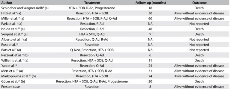
clinical presentation similar to the reported case. Table 37-21 lists
the reports in the literature describing the diferent types of treat-ment for clear cell carcinomas of the abdominal wall that were derived from an endometrioid focus on a previous abdominal scar. Local invasion is an important biological feature for transforma-tion of endometriosis into invasive carcinoma. On the other hand, although lymphatic dissemination may be present, it has only been
reported in three cases.7-9
At the time of the pre-surgical evaluation, it was diicult to make a diagnosis of lymph node involvement. However, the pres-ence of lymph node enlargement in the inguinal region and in the external iliac chain was observed on CT scans. his was investi-gated using computerized tomography with 18-luorodeoxyglu-cose positron emission tomography (FDG-PET).
Presence of a compromised lymph node in the 2-cm external iliac chain was demonstrated, with standardized uptake values (SUV) for the abdominal mass of 4.16 and 2.51 in the iliac lymph node. here were no other signs of FDG uptake.
In our case, lymphadenectomy of the external and inguinal iliac chain was performed, and the metastatic involvement of the lymph node caused by carcinoma was conirmed through histo-logical analysis.
Table 1. Immunohistochemical proile and antigens investigated
Antigen Result
D AE1/AE3 Positive
CD 34 Negative
CK 7 Positive
CK 20 Negative
Estrogen receptor Negative
WT-1 Negative
Vimentin Negative
Table 2. Search of the literature in medical databases for cases of degeneration of abdominal wall endometriosis for clear cell carcinoma. (Search was conducted on April 14, 2017)
Database Search strategies Papers found Reports of cases with
lymphatic dissemination
MEDLINE (via PubMed) endometriosis and cell transformation and adenocarcinoma and
abdominal wall “case reports” [publication type] 17 2
Embase (via Elsevier) endometriosis and cell transformation and adenocarcinoma and
abdominal wall “case reports” [publication type] 0 0
LILACS (via Bireme) endometriosis and cell transformation and adenocarcinoma and
Radical resection is considered to be the primary treatment for endometrioid carcinoma of the wall. Carboplatin-based che-motherapy and radiation therapy schemes have been proposed
without any evidence of improved prognosis or survival.7
Due to the rarity of this tumor, the long-term survival fol-lowing treatment is unknown. However, some recent reports have shown that aggressive radical surgery with total tumor excision with free margins, together with lymphadenectomy of the inguinal and iliac chains may be beneicial for these patients’ disease-free survival.
CONCLUSION
Malignant transformation to clear cell carcinoma from a focus of endometriosis on the abdominal wall is a rare and poorly understood complication. Most recent studies have shown that aggressive surgical resection with safety margins associated with lymphadenectomy is still the most efective treatment with the highest survival rates. he role of adjuvant therapy remains unclear and therefore further studies to assess the long-term ben-eits are required.
In our case, lymphadenectomy of the external and inguinal iliac chain was performed, and the metastatic involvement of the lymph node caused by carcinoma was conirmed through histo-logical analysis.
REFERENCES
1. Blanco RG, Parithivel VS, Shah AK, et al. Abdominal wall endometriomas.
Am J Surg. 2003;185(6):596-8.
2. Zhao X, Lang J, Leng J, et al. Abdominal wall endometriomas. Int J
Gynaecol Obstet. 2005;90(3):218-22.
3. Omranipour R, Najai M. Papillary serous carcinoma arising in abdominal
wall endometriosis treated with neoadjuvant chemotherapy and
surgery. Fertil Steril. 2010;93(4):1347.e17-8.
4. Shalin SC, Haws AL, Carter DG, Zarrin-Khameh N. Clear cell
adenocarcinoma arising from endometriosis in abdominal wall cesarean
section scar: a case report and review of the literature. J Cutan Pathol.
2012;39(11):1035-41.
5. Sampson JA. Endometrial carcinoma of the ovary arising in endometrial
tissue in that organ. American Journal of Obstetrics & Gynecology.
1925;9(1):111-4. Available from:
http://www.ajog.org/article/S0002-9378(25)90949-0/abstract. Accessed in 2017 (Jul 14).
6. Scott RB. Malignant changes in endometriosis. Obstet Gynecol.
1953;2(3):283-9.
7. Bats AS, Zafrani Y, Pautier P, Duvillard P, Morice P. Malignant transformation
of abdominal wall endometriosis to clear cell carcinoma: case report
and review of the literature. Fertil Steril. 2008;90(4):1197.e13-6.
8. Schineber D, Wagner-Kolb D. [Malignant transformation of extragenital
endometriosis]. Geburtshilfe Frauenheilkd. 1986;46(9):658-9.
9. Hitti IF, Glasbergg SS, Lubicz S. Clear cell carcinoma arising in extraovarian
endometriosis: report of three cases and review of the literature. Gynecol
Oncol. 1990;39(3):314-20.
10. Miller DM, Schouls JJ, Ehlen TG. Clear cell carcinoma arising in
extragonadal endometriosis in a caesarean section scar during
pregnancy. Gynecol Oncol. 1998;70(1):127-30.
11. Park SW, Hong SM, Wu HG, Ha SW. Clear cell carcinoma arising in a
Cesarean section scar endometriosis: a case report. J Korean Med Sci.
1999;14(2):217-9.
12. Ishida GM, Motoyama T, Watanabe T, Emura I. Clear cell carcinoma
arising in a cesarean section scar. Report of a case with ine needle
aspiration cytology. Acta Cytol. 2003;47(6):1095-8.
Table 3. Reported cases of clear cell carcinoma of the abdominal wall derived from focus of endometriosis
Author Treatment Follow-up (months) Outcome
Schineber and Wagner-Kolb8 (a) HTA + SOB, R-Ad, Progesterone 18 Death
Hitti et al.9 (a) Resection, HTA + SOB 30 Alive without evidence of disease
Miller et al.10 (a) Resection, HTA + SOB, R-Ad, Q-Ad 60 Alive without evidence of disease
Park et al.11 (a) Resection, R-Ad NA Not reported
Ishida et al.12 (a) Resection, R-Ad 48 Death
Sergent et al.13 (a) HTA + SOB, Q-Ad 9 Death
Alberto et al.14 (a) Resection, Q-Ad, R-Ad NA Not reported
Rust et al.15 Resection NA Not reported
Bats et al.7 (a) Q-Neo, Resection, HTA + SOB NA Not reported
Razzouk et al.16 (b) Resection, Q-Ad 6 Death
Williams et al.17 (a) Resection, HTA + SOB, Q-Ad 11 Death
Yan et al.18 Resection, Q-Ad 24 Alive without evidence of disease
Mert et al.19 (a) Resection, HTA + SOB, R-Ad 31 Alive without evidence of disease
Markopoulos et al.20 (b) Resection, HTA + SOB 24 Alive without evidence of disease
Gücer et al.21 (b) Resection, HTA + SOB, Q-Ad, R-Ad, Progesterone 20 Death
Present case Resection 8 Alive without evidence of disease
13. Sergent F, Baron M, Le Cornec JB, et al. Malignant transformation of
abdominal wall endometriosis: a new case report]. J Gynecol Obstet
Biol Reprod (Paris). 2006;35(2):186-90.
14. Alberto VO, Lynch M, Labbei FN, Jefers M. Primary abdominal wall clear
cell carcinoma arising in a Caesarean section scar endometriosis. Ir J
Med Sci. 2006;175(1):69-71.
15. Rust MM, Susa J, Naylor R, Cavuoti D. Clear cell carcinoma in a
background of endometriosis. Case report of a inding in a midline
abdominal scar 5 years after a total abdominal hysterectomy. Acta
Cytol. 2008;52(4):475-80.
16. Razzouk K, Roman H, Chanavaz-Lacheray I, et al. Mixed clear cell and
endometrioid carcinoma arising in parietal endometriosis. Gynecol
Obstet Invest. 2007;63(3):140-2.
17. Williams C, Petignat P, Belisle A, Drouin P. Primary abdominal wall clear
cell carcinoma: case report and review of literature. Anticancer Res.
2009;29(5):1591-3.
18. Yan Y, Li L, Guo J, Zheng Y, Liu Q. Malignant transformation of an
endometriotic lesion derived from an abdominal wall scar. Int J Gynaecol
Obstet. 2011;115(2):202-3.
19. Mert I, Semaan A, Kim S, Ali-Fehmi R, Morris RT. Clear cell carcinoma
arising in the abdominal wall: two case reports and literature review.
Am J Obstet Gynecol. 2012;207(2):e7-9.
20. Markopoulos C, Gogas H, Eleftheriou G, Floros D. Endometrioid
carcinoma arising in a scar of caesarean section. Case report. Eur J
Gynaecol Oncol. 1996;17(6):520-1.
21. Gücer F, Reich O, Kömetter R, Pieber D. Endometroid carcinoma arising
with a scar endometriosis. Eur J Gynaecol Oncol. 1997;18(1):42-3.
Conlict of interest: None
Sources of funding: None
Date of irst submission: April 9, 2017
Last received: April 22, 2017
Accepted: April 30, 2017
Address for correspondence:
João Kleber de Almeida Gentile
Seção de Técnica de Cirurgia Digestiva, Hospital do Servidor Público
Municipal (HSPM-SP)
Rua Castro Alves, 60
São Paulo (SP) — Brasil
CEP 01532-000
Tel. (+55 11) 3726-8591