www.elsevier.com/locate/breast
T H E
B R E A S T
CASE REPORT
Secretory breast carcinoma
F
case report and
review of the medical literature
N.M. Costa
a,*, H. Rodrigues
a, H. Pereira
b, F. Pardal
c, E. Matos
daMedical Oncology Department, Portuguese Institute of Oncology, Centre of Oporto, Rua Dr. Antonio!
Bernardino de Almeida, Porto 4200-072, Portugal
bRadiotherapy Department, Portuguese Institute of Oncology, Centre of Oporto, Portugal cSurgical Pathologist, Braga, Portugal
dOncology Unity of S. Jose Hospital, Fafe, Portugal
Summary Secretory breast carcinoma (SBC) is a rare type of invasive breast cancer. Since little is known about the biology of this rare tumour, it is useful to report every such case, in order to make as much information as possible available in the medical literature. We present the case of an 18-year-old woman with a SBC treated by mastectomy (Madden) and axillary node dissection (stage pT3N1M0)1 followed by chemotherapy (FEC2 regimen) and radiotherapy. The patient has meanwhile completed 4 years of follow-up with no evidence of recurrence. The authors review the literature and summarize relevant findings concerning definition, pathology, clinical picture, treatment, and follow-up.
&2004 Elsevier Ltd. All rights reserved.
Introduction
More than 95% of breast malignancies arise from the epithelial elements of the breast and are therefore carcinomas. Breast carcinomas can be divided into two major groups: (1) carcinoma in situ, in which the tumour cells remain confined to the ducts; and (2) infiltrating carcinoma, whose tumour cells invade the breast stroma and there-fore, have the potential to metastasize.
SBC is an extremely rare type of breast cancer, accounting for less than 1% of all infiltrating breast
carcinomas. It was originally termed ‘juvenile breast cancer’ by McDivitt and Stewart in 1966, because the only known patients were children or very young women.1–3However, subsequent studies
have included many adult patients, including a substantial number of postmenopausal women, so much so that approximately two-thirds of the almost 100 published cases have been in adults. Consequently, the term juvenile breast cancer has been replaced by the more descriptive ‘secretory breast cancer’ (SBC).3,4
The tumour can occur anywhere in the breast. It frequently presents as a mobile, well-circum-scribed, subareolar mass, sometimes simulating fibroadenoma.3,5
Although it is well circumscribed macroscopi-cally, there may be foci of invasion in the surrounding breast tissue and associated ductal carcinoma in situ (DCIS), which can be responsible
ARTICLE IN PRESS
KEYWORDS
Infiltrating carcinomas of the breast;
Juvenile breast cancer; Secretory carcinoma
*Corresponding author. Tel.: þ351-22-5084000x2305; fax:
þ351-22-5084008.
E-mail address:[email protected],
[email protected], [email protected] (N.M. Costa).
1American Joint Commission on Cancer (1997).
2FEC
F5-fluorouracil (5-FU)þepirubicinþcyclophosphamide.
0960-9776/$ - see front matter&2004 Elsevier Ltd. All rights reserved.
doi:10.1016/j.breast.2004.01.005
for local recurrence after incomplete excision.3,4 Three histological patterns are seen, in varying combinations: tubular, compact and honeycomb.3 The tumour is typically composed of cells with abundant granular eosinophilic cytoplasm arranged around small gland-like spaces. The spaces and the cells themselves contain recognizable material.1 When studied microscopically, SBC proves to be a variant of invasive ductal carcinoma with unusual structural features.4
There is no known hormonal abnormality that would explain the prominent secretory activity in this type of carcinoma. It is normally negative for oestrogen and progesterone receptors.4
It is necessary to perform a surgical biopsy to establish the diagnosis of SBC, but the lesion is sometimes suspected on examination of an aspira-tion cytology specimen.4,6
Case report
The authors present the case of a 18-year-old woman resident in Portugal, who had been followed in the surgical unit of her local hospital from the age of 15 for an occasional brownish discharge from the left nipple. At the time of her first presentation she had undergone mammary echography, which documented cystic formation with a probable intracystic papilloma, galactography, which showed a normal picture, cytological investigation (nega-tive) and bacteriological examination of the ma-millary discharge (negative), and removal of an aspiration cytology specimen, which proved to be compatible with fibrocystic disease. Some months later galactography was repeated, showing: ‘‘y ac-centuated ductal ectasia in the main channels,
y[and] small nodular images especially in the terminal ducts, suggesting intraductal papillomas.’’ All of this was consistent with a diagnosis of juvenile papillomatosis.
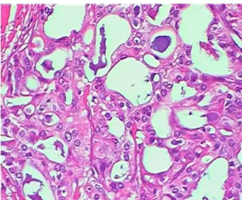
Two years later the patient noted a nodule in the inferior internal quadrant of the left mammary gland. Examination of a biopsy taken from the lesion revealed an infiltrating secretory carcinoma. She was admitted to the local hospital for a left modified radical mastectomy (Madden). Macro-scopic examination of the operative specimen revealed ‘‘ybreast with inverted nipple. (y) in the subareolar area, there was observed a nodule of circinated boards, 3 cm in diameter, white in colour and of a firm consistency’’; the microscopic findings confirmed a SBC (Fig. 1) measuring 5.5 cm in its largest diameter. During axillary node dissec-tion eight lymph nodes were isolated, one of which
contained metastases. There were no signs of venous invasion; but there were visible subareolar papillomatosis lesions. Examination for oestrogen and progesterone receptors was negative. An analysis for Her2/neu expression was not per-formed.
The patient received adjuvant chemotherapy with 5-FU 500 mg/m2 i.v. on day 1, epirubicin
75 mg/m2 i.v. on day 1 and cyclophosphamide
500 mg/m2 i.v. on day 1 of each 3-week cycle for
six cycles, after which she underwent radiotherapy, whole breast and drainage chains, with a total dose of 50 Gy.
After completing a 4-year follow-up with no evidence of local or distant recurrence, the patient went on reconstructive surgery, with an excellent cosmetic outcome.
Discussion
Surgery is still considered the most appropriate treatment for primary SBC.1,3There is some degree of consensus about the extent of surgery, as the principal management problems are late local chest wall recurrence and regional axillary lymph node spread.3Whenever possible, prepubertal girls should be treated initially by wide local excision. Preservation of the breast bud should be at-tempted, but this is not always possible, so that breast development can be impaired. Breast tissue peripheral to the tumour should be sampled generously and examined for intraductal foci, since these can indicate an increased risk of local recurrence. Mastectomy may be necessary in the
ARTICLE IN PRESS
Figure 1 Solid and acinar neoplastic structures with abundant luminal secretion.
case of large primary tumours or to control recurrent disease in the breast.4Axillary dissection should be performed whenever the clinical findings suggest nodal involvement, since late recurrence in the chest wall and axillary lymph nodes is well documented. It has been observed that nodal metastases in children tend to be small and undetectable in clinical examination.4
Of the cases reported thus far in the literature, postoperative radiotherapy has been used in at least two, as has adjuvant chemotherapy. There is too little evidence to justify recommending either approach over the other in the management of primary SBC.3,4 Since systemic metastases are ex-tremely rare, even in patients with axillary node metastases, there is no evidence to suggest that systemic adjuvant therapy would be beneficial for such patients.3,4 SBC has an indolent nature and appears to be nonresponsive to various chemotherapy protocols.3
Some authors believe that local recurrence is more likely to be the result of incomplete excision than of inherent aggressiveness. Inadequate exci-sion often results from a desire to preserve breast tissue. Early wide local excision can ultimately result in conservation of more breast tissue.4 However, there are several reports of late recur-rences and distant metastases, indicating the need for long-term follow-up of these patients.
SBC has a better prognosis than the more usual form of ductal carcinoma.1Tumour size is a strong determinant of outcome: tumours under 2 cm have not usually recurred.5 Other features that ensure an excellent prognosis include young age and the absence of stromal invasion at the periphery of the lesion.7
Recently, three cases of a new SBC entity, acinic cell-like breast carcinoma, were reported. They were typical of SBC, and were clinically, histologi-cally and immunohistochemihistologi-cally analogous to acinic cell carcinoma of the salivary gland; prob-ably they are identical lesions.8
Conclusion
The current case illustrates an unusual presenta-tion of this rare type of breast carcinoma: a large lesion with lymph node involvement at the time of mastectomy and none of the clinicopathological findings that are thought to confer a good prog-nosis. This was crucial in our decision on adjuvant treatment despite the tumour’s biological indo-lence.
Earlier publications on such tumours include few papers that specify the type of chemotherapy and radiotherapy given, when applicable, and results obtained with these. Like Herz et al.,3we too think that it is useful to report every case of this rare tumour, in order to pool as much information as possible about its biology.
References
1. Gallager HS. Pathologic types of breast cancer: their
prognoses.Cancer1984;53:623–9.
2. McDivitt RW, Stewart FW. Breast carcinoma in children.J Am
Med Assoc1966;195:388–90.
3. Herz H, Cooke B, Goldstein D. Metastatic secretory breast cancer. Non-responsiveness to chemotherapy: case report
and review of the literature.Ann Oncol2000;11:1343–7.
4. Rosen PP, Cranor ML. Secretory carcinoma of the breast.Arch
Pathol Lab Med1991;115:141–4.
5. Feiner H, Moezzi M. Pathology of special forms of breast
cancer. In: Rosen D, editor. Breast cancer. Philadelphia:
Churchill Livingstone; 1999, p. 116.
6. D’Amore ESG, Maisto L, Gatteschi MB, Toma S, Canavese G. Secretory carcinoma of the breast: report of a case with
fine needle aspiration biopsy. Acta Cytol 1986;30:309–12
(abstr).
7. Simpson JF, Wilkinson EJ. Malignant neoplasia of the breast:
Infiltrating carcinomas. In: Kirby I, Copeland III E, editors.The
breast, comprehensive management of benign and malignant diseases. 2nd ed. Philadelphia: WB Saunders; 1998, p. 289. 8. Hirokawa M, Sugihara TS, Monobe Y, Kudo H, Sano N, Sano T.
Secretory carcinoma of the breast: a tumour analogous to
salivary gland acinic cell carcinoma. Histopathology 2002;
40:223–9.