RESUmo
O objeivo deste arigo foi ideniicar casos de risco para úlcera por pressão (UP), em pacientes críicos, a parir da escala de Bra
-den e de fotograias digitais. Uma amostra de 42 sujeitos foi avaliada durante 15 dias entre março a junho de 2009 em Fortale
-za, Brasil. No total foram ideniicadas 47 lesões, sendo 23 (48,9%) com estágio I e 24 (51,1%) com estágio II. Conclui-se que é necessário que o enfermeiro que ainda não uiliza usualmente tecnologias na avaliação e tratamento procure se familiarizar com isso, a im de diminuir as repercussões ne
-gaivas deste problema de saúde coleiva.
dEScRitoRES
Úlcera por pressão
Unidades de Terapia Intensiva Medição de risco
Escalas Fotograias
Cuidados de enfermagem
Using the Braden Scale and photographs
to assess pressure ulcer risk
*O
riginal
a
r
ticle
ABStRAct
The objecive of this paper was to idenify criically ill paients at risk for pressure ul
-cer (PU) using the Braden scale and digital photographs. A sample of 42 subjects was evaluated for 15 days, between March and June 2009, in Fortaleza, Brazil. A total of 47 lesions were ideniied, 23 (48.9%) as stage I and 24 (51.1%) as stage II. It is necessary for nurses to become familiar with and adopt the technologies used to assess and treat PU in order to lessen the negaive ef
-fects of this public health problem.
dEScRiPtoRS
Pressure ulcer Intensive Care Units Risk assessment Scales
Photographs Nursing care
RESUmEn
El objeivo de este arículo fue ideniicar casos de riesgo de úlcera por presión (UP) en pacientes críicos a través de la escala de Braden y de fotograías digitales. Una muestra de 42 sujetos fue evaluada durante quince días entre marzo y junio de 2009 en Fortaleza-Brasil. En total, fueron ideniica
-das 47 lesiones, resultando 23 (48,9%) en estado I y 24 (51,1%) en estado II. Es nece
-sario que el enfermero que aún no uiliza con habitualidad tecnologías de evaluación y tratamiento intente familiarizarse con ello, a efectos de minimizar las repercusiones ne
-gaivas de este problema de salud coleciva.
dEScRiPtoRES
Úlcera por presión
Unidades de Cuidados Intensivos Medición de riesgo
Escalas Fotograías
Atención de enfermería thiago moura de Araújo1, márcio Flávio moura de Araújo2, Joselany Áio Caetano3 O usO da escala de Braden e fOtOgrafias na avaliaçãO dO riscO para úlceras pOr pressãO
usO de la escala de Braden y fOtOgrafías en la evaluación del riesgO para úlceras pOr presión
*from the dissertation “acurácia de escalas para avaliação de risco para úlcera por pressão”, post-graduate nursing program of the federal university of ceará, 2009. 1phd student of the post-graduate nursing program of the federal university of ceará. professor of the nursing course of the federal
university of Maranhão. imperatriz, Ma, Brazil: [email protected] 2phd student of the post-graduate nursing program of the federal university
of ceará. professor of the nursing course of the federal university of Maranhão. imperatriz, Ma, Brazil. [email protected] 3professor of the
intRodUction
In Brazil, the adopion of technological resources in nursing care has been an increasing fact since the late 1960’s with the scieniic grounding of the profession. Bra
-zilian nurses who study the relaionships between tech
-nology and care, conceive the irst as the solidiicaion of quoidian experience and research in the development of scieniic knowledge that culminates in the construcion of material products, or not, with therapeuic goals. Thus, there is a disassociaion of the prevailing idea of technol
-ogy and materiality that becomes understood as the ap
-plicaion of skills and abiliies in the fusion of processes that conceive products and/or knowledge(1).
In the treatment and prevenion of pressure ulcers (PUs), for example, there are many technologies available for the nurse, especially in hospitals. There are constructs and/or equipment, such as biological dressings, topical substances, matresses, photographs, etc. There is struc
-tured clinical knowledge that directs this work, such as protocols and PU risk evaluaion scales, and
inally there is the interacion between the nurse and paient. However, much of this is sill onerous and is already used with the pa
-ient who presents skin lesions.
The recogniion of individuals vulner
-able to PUs not only depends on the clini
-cal skills of the health professional, but also on the use of accurate measurement instruments to assist in the ideniicaion of individuals at risk, examples of which are, scales, protocols and photographs of the skin of the paients(2-3). The diagnosis
and evaluaion of PUs based solely on the subjecivity of health professionals through their observaions and documentaion is inaccurate. There are results of studies
which show discrepancies in the ideniicaion process of cicatrizaion with granulaion, ibrin, exudate or ne
-crosis or even the lesion size. There are also diferences of up to 30% in the diagnosis of PUs between healthcare professionals(4).
As highlighted by some studies, digital photography is an accurate technology, important in this aspect because it facilitates the diicult task of measuring, describing and documening the skin lesions of the paients. It allows the electronic transmission of images and the consultaion of specialist professionals in distant locaions. Furthermore, it is an economical technology that can moivate the pa
-ient in situaions of improvement and has a greater im
-pact than the writen word in maters of juridical claims for healthcare(5-6).
The risk evaluaion scales for PUs are also an innova
-ive technology and have presented signiicant results for this problem, although focused on local experiences. From
a literature review, it was possible to encounter more than 40 PU risk scales worldwide, with the most widely used in the Americas and Europe being the scales of Norton, Gos
-nell, Braden and Waterlow. These instruments address intrinsic and extrinsic factors of the paients related to lesion development. These aspects help the nurse in the measurement of the risk and the planning of care directed toward the risk factors of each paient(7). However, their
use should be regular, not limited only to the admission of the paient, because the risk is coninuous and early iden
-iicaion of these paients allows the implementaion of prevenive measures that can reduce PU incidence by up to 50%(7).
Nurse researchers admit that the technology issue is sill incipiently addressed in nursing pracice in Brazil(1).
Added to this, longitudinal studies on the use of technolo
-gies, such as photographs, together with risk scales for PUs, are also scarce. In the case of the photographic doc
-umentaion of wounds, for example, a review of the lit
-erature found that among 43 aricles, only one had been developed in Brazil(8). Therefore, the aim
of this study was to idenify PU risk cases in criically ill paients in northeastern Bra
-zil through the use of the Braden scale and digital photographs.
mEtHod
Study design and paricipants
An exploratory and longitudinal study was developed in three intensive care units (ICU) of a health insitute, a reference in ur
-gent and emergency care in northeastern Brazil. The study was conducted from March to June 2009, with all paients admited, during the study period, to the three adult ICUs of the insituion, who met the following inclusion criteria: being over 18 years of age; not presening PU on admission; having spent up to 48 hours hospitalized in the ICU, prior to data collecion. In turn, the exclusion criteria were as follows: being a hemodynamically unstable pa
-ient; having a medical diagnosis of brain death; and hav
-ing a prognosis of leav-ing the ICU within iteen days.
Populaion and sample
Because the populaion was comprised of paients over 18 years, the pediatric ICU was not integrated into the research, the reference for calculaing the populaion being 23 beds. According to the center of epidemiologi
-cal surveillance of the insituion, in 2008 a total of 432 adult paients in the three ICUs was recorded. For the cal
-culaion of the sample a formula for longitudinal before and ater studies was used, with the conidence interval of 95%. Ater performing the calculaions, a sample of 42 paients for the study populaion was ideniied.
nurse researchers admit that the technology issue is still
To arrive at the 42 paients evaluated, the inclusion and exclusion criteria were taken into account, and dur
-ing the four months there were 83 inpaients, 11 were ad
-mited with PU, two were younger than 18 years, 10 died before compleing the minimum of ten days and 18 were transferred before compleing the minimum of ten days.
Instruments
Two instruments were used in the data collecion, the irst was a form with sociodemographic and clinical data, and the second the translated Braden Scale, adapted and validated in Brazil. It is composed of six subscales which are: sensory percepion, skin moisture, acivity, mobility, nutriional status, fricion and shear. All are scored from 1 to 4, with the excepion of fricion and shear, where the measure ranges from 1 to 3. Total scores range from 6 to 23. Paients achieving a score of 16 or more are consid
-ered low risk for the development of PUs; between 11 and 16, indicates a moderate risk and under 11 a high risk, as this shows inadequate funcioning of the evaluated pa
-rameters(10) .
For the evaluaion of the PUs encountered the classi
-icaion of the Naional Pressure Ulcer Advisory Panel was considered(11): Stage I - redness of intact skin that does
not whiten ater removing the pressure; Stage II - skin loss involving the epidermis, dermis or both. The ulcer is su
-pericial and presents clinically as an abrasion or shallow crater; Stage III - full thickness skin loss, involving damage or necrosis of subcutaneous issue that may deepen, not reaching the fascia. The ulcer presents clinically as a deep crater; Stage IV - full thickness skin loss with extensive de
-strucion, issue necrosis or damage to muscle, bone or supporing structures such as tendons or joint capsules.
Data collecion
The clients who fulilled the criteria for sample inclu
-sion were monitored for 15 consecuive days. Evaluaions were performed once a day, always in the matuinal pe
-riod, the moment at which the applicaion of dressings and cleaning of the paients occurred. At the ime of the irst visit, each client had their clinical data recorded on a form. In turn, the data concerning the skin and the Braden scale were invesigated in all 15 visits performed for each paient. In those cases where the 15 visits were not concluded, for reasons of death or transfer, they only remained in the study if at least 10 visits had already been carried out.
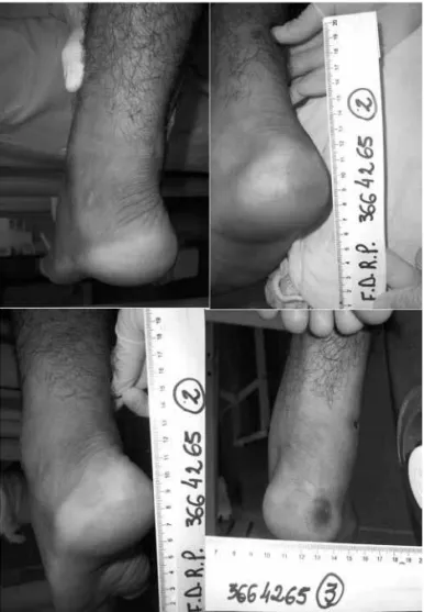
Every ive days digital photographs were taken of a set of anatomical regions of the paients, ater cleaning the wound and before applying new dressings, namely: occip
-ital, scapular, elbows, sacrum, trochanter, calcaneus and malleolus. It is noteworthy that images were only taken of these regions, regardless of whether the skin was intact or not (Figure 1). The camera used had a resoluion of 12 mega pixels and in order to produce a correct image of the
wound without any distorion, the photos of all the above menioned areas were taken in the perpendicular posi
-ion with the lash func-ion acive. The photographs taken were stored on a note book for their later consideraion. Together, two nurses who work with criically ill paients studied the photographs and reached a consensus on the meaning of images.
The data found in the images were not recorded in the medical records of the paient. However, as an ethi
-cal issue, any alteraions detected during the study were reported to the nurses of the evaluaing ICU.
Data organizaion and analysis
The data were double entered and stored in a data
-base using the Excel program, then exported to the Epi Info version 6.04 program for the elaboraion of the ab
-solute frequency and percentage, as well as for measures of central tendency. The Bartlet’s test was employed in the evaluaion of the homogeneity of the data, and a con
-idence level of 95% was used. To compare percentage diferences the Fischer test was carried out, while ANOVA was used to compare the means.
Ethical aspects
The study was conducted ater approval by the research ethics commitee of the insituion, under to pro
-tocol No. 86145/08. In the case of an unconscious paient consent was obtained from the relaives and guardians.
RESULtS
The mean length of monitoring was 14 days (SD±3.6), with 32 (76.2%) paients monitored for 15 days. The re
-maining seven (16.7%) subjects were monitored for 10 days because of transfer to another hospital sector and/ or death. Of the 42 paients evaluated, most were young adults, 13 (31%) between 18 and 25 years of age, followed by 11 (26.2%) between 36 and 46 years of age. The mean age was 35.3 (SD±4.7) years. There was a predominance of males with 34 (81%).
The majority of paients admited to the ICU and in
-cluded in this study came from the anesthesia recovery and emergency rooms, with 25 (59.5%) and 14 (33.3%), respecively. The most common clinical condiions veriied at the ime of admission were neurological dysfuncion, with the predominant traumaic brain injury, 26 (61.9%), followed by surgery, 11 (26.2%), mainly exploratory lapa
-rotomy and neurosurgery. The use of mechanical veni
-laion, vasoacive drugs and sedaion were detected in 78.6%, 31% and 69% of the paients, respecively.
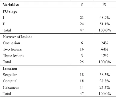
24 (51.1%) as stage II. Regarding the number of lesions per paient, the distribuion was: six (1PU), sixteen (2 PU), and only three (3 PU) (Table 1).
Table 1 - Distribution of the characteristics of the PU according to stage, number and location of lesions of the patients. Fortaleza--Brazil, 2009
Variables f %
PU stage
I 23 48.9%
II 24 51.1%
Total 47 100.0%
Number of lesions
One lesion 6 24%
Two lesions 16 64%
Three lesions 3 12%
Total 25 100.0%
Location
Scapular 18 38.3%
Occipital 18 38.3%
Calcaneus 11 24.4%
Total 47 100.0%
Regarding the locaion of the lesions, the sacral and occipital regions presented the same frequency of 18 (38.3%) paients. The calcaneus region presented a fre
-quency of 11 (23.4%) cases. The PUs in this region were not divided between right and let calcaneus. Among paients who developed two lesions, four paients had lesions in the sacral and calcaneus regions; nine in the sacral and occipital; and three in the calcaneus and oc
-cipital (Table 1). Among these, 88% of the cases occurred in males, however, this was not staisically signiicant according to Fischer’s test (p=0.156). The mean ages of subjects with PU absent or present were similar, thus, there was no staisically signiicant diference in this variable (p=0.918). The same trend was observed when comparing the mean scores of the Braden Scale between these subjects (p=0.709) (Table 2).
In the item sensory perception of the Braden scale, the response completely limited presented a percent
-age above 23.8% in the first 10 days. Following this there was a decrease in this percentage up to the 15th day, when the values started to increase again. In turn, the response very limited to the item sensory percep
-tion presented the highest percentage among the par
-ticipants throughout the entire evaluation, with the minimum and maximum values of 15 (39%) and 20 (68.8%), respectively. The percentage of subjects with sensory perception completely limited and very limit-ed decreased during the study, between the first and
last days of evaluation, as shown by the intervals of 14 (33.3%) to 9 (28.1%) and 20 (47.6%) to 13 (40.6%), respectively.
In the item moisture, 32 (76.2%) paients were clas
-siied as occasionally moist throughout the enire inves
-igaion. The subjects evaluated had litle variaion from the irst unil the tenth day, as shown by the interval: 28 (66.7%) and 32 (76.2%) individuals, respecively. No paient presented an absence of moisture. In the item acivity in the Braden Scale it was found that 90% of the subjects were conined to bed. In the inal ive days there was a decrease in these values. In the quesion of mobility it was observed that 12 (28.6%) paients were
completely immobile during the irst 10 days of evalu
-aion, with a reducion of this percentage over the last ive days. Simultaneously, in this inal phase there was also a small increase in the paients with slight
limita-ion, moreover, only two were categorized as no
limita-ion, with the ability to make major changes in posiion without assistance.
In the subjects investigated there was a decrease in the frequency of those with very poor nutrition and
probably inadequate nutrition between the first and
twelfth days as shown in the variations of 15 (35.7%) to 1 (2.8%) and 13 (31%) to 6 (18.7%) respectively. Con
-sequently, the subjects classified as adequate nutrition
jumped from 14 (33.3%) to 29 (87.8%). In the item of friction and shear of the skin the response problem
presented the highest frequency among those sur
-veyed throughout the entire monitoring period (69% to 87.5%). Between the first and last days of evaluation there was a fall of two thirds of patients with potential
problem, from 13 to 4 individuals. Only 2.4% of subjects
presented no apparent problem for skin friction and shear.
Table 2 - Distribution of gender, age, time to onset of PU and Braden Scale scores, according to the presence of PU. Fortaleza-Brazil, 2010
Variables
Presence of PU
p
Yes No
f % f %
Gender
Male 22 88.0 12 70.6 0.156*
Female 03 12.0 05 29.4
Age (years) X= 35.0 DP (±13.8) X = 35.4 DP (±11.6) 0.918**
Days hospitalized X =14.2 DP (±1.7) X = 13.7 DP (±2.1) 0.396**
Escores na Escala de
Braden (pontos) X =12.2 DP (±1.6) X = 12.4 DP (±1.6) 0.709**
X= Mean; dp= standard deviation *fisher’s test ** anOva test
In general, the paients presented Braden Scale scores with means ranging from 11.6 to 12.5 and from 9.1 to 16.7 per day and per paient, respecively. From this, one pa
Figure 1 - Evolution of the calcaneus region of a critical patient evaluated by digital photographs and the Braden Scale on the 1st, 5th, 10th and 15th day - Fortaleza, Brazil, 2010
diScUSSion
During the reading of these data, it is important to note that the version of the Braden scale adopted in this research has low sensiivity (31.2%) but high speciicity (88.2%)(12). Therefore, iniially it may be thought that this
percentage could be higher, however, the high speciicity of the instrument, coupled with the simultaneous use of digital photographs, ensure that there was a more accu
-rate skin assessment and compleion of the Braden scale. Thus, the data found depict faithfully the situaion of the insituion in quesion. Addiionally, the literature has stressed that relevant clinical disagreements of up 58.4% occur between nurses that use only a physical examina
-ion and those who adopt a physical examina-ion and digital photo images in their consultaions with paients with wounds. Therefore, the nurses who do not use digital images in their evaluaion, either in the domicile or the hospital, are at risk of underesimaing the presence of skin lesions(13).
The data source and the methods of measurement are oten the reason for the divergence of prevalence and in
-cidence of PUs among the studies. For example, publica
-ions based on analysis of medical records, certainly pres
-ent an underesimated frequency of PUs, in addiion, the adopion of diferent scales can cause confusion between the results(14). Another important detail is the frequency of
the applicaion of the PU risk scales. Some authors found issue tolerance to pressure to be a pathophysiological aspect of the evoluion, which in the bones and muscles are respecively 30 and 2 hours(9). Therefore, it is possible
for signiicant changes to occur in these lesions within 30 hours, making daily monitoring ideal, especially in crii
-cally ill paients. However, in the reality of many health insituions there is an increase in the coningent of pa
-ients while the number of nurses is decreasing, leading to the delegaion of pressure ulcer risk assessment to other professionals, without the necessary skills and competen
-cies. This could result in the skin analysis and the illing of such instruments being performed incorrectly, with a con
-sequent impact on the paient’s health. For this reason it is recommended that the applicaion of PU risk scales oc
-curs at least every other day, so that changes detected can be corrected in a imely manner.
The method considered the gold standard for the diag
-nosis of cases of PU is interobserver evaluaion, although, the reliability decreases in the classiicaion of the lesions, especially in the gluteal and hip regions(14). However, this
is someimes made diicult due to a scarcity of health professionals proicient in the area. A fact that could be circumvented with the use of photos shared between experts on the topic. Furthermore, this technology pres
-ents many advantages such as: accuracy in the analysis of the depth, coloraion and posiion of the borders of the lesions over ime; it serves as a record of the evoluion of the therapeuic process; and it reduces the need for handling the lesion during examinaion. However, some peculiariies should be considered, namely: the techno
-logical precision of the photo requires addiional costs in training, equipment and storage, as well as in consultaion with experts in inalizing the diagnosis(4,15).
To make the use of photography viable in the study and intervenion of PUs it is also necessary that some as
-pects are reformulated: there should be standardizaion of anatomical zones to be photographed, health profession
-als must undergo training to guarantee the precision of the images, and they should also use similar equipment(6).
On the same theme, the Wound, Ostomy and Coninence Nurses Society (WOCN) states that the use of digital pho
-tography is not a new resource in the documentaion of injuries, it is even banal, but nevertheless can be an im
-portant complement since it is supported by valid proto
-cols that address ethical aspects, skills and abiliies in the technique and handling of these arsenals(16). However, as
previously stated, in Brazilian hospitals the adopion of this technology is sill not rouine for documentaion of the evoluion of skin lesions. According to Brazilian nurs
-es, the main diiculies of producing and applying assis
and even curriculum problems(1). Thus, nurses can en
-counter another drawback: the lack of access for nurses to training and administraive support for the adopion of new technologies, which raises the costs of adopion of these resources. Therefore, as this interferes with their care pracices, nurses must learn how to work with new technologies, given their ime, knowledge and demand and then combine these resources with their quoidian clinical pracice without reducing the direct contact with the paient.
With regard to the spread of use, the Braden scale has a diferent posiion to digital photos, since its adopion is already rouine in Brazilian insituions. Thus, the results of this and other studies conducted in Brazil conirm that the scores obtained for the Braden scale can assist nurses, from the iniial evaluaion upon admission, in the idenii
-caion of paients at higher risk for developing PUs, so that eforts be made to implement the recommended preven
-ion measures(10,17-20).
The fact is that in this study of the 34 subjects with some degree of risk, only 25 developed PUs during the study period. This reinforces that it is possible to block the genesis through prevenive acions, such as PU risk scales and digital photographs, thereby reducing the eco
-nomic and clinical burden of the insituions and of the paients, respecively. For this, the healthcare insituions should seek to ensure the supplies necessary for the work of all the healthcare professionals involved in wound care. The nurse, in turn, should aim to become familiar with the new technologies in wound care, such as the PU risk scales and digital photographs, and to uilize them in their quoidian. Quesions such as the frequency of recording and anatomical points necessary, imes and the inluence of structural and human resource factors should be focus of new studies in this area to clarify the use of more accu
-rate and reproducible pressure ulcer risk scales and digital photographs in prevenion.
concLUSion
This study has some limitaions that should be listed. Firstly, the ime set for monitoring of the paients pre
-vented the monitoring of the outcome of paients with low risk for developing PUs; secondly, there was no com
-parison between the results obtained during the data collecion and the notes in the medical records of the paients in order to clarify similariies and diferences regarding the skin of the subjects; and, inally, the evalu
-aions of the digital photos were performed by only two nurses. However, the results of this study conirm the indings of the Brazilian and foreign literature: the adop
-ion of digital photos and the Braden Scale is an adjunct in the work of ideniicaion and prevenion of PUs in criically ill paients.
It is important to highlight that the version of the Braden scale adopted in this study has low sensiivity but high speciicity. However, the high speciicity of the instru
-ment, coupled with simultaneous use of digital photos, ensures that the data found faithfully portray the situa
-ion of the ICU of the insitu-ion evaluated in rela-ion to pressure ulcers.
Today there are many technologies available to the nurse to prevent the emergence of PUs, however, many of them are only parially accessible in many countries, as is the case in Brazil. Therefore, it is necessary that nurses, not only of Brazil but also of other countries, who sill do not usually use these resources in the whole of their terri
-tory, seek to become familiar with these technologies for diagnosing and coping with PUs. Furthermore, their use should be coninuous and systemaic not restricted only to the admission of the paient. It is recommended that further research be developed with a view to divulge and suggest improvements or miigaion of costs in the use of these technologies, so that inally there is a spread of these technologies.
REFEREncES
1. Rocha PK, Prado ML, Wal ML, Carraro TE. Care and technol -ogy: approaches through the Care Model. Rev Bras Enferm. 2008;61(1):113-5.
2. Gould D, Goldstone L, Kelly D, Gammom J. Examining the va -lidity of pressure ulcer risk assessment scales: a replicaion study. Int J Nurs Stud. 2004;41(3):331-9.
3. Rocha ABL, Barros SMO. Avaliação de risco de úlcera por pressão: propriedades de medida da versão em português da escala de Waterlow. Acta Paul Enferm. 2007; 20(2):143-50.
4. Dufrene C. Photography as an adjunct in pressure ulcer docu -mentaion. Crit Care Nurs Q. 2009;32(2):77-80.
5. Wild T, Prinz M, Fortner N, Krois W, Sahora K, Stremitzer S, et al. Digital measurement and analysis of wounds based on colour segmentaion. Eur Surg. 2008; 40(1):5-10.
6. Baumgarten M, Margolis DJ, Selekof JL, Moye N, Jones PS, Shardell M. Validity of pressure ulcer diagnosis using digital photography. Wound Rep Reg. 2009;17(2):287-90.
7. Borges EL, Saar SRC, Magalhães MBB. Feridas: como tratar. 2ª ed. Belo Horizonte: Coopmed; 2008.
9. Cardoso MCS, Caliri MHL, Hass VJ. Prevalência de úlcera de pressão em pacientes críicos internados em um hospital uni -versitário. REME Rev Min Enferm. 2004;8(2): 316-20.
10. Paranhos WY, Santos VLCG. Avaliação do risco para úlcera de pressão por meio da Escala de Braden na língua portuguesa. Rev Esc Enferm USP. 1999;33(n.esp):191-204.
11. Agency for Health Care Policy and Research. Pressure ulcers in adults: predicion and prevenion. Clinical Pracice Guide -line, 3. Decubitus. 1992;5(3):26-30.
12. Araújo TM, Araújo MFM, Cavalcante CS, Barbosa Júnior GM, Caetano JA. Acurácia de duas escalas de avaliação de risco para úlcera por pressão em pacientes críicos. Rev Enferm UERJ. 2011;19(3):381-5.
13. Buckley KM, Adelson LK, Agazio JG. Reducing the risks of wound consultaion: adding digital images to verbal reports. J Wound Ostomy Coninence Nurs. 2009;36(2):163-70.
14. Stausberg J, Lehmann N, Kroger K, Maier I, Niebel W. Reli -ability and validity of pressure ulcer diagnosis and grading: an image-based survey. Int J Nurs Stud. 2007; 44(8):1316-23.
15. Localio AR, Margolis D, Kagan SH, Lowe RA, Kinosian B, Ab -buhl S, et al. Use of photographs for the ideniicaion of pressure ulcers in elderly hospitalized paients: validity and reliability. Wound Rep Reg. 2006;14(4):506-13.
16. Demarest L, Acoraci LR. Choosing and using a digital camera in home care. Home Healthcare Nurse. 2004;22(1):61-3.
17. Rogenski NM, Santos VLCG. Estudos sobre a incidência de úlceras por pressão em um hospital universitário. Rev Laino Am Enferm. 2005;13(4):474-80.
18. Fernandes LM, Caliri MHL. Using the Braden and Glasgow scales to predict pressure ulcer risk in paients hospi -talized at intensive care units. Rev Laino Am Enferm. 2008;16(6):947-52.
19. Gomes FSL, Bastos MAR, Matozinhos FP, Temponi HR, Ve -lásques-Meléndez. Risk assessment for pressure ulcer in criical paients. Rev Esc Enferm USP [Internet]. 2011 [cited 2011 May 15];45(2):313-8. Available from: htp://www.sci -elo.br/pdf/reeusp/v45n2/en_v45n2a01.pdf