Claudio Calazan do CarmoI Ronir Raggio LuizII
I Serviço de Oncologia Clínica. Hospital do Câncer II. Instituto Nacional de Câncer. Rio de Janeiro, RJ, Brasil
II Departamento de Bioestatística. Instituto de Ensino em Saúde. Universidade Federal do Rio de Janeiro. Rio de Janeiro, RJ, Brasil Correspondence:
Claudio Calazan do Carmo Instituto Nacional de Câncer R. Equador 831 – Santo Cristo 22220-040 Rio de Janeiro, RJ, Brasil E-mail: [email protected] Received: 7/25/2010 Approved: 3/15/2011
Artigo disponível em português e inglês em: www.scielo.br/rsp
Survival of a cohort of women
with cervical cancer diagnosed
in a Brazilian cancer center
Sobrevida de mulheres com câncer
de colo uterino diagnosticadas em
um centro brasileiro
ABSTRACT
OBJECTIVE: To assess overall survival of women with cervical cancer and describe prognostic factors associated.
METHODS: A total of 3,341 cases of invasive cervical cancer diagnosed at the Brazilian Cancer Institute, Rio de Janeiro, southeastern Brazil, between 1999 and 2004 were selected. Clinical and pathological characteristics and follow-up data were collected. There were performed a survival analysis using Kaplan-Meier curves and a multivariate analysis through Cox model.
RESULTS: Of all cases analyzed, 68.3% had locally advanced disease at the time of diagnosis. The 5-year overall survival was 48%. After multivariate
analysis, tumor staging at diagnosis was the single variable signifi cantly
associated with prognosis (p<0.001). There was seen a dose-response relationship between mortality and clinical staging, ranging from 27.8 to 749.6 per 1,000 cases-year in women stage I and IV, respectively.
CONCLUSIONS: The study showed that early detection through prevention programs is crucial to increase cervical cancer survival.
Cervical cancer is one of the most common cancers worldwide, the sixth in incidence and the third leading
cause of death among women .16 Most cases (80%)
are reported in developing countries in Africa, Latin America and the Caribbean, and they are relatively
uncommon in Europe, Israel and the US.a In Brazil,
cervical cancer is the second leading cause of death due to cancer in women. The Brazilian Ministry of Health
estimated approximately 18,430new cases in 2010b with
4,315 deaths confi rmed in 2008.c
The major prognostic factor of cervical cancer is tumor staging. Tumor staging according to the International
Federation of Gynecology and Obstetrics11 (FIGO)
staging system is directly associated with risk of recur-rence and overall survival. It assesses the primary tumor size, local extension, invasion of adjacent structures, and presence of distant lesions. However, the FIGO staging system does not assess several other factors such as age, skin color, socioeconomic condition and histopathological type of tumor, which have also been reported as prognostic factors.7,d
RESUMO
OBJETIVO: Avaliar a sobrevida global de mulheres com câncer de colo uterino
e identifi car fatores prognósticos relacionados.
MÉTODOS:Todos os 3.341 casos tratados no Instituto Nacional de Câncer, Rio de Janeiro, RJ, entre 1999 e 2004 foram selecionados e seus dados clínicos, anatomo-patológicos e de seguimento foram coletados. Foram utilizados a curva de Kaplan-Meier e o modelo de Cox para avaliação da sobrevida e para análise logística múltipla, respectivamente.
RESULTADOS: Dos casos estudados, 68,3% apresentavam doença localmente avançada. A sobrevida global em cinco anos foi de 48%. Após análise múltipla, o estádio clínico ao diagnóstico foi a única variável signifi cativamente associada com o prognóstico (p < 0,001). Mortalidade e o estádio clínico da doença apresentaram efeito dose-resposta, variando de 27,8 por 1.000 casos-ano a 749,6 por 1.000 casos-ano em mulheres com estádio I e IV, respectivamente.
CONCLUSÕES: Os resultados mostram que a detecção precoce por meio de programas de prevenção é fundamental para aumentar a sobrevida em câncer cervical.
DESCRITORES: Neoplasias do Colo do Útero. Detecção Precoce de Câncer. Diagnóstico Tardio. Análise de Sobrevida.
INTRODUCTION
a Ferlay J, Parkin DM, Pisani P. GLOBOCAN 2002. Cancer Incidence, Mortality and Prevalence Worldwide. Lyon: International Agency for Research on Cancer; 2004.
b Ministério da Saúde, Secretaria Nacional de Assistência a Saúde, Instituto Nacional de Câncer. Estimativa da incidência e mortalidade por câncer no Brasil. Rio de Janeiro; 2007.
c Ministério da Saúde. DATASUS. 1. Sistema de Informação de Mortalidade (SIM). Indicadores e Dados Básicos - Brasil - 2008. IDB-2008. Brasília; 2009.
d American Joint Committee on Cancer. Manual for staging of cancer: cervix uteri. 3. ed. Philadelphia: Lippincott; 1998.
e International Agency for Research on Cancer. IARC confi rms effi cacy of cervix cancer screening for women 25-65 in reducing mortality. Lyon; 2004. [cited 2011 Mar 15]Available from: http://www.iarc.fr/en/media-centre/pr/2004/pr151.html
f Ministério da Saúde. DATASUS. 1. Sistema de Informação de Mortalidade (SIM). Indicadores e Dados Básicos - Brasil - 2005. IDB-2005. Brasília; 2006. [cited 2011 Mar 15] Available from: http://tabnet.datasus.gov.br/cgi/idb2005/matriz.htm
There is a clear relationship between the extension of disease at diagnosis (tumor staging) and disease-associated mortality. This is the basis for screening programs,10 which can effectively detect pre-invasive or
early stage disease and thus reduce associated morbidity and mortality.1,11,e
The present study aimed at analyzing overall survival of women diagnosed with cervical cancer and describing prognostic factors associated.
METHODS
The Brazilian National Cancer Institute (Instituto
Nacional do Câncer – INCA) is a reference center for cancer prevention and control in Brazil. The INCA serves both the metropolitan area of the city of Rio de Janeiro and the rest of the state. In 2003, of 896 cases of cervical cancer diagnosed in the city of Rio de Janeiro, and 2010 cases in the state,f 597 patients were attended
A retrospective study with data obtained from INCA registries was conducted. All cases of primary cervical tumors diagnosed from 1999 to 2004 were assessed.
Since 1999 the Hospital do Câncer 2 (HC2), one of
the four INCA units, is a reference center for all gyne-cologic cancer cases.
The following variables were analyzed: age; tumor staging; histopathological type of tumor; level of education; skin color; and year of diagnosis.
Age was divided into two groups: <65 years and ≥65
years. Level of education was categorized as 11 or more years of schooling and less than 11 years of schooling. Skin color was categorized as white and non-white.
Cases were classifi ed according to the FIGO staging
system, stages I to IV. Histopathologically tumors were
classifi ed as: squamous cell carcinoma;
adenocarci-noma; and other types.
The chi-square test was used to assess the association between tumor staging and the independent variables.
For estimating survival, all deaths during the study period were considered events; the remaining cases were censored at the last hospital visit on November 2006. Data on deaths were collected from the hospital registry and the Brazilian Mortality Database (SIM). Kaplan-Meier curves were used to analyze overall survival and the log-rank test was used for comparing survival curves.
The association between survival (dependent variable) and independent variables (staging, histopathological type of tumor, age, skin color, level of education and
year of diagnosis) was assessed through Cox
propor-tional hazards model. The proporpropor-tionalhazard
assump-tion was assessed by visual inspecassump-tion of the log minus logplot proportional hazards test. The case-fatality rate at each stage was calculated based on the number of deaths per person-time (incidence density).
The study was approved by INCA Ethics Commitee on Research (Process nº 52/2006).
RESULTS
A total of 3,341 patients with primary cervical tumors were attended at INCA during the study period, of which 68 were excluded due to missing information. The maximum follow-up time was 94.0 months (median 28.1 months).
Most patients at early stage (I) were treated with surgery as single therapy (69.5%) followed by local radiation therapy (22.5%). Only 18% of these patients were treated with radiation therapy only. This standard of care did not change along the period analyzed.
Patients with locally advanced disease (FIGO stages II and III) were treated with radiation therapy (69.8%). Combined radiation therapy and chemotherapy (weekly cisplatin) was used in 21% and it is important to note that beginning in 2004 this standard of care changed (78.5% received combined treatment).
Table 1 shows case distribution according to clinical and epidemiological characteristics by stage. Overall, missing data were minimal (age in one patient, skin color in 2, and level of education in 4 patients). Most
Table 1. Clinical and epidemiological characteristics of cervical cancer cases according to stages. Rio de Janeiro, Southeastern
Brazil, 1999–2004. (N = 3,341)
Variable Total
Tumor staging p-value
I II III IV
n % n % n % n % n % X2
Age (years)
<65 2,817 81.9 842 89.4 905 79.9 950 78.1 120 80.5
<0.001
≥65 623 18.1 100 10.6 228 20.1 266 21.9 29 19.5
Skin color
White 1,909 55.5 535 56.9 647 57.1 644 53.0 83 55.7
0.168
Non-white 1,530 44.5 406 43.1 486 42.9 572 47.0 66 44.3
Schooling (years)
<11 3,114 90.6 811 86.3 1012 89.4 1154 94.9 137 91.9
<0.001
≥11 323 9.4 129 13.7 120 10.6 62 5.1 12 8.1 Histological type of tumor
Squamous cell carcinoma 2,908 84.5 757 80.4 944 83.3 1084 89.1 123 82.6
<0.001
Adenocarcinoma 414 12.0 146 15.5 146 12.9 102 8.4 20 13.4
patients were white, <65 years of age, and poorly educated. The most prevalent histological type of tumor was squamous cell carcinoma (84.5%). Locally advanced disease (stages II and III) was seen in 68.3% of the cohort. Most cases were followed-up for at least two years (80.7%), and 1,499 deaths were recorded (50.1% of cases). Deaths occurred more often in the
fi rst two years of follow-up (71.2%) and at stages II or III (84.9% of all deaths).
The chi-square analysis showed that the number of patients with 11 years of schooling or more was signifi -cantly greater at stages I and II (p<0.001), although they accounted for a small proportion of the cohort. Despite slight predominance of white patients, no statistically
signifi cant difference was seen in the distribution of
cases by stage (p=0.168). Squamous cell carcinoma was most commonly seen at FIGO stage III while adenocarcinoma and adenosquamous carcinoma (plus other histological types) were more often seen at early stages (p<0.001). Year of diagnosis was not statistically signifi cant (p=0.144) in this analysis.
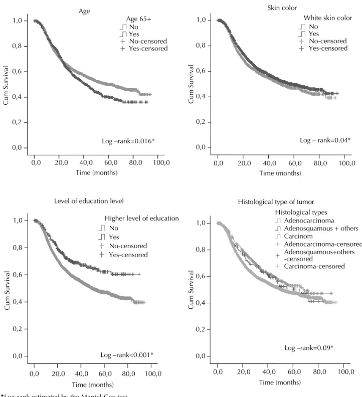
Figures 1 and 2 show the Kaplan-Meier curves. The median survival for the entire cohort was 53.4 months (95% CI 47.65;59.20) and the 5-year overall survival
was 48%. A signifi cant difference was found in median
survival between the two age groups studied (62.6 vs. 41.3 months, higher in the younger age group; p=0.016;
Fig. 1.A). Although not signifi cant, non-white women
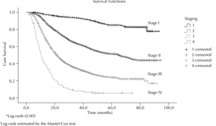
showed better survival compared with white ones (56.43 vs. 49.46 months; p=0.04; Fig. 1.B). As for level of education (Fig. 1.C), those with 11 years of schooling or more showed a signifi cantly better survival (median survival has not yet been estimated for this subgroup). The median survival for those with lower education was 49.9 months, p<0.001. The histological type of tumor (Fig. 1.D) was not statistically different in terms of survival as survival curves were very similar (p=0.09). There was a clear relationship between tumor staging at diagnosis and survival (Fig. 2), i.e., greater survival rates were found at stage I (no median survival estimated), and progressively reduced at more advanced stages (65.6 months, 21.7 months and 10.4 months for stages II, III and IV, respectively; p<0.001).
In the multiple logistic analysis using Cox model
only tumor staging remained statistically signifi cant
(p<0.001) (Table 2) while all other variables from the univariate analysis lost their statistical signifi cance.
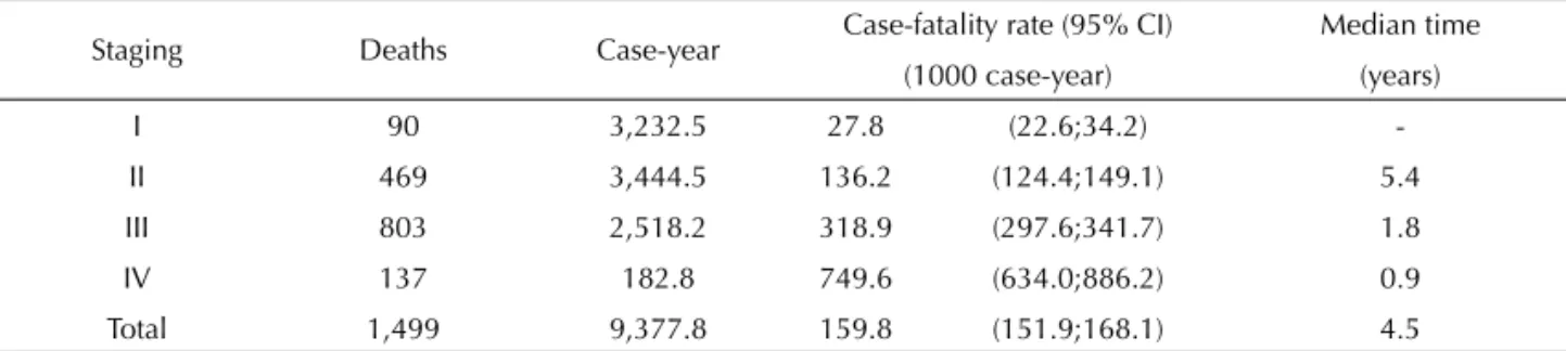
The case-fatality rate in Table 3 showed a dose-response effect between mortality and clinical stage, ranging from 27.8 to 749.6 per 1,000 cases-year in women at stages I and IV, respectively.
Table 2. Cox models for mortality of the cohort of women
with invasive cervical cancer. Rio de Janeiro, Southeastern Brazil, 1999–2004.
Characteristics
Univariate Multiple logistic analysis
Cox model Cox model 1
HR p-value HRadj p-value Age (years)
<65 1 1
≥65 1.17 0.016 0.95 0.399
Skin color
White 1 1
Non-white 1.11 0.040 1.09 0.107
Schooling (years)
<11 1 1
≥11 0.65 <0.001 0.86 0.172
Hystological type of
tumor 0.012 0.881
Squamous cell
carcinoma 1 1
Adenocarcinoma 0.79 0.004 0.96 0.637
Adenosquamous
and others 0.87 0.354 0.97 0.836
Tumor staging <0.001 <0.001
Stage I 1 1
Stage II 4.90 <0.001 4.90 <0.001
Stage III 11.44 <0.001 11.33 <0.001
Stage IV 28.96 <0.001 28.91 Year of diagnosis 0.995 0.778 0.998 0.927
Table 3. Case-fatality rate adjusted for tumor staging in women with invasive cervical cancer. Rio de Janeiro, Southeastern
Brazil, 1999–2004.
Staging Deaths Case-year Case-fatality rate (95% CI) Median time
(1000 case-year) (years)
I 90 3,232.5 27.8 (22.6;34.2)
-II 469 3,444.5 136.2 (124.4;149.1) 5.4
III 803 2,518.2 318.9 (297.6;341.7) 1.8
IV 137 182.8 749.6 (634.0;886.2) 0.9
DISCUSSION
There are few Brazilian studies on survival of women with cervical cancer. A reference center in Paraná, southern Brazil, reported 2,396 cases (1990–1994) with
a 5-year survival of 53.6%.gAnother study conducted
in São Paulo, southeastern Brazil, reported an overall 5-year survival of 66.5% representing a unexpectedly
high survival rate (66.5%) for advanced cases.2
According to the American Cancer Society, the 5-year
overall survival in the US is 72%.h Population-based
studies have reported cancer survival rates of 62.5% in
the United Kingdomi14 and 58% in Australia.1
This scenario is signifi cantly worst in less developed
regions. Although data from the 1980s in Cuba showed
g Liga Paranaense de Combate ao Câncer. Relatório Epidemiológico: RHC 2000 a 2004. Curitiba: Hospital Erasto Gaertner; 2007. h American Cancer Society. Cancer facts and fi gures. Atlanta; 1999.
i London Research Institute. Cancer Research United Kingdom. Cancer Statistics in the United Kingdom. London; 2009. [cited Mar 19] Available from: http://info.cancerresearchuk.org/cancerstats/types/cervix/survival
*Log-rank estimated by the Mantel-Cox test.
100,0 80,0 60,0 40,0 20,0 0,0
Time (months) 1,0
0,8
0,6
0,4
0,2
0,0
Cum Survival
Yes-censored No-censored YesNo Age 65+
100,0 80,0 60,0 40,0 20,0 0,0
Time (months) 1,0
0,8
0,6
0,4
0,2
0,0
Cum Survival
Yes-censored No-censored Yes No
Higher level of education
100,0 80,0 60,0 40,0 20,0 0,0
Time (months) 1,0
0,8
0,6
0,4
0,2
0,0
Cum Survival
Carcinoma-censored Adenosquamous+others -censored
Adenocarcinoma-censored Carcinom
Adenosquamous + others Adenocarcinoma Histological types
100,0 80,0 60,0 40,0 20,0 0,0
Time (months) 1,0
0,8
0,6
0,4
0,2
0,0
Cum Survival
Yes-censored No-censored Yes No
White skin color
Log –rank=0.016* Log – rank=0.04*
Log –rank=0.09* Level of education level
Log –rank<0.001*
Histological type of tumor Skin color Age
Figure 1. Kaplan-Meier survival curves for women with invasive cervical cancer according to age, skin color/ethnicity, level of
a 53.5% 5-year overall survival, lower rates have been reported in some cities in Asia: 46.5% in Bombay, India; 37.5% in Qidong, China; and 24.6% in Rizal, Philippines.1
Low socioeconomic condition has been associated with poor prognosis in cervical cancer.5,7,9 In our study level
of education was a proxy of socioeconomic condition
and there were two major fi ndings: more educated
women were a small minority (9.4%) of the cohort studied and they had a 35% reduction in the risk of death when compared with those less educated. However, this
difference fell a non-signifi cant 14% in the multiple
logistic analysis. This association may be explained by the fact that more educated women usually seek health care and have more knowledge on prevention and thus their disease is detected at an earlier phase with more successful treatment. However, in the multiple logistic analysis, the clinical magnitude of the difference between level of education (HR 0.86) remained after adjustment, despite losing its statistical signifi cance.
Data from hospital registries in Japan have showed that older age is associated with a poorer cancer survival, even after adjusting for tumor stage, and
this fi nding is also associated with more effective
screening at younger ages (<45 years).9 Another study
has argued there are adverse biological factors that
may cause a more aggressive course.11 However, the
North American Surveillance, Epidemiology and End
Results Program (SEER) did not fi nd any signifi cant
difference after adjusting for other causes of death and life expectancy when assessing case-fatality rate due to
cervical cancer in different age groups.8 In the present
study, although advanced age was also associated with a poorer prognosis (HR = 1.17) in the univariate analysis, this association disappeared in the multiple logistic analysis. The high prevalence of locally advanced cancer at advanced ages may have contributed to a poorer survival in the univariate analysis.
A trend towards a worse prognosis has been reported for the histological types adenocarcinoma and adenos-quamous carcinoma compared with sadenos-quamous cell carcinoma,3,11 although this same trend was not seen in
the multiple logistic analysis in our study. One plau-sible explanation may be the small number of patients with adenocarcinoma and adenosquamous carcinoma subtypes, which were most commonly seen at early stages. Another factor possibly associated is the use of more aggressive treatments in these cases (adjuvant radiation therapy), which may have contributed to increased survival. However, this is not completely understood, and the presence of residual confounding cannot be ruled out.
According to the SEER,3 white women are diagnosed
at earlier stages and have a more favorable prognosis when compared with black ones (HR = 1.35). This is mainly attributed to unequal access to health care
and different treatment provided.4,6 In our cohort, in
the multiple logistic analysis there was no signifi cant survival difference between white and non-white women. Given the greater number of white compared with non-white patients, it should be emphasized that, according to the 2003 National Household Survey *Log-rank<0.001
Cum Survival
Survival Functions
Time (months)
Stage I
Staging
Stage II
Stage III
Stage IV 1,0
0,8
0,6
0,4
0,2
0,0
0,0 20,0 40,0 60,0 80,0 100,0
1-censored 2-censored 3-censored 4-censored 4 3 2 1
*Log-rank estimated by the Mantel-Cox test.
Figure 2. Kaplan-Meier survival curves for women with invasive cervical cancer according to tumor staging. Rio de Janeiro,
(PNAD),j white women accounted for 61.4% of the
population in the state of Rio de Janeiro, which may partially explain this difference.
Early screening and tumor staging were the main prog-nostic factors in the population studied. Although age and level of education had an effect in the univariate analysis, it disappeared in the multiple analysis,
sugges-ting an independent effect of staging. This fi nding is
consistent with the literature and confi rms tumor staging as the main prognostic factor in cervical cancer.11 The
prognostic signifi cance of staging becomes more evident when the case-fatality rate at each stage is estimated.
The study limitations are related to the retrospective design and use of secondary data with no further details on treatment, such as delayed or prolonged radiation therapy and surgical procedures. Selection bias is unli-kely to have affected the results because most women in the cohort were followed up for at least two years. Lead-time bias was not likely as approximately 70% of the cases were locally advanced.
This study is probably not representative of the national reality of cervical cancer given that there are wide socioeconomic differences among Brazilian
macroregions (especially the North and Northeast regions).[8] However, the results of our study are
repre-sentative of the metropolitan area of Rio de Janeiro and, to a lesser extent, of the Southeast region. The 48% 5-year overall survival, compared with more developed countries like the US (72%),1 indicates a less favorable
outcome consistent with that of developing countries.1
The low overall survival found at the INCA may be attributed to delayed combined treatment (radiation therapy plus chemotherapy, the standard care for locally advanced disease). With poorer prognosis at more advanced stages, it is remarkable the disproportionate high number of cases with locally advanced stages (85%, 51% and 24% at stages I, II and III, respectively), slightly
lower rates than those reported in the US and UK.2,4
Further studies with longer follow-up are needed to explore improvement in specifi c-stage survival benefi t with the recent introduction of chemotherapy plus radia-tion therapy as the standard care for locally advanced cancer. There is also a need for mass campaigns and prevention programs for adequate cancer management so to achieve a plateau in overall survival similar to that seen in developed countries.
j Instituto Brasileiro de Geografi a e Estatística. Pesquisa Nacional por Amostra de Domicílios 2003. Rio de Janeiro; 2003 [cited 2011 Apr 9]. Available from: http://www.ibge.gov.br/home/estatistica/populacao/trabalhoerendimento/pnad2008/ default.shtm
1. Australian Institute of Health and Welfare. Australasian Association of Cancer Registries. Cancer survival in Australia 1992-1997: geographic categories and socioeconomic status. Canberra; 2000.
2. Coelho FRG, Kowalski LP, Franco IF, Contesine H, Zeferino LC. Análise de sobrevida de uma amostra de 2 mil casos de câncer tratados no Hospital A. C. Camargo de 1980 a 1987. Acta Oncol Bras. 1993;13(1/3):8-16
3. Coker A, Du X, Fang S, Eggleston K. Socioeconomic status and cervical cancer survival among older women: fi ndings from the SEER-Medicare linked data cohorts. Gynecol Oncol. 2006;102(2):278-84.
4. Eddy DM. Screening for cervical cancer. Ann Intern Med. 1990;113(3):214-22.
5. Greenwald HP, Borgatta EF, McCorkle R, Polissar N. Explaining reduced cancer mortality among disadvantaged. Milbank Q. 1996;74(2):215-38.
6. Kim SH, Kim SC, Choi BI, Han MC. Uterine cervical carcinoma:evaluation of pelvic lymph node metastasis
with MR imaging. Radiology. 1994;190(3):807-11.
7. Newmann SJ, Garner EQ. Social inequities along the cervical cancer continuum: a structured review. Cancer Causes Control. 2005;16(1):63-70. DOI:10.1007/s10552-004-1290-y
8. Parkin DM, Pisani P, Ferlay J. Global Cancer Statistics.
CA Cancer J Clin. 1999;49(1):33-64. DOI:10.3322/ canjclin.49.1.33
9. Parkin DM, Whelan SL, Ferlay J, Teppo L, Thomas DB. Cancer incidence in fi ve continents. Lyon: International Agency for Research on Cancer; 2002. (Scientifi c Publications, 155)
10. Peto J, Gilham C, Fletcher O, Matthews FE. The cervical cancer epidemic that screening has
prevented in the UK. Lancet. 2004;364(9430):249-58. DOI:10.1016/S0140-6736(04)16674-9
11. Shepperd JH. Staging announcement: FIGO staging of gynecologic cancers; cervical and vulva. Int J Gynecol Cancer. 1995;5:319-25.
REFERENCES