257
Case Report
Case Report
Revista da Sociedade Brasileira de Medicina Tropical 47(2):257-258, Mar-Apr, 2014 http://dx.doi.org/10.1590/0037-8682-0212-2013
INTRODUCTION
Address to: Dr. Augusto Scardazan Heeren Neto. Rua Evaristo Pires 162,
casa 103, Bangu 21840-040, Rio de Janeiro, RJ, Brasil. Phone: 55 21 9452-4347
e-mail: [email protected] Received 13 October 2013 Accepted 3 February 2014
Skin lesions simulating blue toe syndrome caused
by prolonged contact with a millipede
Augusto Scardazan Heeren Neto
[1],
Fred Bernardes Filho
[2]and
Gustavo Martins
[3][1]. Programa de Pós-Gradução de Ensino e Pesquisa em Entomologia Médica e Acariologia, Instituto Oswaldo Cruz, Fundação Oswaldo Cruz, Rio de Janeiro, RJ. [2]. Programa de Pós-Graduação de Dermatologia, Instituto de Dermatologia Professor Rubem David Azulay, Santa Casa da Misericórdia do Rio de Janeiro, Rio de Janeiro, RJ. [3]. Dermatologista especialista pela Sociedade Brasileira de Dermatologia, Clínica Privada, Ituiutaba, MG.
ABSTRACT
Venomous animals are those that, by means of a hunting and defense mechanism, are able to inject their prey with a toxic substance produced in their bodies, directly from specialized glands (e.g., tooth, sting, spur) through which the poison passes. Millipedes are poisonous animals; they can be harmful to humans, and their effects usually manifest as erythematous, purpuric, and cyanotic lesions; local pain; and paresthesia. Here, we report a case of skin contact with a millipede for 6h resulting in skin lesions similar to blue toe syndrome.
Keywords: Arthropod venom. Arthropods. Hydrogen cyanide.
Millipedes are elongated wormlike animals of the phylum Arthropoda and class Diplopoda, scientifically known as millipede or centipede and popularly known as gongolo. Most have at least 30 pairs of legs, and most of the body segments have 2 pairs of legs1. It has a marked seasonality, and its emergence
occurs mainly during warm and rainy periods. It is found in damp places under leaves, stones, and wood and around moss or soil1,2.
Many species can emit a liquid with an unpleasant odor that is strong enough to kill some insects. They release quinones and other irritants and pigments for their defense, and, in some cases, the presence of hydrogen cyanide has been observed. Millipedes are not considered venomous animals; however, this defense mechanism can be harmful to humans and usually manifests as erythematous, purpuric, and cyanotic lesions; local pain; and paresthesia3,4.
The differential clinical diagnosis should be performed using acute arterial occlusion, of which the frequent causes are embolisms, thrombosis, and traumas. Atheroembolism, or peripheral microembolization, may occur in any arterial area, but, in the lower limbs, it is characterized by the classic presentation of blue toe syndrome. This acute manifestation of digital ischemia is evidenced by the sudden change in
temperature and color of the toe, which initially becomes cold and pale and later acquires a cyanotic aspect5. A key symptom of
acute arterial occlusion is pain, and the sudden onset of arterial occlusion and severity of the resulting ischemia dominate the clinical condition5-7.
Here, we describe a case from the western region of the City of Rio de Janeiro, which has the highest temperatures in the state. The patient accidentally stepped on a millipede in her shoe.
CASE REPORT
A 23-year-old female patient from Bangu, Rio de Janeiro, presented to the emergency room (ER) in Bangu with pain,
paresthesia, and blackened erythematous lesions on the fi rst
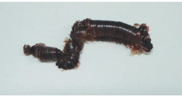
3 toes of her left foot. She denied intermittent claudication. She reported that she experienced the symptoms after crushing a millipede inside her shoe; she remained in contact with the millipede for approximately 6h. The patient presented the
animal that had been trapped in her shoe, which was identifi ed as a millipede. The defi nition of the species was not possible
due to the destruction of the morphological aspects during the crushing (Figure 1).
On physical examination, there was no difference in temperature at the sites of the lesions compared to the contralateral foot, and there were normal peripheral pulses,
which were strong and symmetrical. The capillary refi ll time
in the 3 injured toes was normal, and there was no pallor on elevation of the left foot. Dermatological examination revealed erythematous, cyanotic, and blackened lesions at the distal end of the left hallux in addition to the medial and distal phalanges of the second and third left toes; she had local hyperesthesia (Figures 2 and 3). The patient was told that it was a benign condition and was medicated with analgesics,
258
Heeren Neto AC et al - Millipede contact resulting in simulation of blue toe syndrome
REFERENCES
DISCUSSION
FIGURE 1 - Millipede found crushed inside the patient’s shoe. It was not possible to identify the species due to the destruction of the morphological
characteristics of the animal. FIGURE 2 - Erythematous, cyanotic lesions on the left hallux and blackened erythematous lesions on the second and third toes.
FIGURE 3 - Erythematous blackened lesions on the left toes with a necrotic aspect on the second toe.
Millipede populations can be very high, reaching 30-40 individuals/m2 in some areas1,2. These animals assume a coiled
position when threatened and may release a number of irritants, including quinones and cyanides, that cause conditions ranging from mild local irritation to skin necrosis; the clinical injury is limited to the contact site7-9. Skin lesions may occur in any
individual in the absence of predisposition, simply through
direct contact with the fl uid released by the millipedes7. Usually
a dark reddish or blackish staining of the skin that simulates
infl ammatory or even necrotic lesions is observed10.
In this case, the peculiarity of the lesions and identifi cation
of a millipede inside the shoe assisted with the correct diagnosis. The prolonged contact time with the animal was responsible for the more blackened tonality of the lesions. The cyanotic and blackened aspects looked like a standard ischemic tissue condition, and clinical peripheral vascular disease can often be
a diagnostic challenge, especially if there is no awareness of contact with an animal.
The clinician and, especially, the dermatologist should be alerted to this diagnosis, be aware of whom to consult, and accompany the case until complete resolution.
1. Triplehorn CA, Jonnson NF. Estudo dos insetos. 7th ed. São Paulo:
Cengage Learning; 2011.
2. Sierwald P, Bond JE. Current status of the Myriapod class diplopoda (millipedes): taxonomic diversity and phylogeny. Annu Rev Entomol 2007; 52:401-420.
3. Cardoso JLC, França FOS, Hui FH, Malaque CMS, Haddad Jr V. Animais peçonhentos no Brasil: biologia, clínica e terapêutica dos acidentes. São Paulo: Editora Sarvier; 2003. p. 258-64.
4. Haddad Jr V, Cardoso JLC, Rotta O, Eterovic A. Accidents provoked by Millipede with dermatological manifestations: report of two cases. An Bras Dermatol 2000; 75:471-474.
5. Kauffman P, Netto BM, Presti C, Sitrângulo Jr. CD, Simão E, Ferrari F, et al. Doenças Vasculares das Extremidades – Arterial, Venosa e Linfática. In: Lopes
AC, editor. Tratado de Clínica Médica. São Paulo: Roca; 2006. p. 734-787.
6. Lima CAJ, Cardoso JLC, Magela A, Oliveira FGM, Talhari S, Haddad Jr V. Exogenous pigmentation in toes feigning ischemia of the extremities: a diagnostic challenge brought by arthropods of the Diplopoda Class ("millipedes"). An Bras Dermatol 2010; 85:391-392.
7. Haddad Jr V, Cardoso JL, Lupi O, Tyring SK. Tropical dermatology: Venomous arthropods and human skin: Part II. Diplopoda, Chilopoda, and Arachnida. J Am Acad Dermatol 2012; 67:347.e1-9.
8. Goday Buján JJ, Yanguas Bayona I, Soloeta Arechavala R. Allergic contact dermatitis from cyanamide: report of 3 cases. Contact Dermatitis 1994; 31:331-332.
9. Trébol I, Lasa O, Navajas B, Ratón JA, Díaz-Pérez JL. Allergic contact dermatitis from cyanamide. Dermatitis 2005; 16:32-33.