ABSTRACT
ORIGINAL AR
Vascular Surgery Unit, Department of Surgery, Faculdade de Ciências
Médicas da Irmandade da Santa Casa de Misericórida de São Paulo,
São Paulo, Brazil
INTRODUCTION
It has been estimated that there are approximately 16 million diabetics in the United States, and this disease is one of the main causes of morbidity and mortality in society.1 In Brazil, a multicenter study has
shown that there is a 7.6% overall prevalence of diabetics in the urban population in nine Brazilian state capitals, with ages ranging from 30 to 69 years.2 An epidemiological
study in a Brazilian hospital found that diabetic foot complications were one of the most prevalent causes of hospitalization among diabetic patients.3
Peripheral vascular insufficiency occurs earlier in diabetics than in non-diabetics, and the arteries of the infrapatellar segment are attacked with greater frequency.4,5 The
preva-lence of peripheral vascular disease is about 10% in diabetics, whereas in non-diabetics the prevalence is 2.6%.6
Ischemia may be the cause of or con-tribute towards the progression of trophic lesions in the foot, which form favorable locations for infections to take hold. The coexistence of neuropathy, ischemia and leukocyte immune function disorders in diabetic patients favors the development of severe and extensive infections in the lower limbs that, if not adequately treated, may lead to amputation and death.1
Diabetic patients present a relative risk of having to undergo an amputation over the course of their lives that is 15 to 40 times greater than for non-diabetic individuals. Those with ischemic and infected lesions have a chance of undergoing an amputation of a lower limb that is up to 90 times greater than for those without ischemia or infection.7
In addition to the raised risk of amputation and the higher rates of mortality, only half of the diabetic patients who have undergone an amputation have satisfactory rehabilitation.8
To improve care for diabetics, it is essential to combine diabetes mellitus prevention and control and lesion treatment, together with the set of surgical, antimicrobial and peripheral vascular disease treatments, with the goal of reducing diabetes mellitus-related mortality and morbidity.
OBJECTIVE
To analyze risk factors relating to major amputations (supra and infrapatellar) in dia-betic patients with foot infections.
METHODS
On the basis of a pre-established protocol, data from 99 diabetic patients with infected lesions in their feet were analyzed. These pa-tients were hospitalized within the Vascular Surgery Unit of the Department of Surgery, Faculdade de Ciências Médicas da Irman-dade da Santa Casa de Misericórdia de São Paulo, between March 1999 and November 2001. Sixty of them (69.7%) were men and 30 (30.3%) were women. The patients’ ages ranged from 21 to 90 years, with a mean of 60.2 ± 12.4 years.
Type 1 and 2 diabetic patients were included, whose lesions were grade 3, 4 or 5 according to Wagner’s classification (grade 0: skin lesions absent, hyperkeratosis below or above bony prominences; grade 1: skin and immediate subcutaneous tissue are ulcerated; grade 2: lesions are deeper and may penetrate to tendon, bone or joint capsule; grade 3: deep tissues are always involved, osteomyelitis may be present; grade 4: gangrene of some portion of the toes or forefoot; and grade 5: the entire foot is gangrenous).9 None of these patients
showed signs of acute arterial insufficiency, or had peripheral vascular disease, or were affected by chronic arterial insufficiency for which arterial revascularization would be impossible. The patients’ clinical conditions
CONTEXT AND OBJECTIVE: Diabetic patients present high risk of having to undergo minor or major amputation during their lifetimes, be-cause of ischemia or infection. The aim of this study was to identify and quantify risk factors for major amputation in diabetic patients with foot infections.
DESIGN AND SETTING: Retrospective clinical-surgical trial at the Vascular Surgery Service of Santa Casa de São Paulo.
METHODS: Ninety-nine patients with diabetic foot infections who underwent 129 hospitalizations in the Vascular Surgery Unit were analyzed in accordance with a pre-established protocol to compare two groups of diabetic patients: one that underwent major amputations and the other that underwent minor amputations or debridements. The patients were predominantly male, in their sixth decade of life, and had type 2 diabetes mellitus. Chronic arterial insufficiency, age, diabetes mellitus duration, ascending lymphan-gitis, calcaneal lesions, Wagner’s classification, laboratory tests and different microorganisms in deep tissue cultures were the risk factors evalu-ated in all patients.
Table 1. Frequency distribution of the findings from 118 positive cultures in the foot lesions of 99 patients
Culture findings n %
Gram-positive bacteria 31 26.3
Gram-negative bacteria 39 33.1
Anaerobic bacteria 1 0.8
Mixed 47 39.8
Total 118 100.0
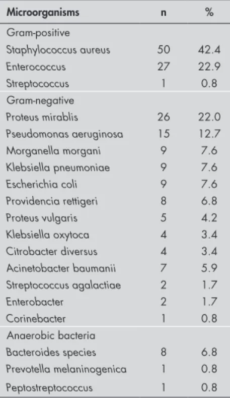
Table 2. Frequency distribution of the mi-croorganisms found in 118 positive cul-tures in the foot lesions of 99 patients
Microorganisms n %
Gram-positive
Staphylococcus aureus 50 42.4
Enterococcus 27 22.9
Streptococcus 1 0.8 Gram-negative
Proteus mirablis 26 22.0
Pseudomonas aeruginosa 15 12.7
Morganella morgani 9 7.6
Klebsiella pneumoniae 9 7.6
Escherichia coli 9 7.6
Providencia rettigeri 8 6.8
Proteus vulgaris 5 4.2
Klebsiella oxytoca 4 3.4
Citrobacter diversus 4 3.4
Acinetobacter baumanii 7 5.9
Streptococcus agalactiae 2 1.7
Enterobacter 2 1.7
Corinebacter 1 0.8 Anaerobic bacteria
Bacteroides species 8 6.8
Prevotella melaninogenica 1 0.8
Peptostreptococcus 1 0.8
were established by means of clinical and arteriographic evaluations. From the physical examination, all these patients were found to present infected lesions at an advanced stage that involved the whole foot. Neuropathy was not investigated in these patients. Most of the patients presented ascending lymphangitis, with hyperemia above the knee.
For these 99 patients, 135 cultures were done, of which 17 (12.6%) tested negative. Specimens for culturing were taken from deep tissue at the time of surgery. Aerobic and anaerobic bacteria were isolated from all speci-mens. The aerobic and facultative strains were inoculated in blood agar. The distribution of the microorganisms with regard to Gram-posi-tive staining is shown in Table 1.
The most important infectious micro-organism was Staphylococcus aureus (42.2%), followed by Enterococcus (22.9%), Proteus mirabilis (22.0%) and Pseudomonas aeruginosa (12.3%) (Table 2).
Two groups of patients were established: one that underwent major amputation and the other that underwent minor amputation or debridement. The following variables were comparatively analyzed in relation to these two groups: gender, age, presence of ascending lymphangitis, lesion location (toes, anterior region of the foot, calcaneum or the entire foot), lesion depth according to Wagner’s clas-sification, presence of chronic arterial insuf-ficiency, duration of diabetes mellitus, use of insulin, and laboratory data (white blood cells, urea, creatinine and glycemia levels).
The data expressed in terms of frequency were analyzed using the chi-squared test (χ2),
adopting a probability of 95% (p ≤ 0.05) for rejection of the null hypothesis. When a statistically significant difference was found between the frequencies compared, the odds ratio for the occurrence of this phenomenon was calculated.
The data expressed in terms of means and their respective standard deviations were analyzed using the Student t test, for which a significance level of p ≤ 0.05 was also adopted.
RESULTS
The patient distribution according to Wagner’s classification showed that 57% of the patients presented Wagner grade 4 le-sions, 25% Wagner grade 5 lesions and 18% Wagner grade 3 lesions. Chronic arterial in-sufficiency was present in 47.5% of the cases. The femoropopliteal segment was affected in 47.5% of the cases, the infrapatellar segment in 20.3% of the cases and the aortic-iliac
segment in 4.0%. With the exception of two patients, all the patients had type 2 diabetes mellitus, and they had had the disease for between one and 35 years, with a mean of 12.4 ± 7.2 years. 41.4% of the patients were on insulin treatment. Associated diseases were observed in 63.6% of the patients, with predominance of systemic arterial hyperten-sion (prevalence of 41.4%).
The mean length of hospitalization was 17.7 ± 16.8 days, for the sample as a whole. Concerning treatment, minor amputations or full debridement were done on 48 patients (49.5%), and these were considered together for comparative analyses. Major amputations were performed on 51 patients. There were no significant differences in treatment type between the men and women.
In comparison with the patients treated with minor amputations, those whose treat-ment required major amputation presented a significantly higher mean age (63.5 ± 10 versus 56.5 ± 13 years, p = 0.0052), and also significantly greater length of time with the diabetes mellitus diagnosis (13.9 ± 6 years versus 10.9 ± 6 years, p = 0.041). The pres-ence of ascending lymphangitis was also seen at a statistically higher frequency (88.2% versus 75%, χ2 = 3.86). This indicated that
the presence of this characteristic during the physical exam led to a probability of supra or infrapatellar amputation that was 2.5 times greater than when it was absent.
The presence of chronic arterial insuf-ficiency without the possibility of revascu-larization, because of advanced gangrenous lesions associated with absence of distal artery opacification in angiography, led to a risk that the patient would need a major amputation that was 4.5 times greater than in the absence of this condition (Table 3).
The presence of lesions located in the calcaneum and grade 5 lesions based on Wagner’s classification led to probabilities that the patients with an infected diabetic foot would need to undergo a major amputation that were, respectively, 10.5 and 3.4 times greater than in the absence of these condi-tions (Table 3).
The serum concentrations of leukocytes (15,629 ± 6,927 mil/u), urea (58.7 ± 45.1 mg/100 ml), creatine (1.5 ± 0.9 mg/100 m) and glucose (255.4 ± 119.5 mg/100 m) displayed homogeneity between the two groups.
The presence of Gram-positive bacteria was significantly more frequent in patients treated by means of major amputations than in those with minor amputations (χ2 = 7.92)
(Ta-ble 4). Nevertheless, no important difference
was observed when the groups were compared with regard to specific microorganisms.
DISCUSSION
Several risk factors for amputations among diabetics have been cited in the lit-erature. However, few distinguish between major and minor amputations. Moss et al.10
observed that the risk factors for amputa-tion were: previous history of foot ulcers, advanced age and high blood pressure. Among 1,370 diabetics with disease incep-tion after the age of 30, the risk factors for amputation were: male gender, history of foot ulcers, elevated levels of glycosidic hemoglobin, proteinuria, and length of time since diabetes mellitus diagnosis.10 On
the other hand, Reiber et al.11 did not find
Table 3. Diabetic patient distribution according to treatment of the foot lesions, in rela-tion to variables studied
Variables Treatment Total
Minor amputation Major amputation
Injured region n % n % n %
Anterior region of the foot 18 37.5 14 27.4 32 32.3
Calcaneum 3 6.3* 21 41.2* 24 24.2
Toes 22 45.8 10 19.6 32 32.3
Entire foot 5 10.4 6 11.8 11 11.2
Total 48 100.0 51 100.0 99 100.0
Wagner’s classification n % n % n %
3 13 27.1 5 9.8 18 18.2
4 35 72.9 21 41.2 56 56.5
5 0 -† 25 49.0† 25 25.2
Total 48 100.0 51 100.0 99 100.0
Peripheral vascular disease n % n % n %
Present 13 27.1‡ 34 66.7‡ 47 47.5
Absent 35 72.9 17 33.3 52 52.5
Total 48 100.0 51 100.0 99 100.0
*χ2 test, minor versus major amputation in relation to calcaneum, χ2 = 18.86, odds ratio (OR) = 10.5; † χ2 test, minor versus
major ampulation in relation to Wagner grade 5, χ2 = 31.56, OR = 3.4; ‡ χ2 test, minor versus major amputation in relation
to peripheral vascular disease χ2 = 17.16, OR = 5.4.
Table 4. Frequency distribution of the findings from 118 positive cultures of the foot lesions of diabetic patients in relation to treatment
Culture findings Minor amputation Major amputation Total
n % n % n %
Gram-positive bacteria 10 16.1* 21 37.5* 31 26.3
Gram-negative bacteria 24 38.7 15 26.8 39 33.1
Anaerobic bacteria 1 1.6 0 - 1 0.8
Mixed 27 43.6 20 35.7 47 39.8
Total 62 100.0 56 100.0 118 100.0
*χ2 Test, minor versus major amputation in relation to Gram-positive bacteria, degrees of freedom = 3, χ2 = 7.92
(signifi-cant), OR = 2.4.
Langer et al.4 observed outcomes from
emergency surgery on diabetic patients with infected and necrotic lesions of the lower limbs. They found that it was possible to save the lower limb of 76% of the patients that did not present chronic arterial insuf-ficiency and 42% of the patients that did present this condition. They also observed that toe lesions that were not associated with chronic arterial insufficiency resulted in limb preservation in 95% of the cases, while such preservation was only achieved in 40% of the cases of calcaneallesion associated with chronic arterial insufficiency.
Calle-Pascual et al.12 found that, among
diabetics, 100% of the major amputations were associated with the presence of peripheral vascular disease, 78% were associated with neuropathy and 24% were associated with infection. On the other hand, in the cases of minor amputations, 62% were associated with peripheral vascular disease, 92% were
associated with neuropathy, and 84% were associated with infection.
The present study only included patients affected by lesions of Wagner grades 3, 4 and 5, thus differing from the majority of other authors,7,9,11,13-18 who also included the
more superficial lesion grades from Wagner’s classification. It is likely that this difference is important, and would explain some of the divergences relating to our culture find-ings and the evolution of our patients with major amputations. Other authors have also reported high occurrence of peripheral vascular disease in diabetics,4,17,19-22 especially
in the leg arteries.
With regard to the treatment type, the 49.5% rate of major amputations was high. This may reflect the characteristics of the group studied: patients with peripheral vas-cular disease, advanced age, advanced grades of lesion depth, no possibility of revascu-larization, elevated white blood cell counts
and ascending lymphangitis, and generally presenting hypertension and other associated diseases. In other words, these were patients with high local and systemic disease severity.
According to the study by Moss et al.,10
male gender was a risk factor for amputa-tions in type 2 diabetics. In our study, all of the patients underwent some type of amputation or full debridement, but males did not present a higher rate of supra and infrapatellar amputations.
Mueller et al.5 found that only one-third
of the diabetics who were candidates for arte-rial reconstruction had distal arteries suitable for revascularization. Our focus was not on the evaluation of diabetics with arterial insufficiency that presented the possibility of revascularization, because such patients present other patterns of lesion evolution that generally have a better prognosis.
Because we were evaluating diabetics with arterial insufficiency that did not pres-ent the possibility of revascularization, as demonstrated by clinical and arteriographic analysis, this allowed us to demonstrate the significant influence of arterial insufficiency on these patients’ evolution, in relation to major amputation. Distal revascularization is the treatment of choice for ischemic legs but, unfortunately, some diabetic patients present clinical conditions (deep infections) and arterial conditions that are too poor to enable vascular bypass.
The cultured microorganism profiles for patients with a infected diabetic foot are fairly diverse and depend on the way the specimen for culturing is collected.5,23,24 In our study,
all of the specimens for culturing were col-lected from deep tissue during the surgical procedure. In agreement with the findings from other authors,5,9,13,14,23,25-27 the rate of
polymicrobial culture findings was high. The bacterium found most frequently through culturing was Staphylococcus au-reus, in agreement with the reports in the literature.9,13,14,23,27 However, the proportion
of Gram-negative microorganisms was high in our study, and this may be attributable to the fact that the lesions of the patients studied were all in Wagner grades 3, 4 and 5. Depending on the lesion type, these are more frequently susceptible to infection by such bacilli.24,28
REFERENCES
complications such as atherosclerotic disease, immune alterations and peripheral neuropa-thies increase in incidence and severity over the course of the disease. Nelson et al.29 and
Moss et al.10 also found that the frequency of
amputations increased with the length of time since diabetes mellitus diagnosis. However, these authors did not distinguish between the rates of major and minor amputations.
Melton et al.20 and Most and Sinnock30
demonstrated that the longer the time since the diabetes mellitus diagnosis, the greater the incidence of peripheral vascular disease. This, in turn, increased the risk of amputation. Most and Sinnock30 also reported an increasing rate
of amputations with age progression among the patients.
The presence of ascending lymphan-gitis as a risk factor for major amputation suggests that our patients presented more severe infectious conditions, with greater phlogiston signs in the lower limbs, thus needing more aggressive surgical procedures. Oyibo et al.16 demonstrated that infected
lesions in diabetics led to a probability of amputation that was 11 times greater than for those without infection.
In our study, the lesions located in the calcaneum and those that were grade 5 in Wagner’s classification (gangrene of the entire
foot) were significantly associated with major amputations. These findings are understand-able, because the lesions were of greater depth and thus harder to treat, and were frequently associated with peripheral vascular disease. Wagner grade 4 and 5 lesions are indeed associated with higher risk of amputation of the lower limbs in the literature.4,9,16,31 In a
study by Armstrong et al.,7 diabetic patients
with ischemic and infected lesions presented probabilities of undergoing amputation of the lower limb that were up to 90 times greater than for those without these conditions.
Lesions located in the calcaneal region are also more difficult to treat, because of the con-stant support needed and the trauma involved, with a high risk of amputation. Langer et al.4
reported limb amputation rates reaching up to 60% for lesions in this location, when associ-ated with peripheral vascular disease.
The presence of peripheral arterial disease has been cited by many authors as a risk factor for amputations in diabetics.4,7,9,11,12,16,17,29,32,33
Our findings indicated that the presence of peripheral vascular disease that did not pres-ent the possibility of revascularization led to a significantly higher rate of major amputations (odds ratio, OR: 5.4). It is irrefutable that the presence of peripheral vascular disease causes problems in the blood flow that is
fundamen-tal for healing and combating severe infections that attack diabetic feet. Calle-Pascual et al.12
reported that 100% of the major amputations in their series were associated with peripheral vascular disease. Further studies in our hos-pital will be necessary in order to determine the impact of revascularization surgery, and the results from vascular exploration in our diabetic patients.
The predominance of Gram-positive bac-teria in cases that require major amputation may be due to the high proportion of methi-cillin-resistant Staphylococcus aureus (MRSA). Such bacteria have high pathogenicity and cause severe tissue damage because of the pro-duction of extracellular enzymes and toxins. Other studies in the literature consulted9,11,13,25
have not place such emphasis in correlations of data from culturing, in relation to infected diabetic foot amputations.
CONCLUSION
Patients with an infected diabetic foot and advanced age, long duration of diabetes mel-litus, ascending lymphangitis, calcaneal lesions and grade 5 lesions according to Wagner’s classification, and affected by chronic arterial insufficiency without the possibility of revas-cularization, present a high risk of needing major amputation.
1. Slovenkai MP. Foot problems in diabetes. Med Clin North Am. 1998;82(4):949-71.
2. Malerbi DA, Franco LJ. Multicenter study of prevalence of diabetes mellitus and impaired glucose tolerance in the urban Brazilian population aged 30-69 yr. The Brazilian Cooperative Group on the Study of Diabetes Prevalence. Diabetes Care.Diabetes Care. 1992;15(11):1509-16.
3. Salgado Filho N, Salgado BJL, Brito LGO, Ferro GAC, Sampaio ALO. Perfil do paciente diabético internado no Hospital Universitário Presidente Dutra, São Luís, Maranhão. Diabetes Clínica. 2001;5(5):333-8. Available from URL: http://www.atlanticaeditora.com.br/pagina. asp?cop=37. Accessed in 2006 (Jan 31).
4. Langer B, Aguiar ET, Wolosker N. Complicações vasculares do diabetes. In: Wajchenberg BL, editor. Tratado de EndocrinologiaIn: Wajchenberg BL, editor. Tratado de EndocrinologiaTratado de Endocrinologia Clínica. São Paulo: Roca; 1992. p. 787-97.
5. Mueller MP, Wright J, Klein SR. Diabetes and peripheral vas-cular disease. In: Veith FJ, Hobson RW, Williams RA, Wilson S, editors. Vascular surgery: principles and practice. 22nd ed. New York: McGraw Hill; 1994. p. 514-22.
6. Brasil. Ministério da Saúde. Diabetes mellitus como problema de saúde pública. In: Brasil. Ministério da Saúde. Manual de Diabetes. 2a ed. Brasília. Ministério da Saúde; 1993. p. 7-10. 7. Armstrong DG, Lavery LA, Harkless LB. Validation of a
dia-betic wound classification system. The contribution of depth,m. The contribution of depth, infection, and ischemia to risk of amputation. Diabetes Care. 1998;21(5):855-9.
8. Cameron HC, Lennard-Jones JE, Robinson MP. Amputations in the diabetic outcome and survival. Lancet. 1964;18:605-7. 9. Pittet D, Wyssa B, Herter-Clavel C, Kursteiner K, Vaucher J,
Lew PD. Outcome of diabetic foot infections treated conser-vatively: a retrospective cohort study with long-term follow-up. Arch Intern Med. 1999;159(8):851-6.
10. Moss SE, Klein R, Klein BE. The prevalence and incidence of lower extremity amputation in diabetic population. Arch Intern Med. 1992;152(3):610-6.
11. Reiber GE, Pecoraro RE, Koepsell TD. Risk factors for amputa-tion in patients with diabetes mellitus. A case-control study. Ann Intern Med. 1992;117(2):97-105.
12. Calle-Pascual AL, Garcia-Torre N, Moraga I, et al. Epidemiology of nontraumatic lower-extremity amputation in area 7, Madrid, between 1989 and 1999: a population-based study. Diabetes Care. 2001;24(9):1686-9.
13. Lipsky BA, Pecoraro RE, Larson SA, Hanley ME, Ahroni JH. Outpatient management of uncomplicated lower-ex-tremity infections in diabetic patients. Arch Intern Med. 1990;150(4):790-7.
14. Goldstein EJ, Citron DM, Nesbit CA. Diabetic foot infec-tion. Bacteriology and activity of 10 oral antimicrobial agents against bacteria isolated from consecutive cases. Diabetes Care. 1996;19(6):638-41.
15. Armstrong DG, Nguyen HC. Improvement in healing with aggressive edema reduction after debridement of foot infection in persons with diabetes. Arch Surg. 2000;135(12):1405-9.
16. Oyibo SO, Jude EB, Tarawneh I, Nguyen HC, Harkless LB, Boulton AJ. A comparison of two diabetic foot ulcer classification systems: the Wagner and the University of Texas wound classification systems. Diabetes Care. 2001;24(1):84-8.
17. Muller IS, de Grauw WJ, van Gerwen WH, Bartelink ML, van Den Hoogen HJ, Rutten GE. Foot ulceration and lower limb amputation in type 2 diabetic patients in dutch primary health care. Diabetes Care. 2002;25(3):570-4.
18. Lavery LA, Armstrong DG, Wunderlich RP, Tredwell J, Boulton AJ. Diabetic foot syndrome: evaluating the prevalence and incidence of foot pathology in Mexican Americans and non-Hispanic whites from a diabetes disease management cohort. DiabetesCare. 2003;26(5):1435-8.
19. Kannel WB, McGee DL. Diabetes and cardiovascular disease. The Framingham study. JAMA. 1979;241(19):2035-8. 20. Melton LJ 3rd, Macken KM, Palumbo PJ, Elveback LR.
Inci-dence and prevalence of clinical peripheral vascular disease in a population-based cohort of diabetic patients. Diabetes Care. 1980;3(6):650-4.
21. Bendick PJ, Glover JL, Kuebler TW, Dilley RS. Progression ofProgression of atherosclerosis in diabetics. Surgery. 1983;93(6):834-8. 22. Brasil. Ministério da Saúde. Epidemiologia do diabetes mellitus.
In: Brasil. Ministério da Saúde. Manual de Diabetes. 2a ed. Brasília. Ministério da Saúde; 1993. p. 17-24.
AUTHOR INFORMATION
Vanessa Prado dos Santos, MD. Assistant in the Vascular Surgery Unit, Surgery Department, Faculdade de Ciências Médicas da Irmandade da Santa Casa de Misericórdia de São Paulo, São Paulo, Brazil.
Denise Rabelo da Silveira, MD. Assistant professor in the Vascular Surgery Unit, Surgery Department, Faculdade de Ciências Médicas da Irmandade da Santa Casa de Misericórdia de São Paulo, São Paulo, Brazil.
Roberto Augusto Caffaro, MD. Associate professor and Head in the Vascular Surgery Unit, Surgery Department, Facul-dade de Ciências Médicas da IrmanFacul-dade da Santa Casa de Misericórdia de São Paulo, São Paulo, Brazil.
Address for correspondence:
Vanessa Prado dos Santos
Rua Martinico Prado, 128 — Apto. 72 — Higienópolis São Paulo (SP) — Brasil — CEP 01224-010 Tel. (+55 11) 3226-7273
E-mail: [email protected]
Copyright © 2006, Associação Paulista de Medicina
RESUMO Fatores de risco para amputações maiores primárias em pacientes diabéticos
CONTEXTO E OBJETIVO: Os pacientes diabéticos apresentam risco elevado de amputação ao longo da vida, devido a isquemia ou infecção. Objetivou-se identificar e quantificar os fatores de risco para amputações maiores em pacientes diabéticos, com lesões infectadas de extremidades.
TIPO DE ESTUDO E LOCAL: Estudo clínico-cirúrgico retrospectivo realizado na Disciplina de Cirurgia Vascular da Santa Casa de São Paulo.
MÉTODOS: Analisamos comparativamente 99 pacientes (129 internações) com o diagnóstico de pé di-abético infectado, divididos em dois grupos, de acordo com o tipo de procedimento cirúrgico. No grupo 1, pacientes submetidos a amputações maiores e, no grupo 2, pacientes com amputações menores. Os pacientes eram predominantemente do sexo masculino, na sexta década de vida e diabéticos tipo 2. Fatores de risco analisados: idade, presença de insuficiência arterial, tempo de diagnóstico do diabetes, linfangite ascendente, lesões no calcâneo, classificação de Wagner, exames laboratoriais e presença de diferentes microorganismos na cultura.
RESULTADOS: Fatores de risco que se correlacionaram significativamente com amputações maiores: idade e linfangite ascendente (razão de chances, RC: 2,5), lesão do calcâneo (RC: 10,5), classificação de Wagner grau 5 (RC: 3,4), insuficiência arterial crônica sem possibilidade de revascularização (RC: 5,4), tempo do diagnóstico de diabetes e presença de bactérias Gram-positivas na cultura. Leucograma, glicemia, uréia e creatinina não se correlacionaram a risco aumentado de amputações maiores.
CONCLUSÕES: Idade, tempo de diagnóstico do diabetes, infecção por Gram-positivos, linfangite ascen-dente, lesões de calcâneo e grau 5 de Wagner e insuficiência arterial constituem fatores de risco para amputações maiores em pacientes diabéticos.
PALAVRAS-CHAVE: Diabetes mellitus. Pé diabético. Amputação. Linfangite. Infecções bacterianas.
24. Mayall RC, Mayall AC, Mello AV, Mayall JC, Mayall LC. Pé diabético. In: Maffei FHA, Lastória S, Yoshida WB, Rollo HA,In: Maffei FHA, Lastória S, Yoshida WB, Rollo HA, editors. Doenças vasculares periféricas. Rio de Janeiro: Medsi; 1995. p. 1277-302.
25. Louie TJ, Bartlett JG, Tally FP, Gorbach SL. Aerobic and anaerobic bacteria in diabetic foot ulcers. Ann Intern Med. 1976;85(4):461-3.
26. Sharp CS, Bessmen AN, Wagner FW Jr, Garland D, Reece E. Microbiology of superficial and deep tissues in infected diabetic gangrene. Surg Gynecol Obstet. 1979;149(2):217-9. 27. Pathare NA, Bal A, Talvalkar GV, Antani DU. DiabeticDiabetic
foot infections: a study of microorganisms associated with the different Wagner grades. Indian J Pathol Microbiol. 1998;41(4):437-41.
28. van der Meer JW, Koopmans PP, Lutterman JA. Antibiotic therapy in diabetic foot infection. Diabet Med. 1996;13(Suppl 1):S48-51.
29. Nelson RG, Gohdes DM, Everhart JE, et al. Lower-extrem-ity amputations in NIDDM. 12-yr follow-up study in Pima Indians. Diabetes Care. 1988;11(1):8-16.
30. Most RS, Sinnock P. The epidemiology of lower extrem-ity amputations in diabetic individuals. Diabetes Care. 1983;6(1):87-91.
31. Lange E. Current surgery/drug combination treatment of diabetic gangrene of the foot. Infection. 1991;19(Suppl 6): S351-4.
32. Williams HT, Hutchinson KJ, Brown GD. Proceedings: Gan-grene of the feet in diabetics. Arch Surg. 1974;108(4):609-11.
33. Fratezi AC, Albers M, De Luccia ND, Pereira CA. Outcome andOutcome and quality of life of patients with severe chronic limb ischaemia: a cohort study on the influence of diabetes. Eur J Vasc Endovasc Surg. 1995;10(4):459-65.
Acknowledgments: We are grateful to the Support Center for Scientific Publications of Faculdade de Ciências Médicas das of Faculdade de Ciências Médicas da Irmandade da Santa Casa de Misericórdia de São Paulo for editorial assistance.
Sources of funding: None
Conflict of interest: None
Date of first submission: October 10, 2004
Last received: March 20, 2006