Members of Pleurobranchidae are generally characterized by body oval to elongated with centralized rhinophores, inter-nalized plate-like shell, gill exposed on the right side of the body and veil trapezoidal. The family is currently composed of seven genera: Bathyberthella Willan, 1983; Berthella Blainville, 1824; Berthellina Gardiner, 1936; Boreoberthella Martynov & Schrödl, 2009; Pleuredhera Ev. Marcus & Er. Marcus, 1970; Pleurobranchus Cuvier, 1805; and, Tomthompsonia Wägele & Hain, 1991. Berthella and Berthellina have an intricate taxonomic history due to their similarities in external morphology and consequent misidentifications through the years (e.g., GARDINER 1936). The elongated teeth with numerous subterminal den-ticles in Berthellina clearly distinguish these two genera. Berthella resembles Berthellina in the external morphology, but in charac-ters of the radula, jaws and reproductive system, it is more similar to Pleurobranchus (WILLAN 1983).
Berthella is one of the most species-rich genus of Pleurobranchidae, with 29 valid species, while Berthellina pre-sents only 13 valid species. Berthella and Berthellina generally occur in tropical and warm temperate waters and range from
the intertidal zone to moderate subtidal depths (WILLAN 1987). These genera are well known because some of their species are widespread geographically and rather common (e.g., Berthella stellata (Risso, 1826) and Berthellina citrina Ruppell & Leuckart, 1828). However, Berthella and Berthellina are not well-estab-lished clades, following recent phylogenetic studies that have failed to support them, and according to which species of these two genera cluster in a large polytomy inside Pleurobranchidae (MARTINOV & SCHRÖDL 2009). Berthella currently comprises five valid species recorded from the western Atlantic, of which only Berthella agassizii (MacFarland, 1909) and Berthella stellata are recorded from Brazil (GARCÍA et al. 2008, RIOS 2009). Berthella tupala Er. Marcus, 1957 was described from São Paulo, Brazil, but it is currently considered a junior synonym of B. stellata. Besides that, PADULA et al. (2012) recorded an undescribed morphotype of Berthella from Alagoas, Brazil.
Two species of Berthellina are recorded from the western Atlantic: Berthellina circularis (Mörch, 1863) and Berthellina quadridens (Mörch, 1963), both originally described from St. Thomas and recorded from Brazil, respectively from
Pernam-Taxonomic review of
Berthella
and
Berthellina
(Gastropoda:
Pleurobranchoidea) from Brazil, with description of two new species
Juliana Alvim
1,*& Alexandre Dias Pimenta
11Departamento de Invertebrados, Museu Nacional, Universidade Federal do Rio de Janeiro. Quinta da Boa Vista,
São Cristóvão, 20940-040 Rio de Janeiro, RJ, Brazil.
*Corresponding author. E-mail: [email protected]
ABSTRACT. Berthella currently comprises five valid species recorded from the western Atlantic, of which only Berthella agassizii (MacFarland, 1909) and Berthella stellata (Risso, 1826) have been recorded from Brazil. Two species of Berthellina occur in Brazil: Berthellina quadridens (Mörch, 1963) and Berthellina circularis (Mörch, 1863); however, based on a literature review and the anatomical study presented here, we only recognize B. circularis as occurring in the country. This study gives a detailed re-description of B. agassizii, B. stellata and B. circularis based on material from Brazil, and adds two new species to the genus, from the Brazilian coast, Berthella punctata sp. nov. and Berthellinaignissp. nov.
Berthellina circularis and B. quadridens are considered distinct species based on the denticulation of the radular teeth. B. quadridens presents the majority of teeth with two denticles, rarely three-four denticles, while in B. circularis from Brazil the radular teeth are highly denticulate with up to 17 denticles. Berthella and Berthellina do not form a monophyletic group, and cluster in a large polytomy within Pleurobranchidae. A detailed anatomical study is performed to improve the understanding of the evolutionary history of Pleurobranchidae. The comparison with available data on the odontophoric muscles of Pleurobranchoidea shows that Berthella and Berthellina present a pair of the muscle m10v that connects the mj with the ventral portion of the m5, while in Pleurobranchus and Pleurobranchaea the m10v connects the mj with the ventral portion of the m4; Berthella and Berthellina do not present the muscle m10d, which is present in Pleurobranchus and Pleurobranchaea. Based on these characters, Berthella seems to be more closely related to Berthellina than to Pleurobranchus.
cal delimitation of these species. Most of them were described based on one or two preserved specimens, and subsequent records only expanded their geographical distribution, without adding information on their morphology. Moreover, two new species from Brazil are described, one from each genus.
MATERIAL AND METHODS
Descriptions of the external morphology were based on living and preserved specimens. Dissections and drawings of the digestive system, including intrinsic and extrinsic muscles of the odontophore, the reproductive system, the nervous sys-tem and the circulatory syssys-tem were performed under a stere-omicroscope with a drawing tube.
The radula and jaw were cleaned in 10% potassium hy-droxide (KOH), subsequently rinsed in water and mounted for examination in a Jeol JSM-6390LV scanning electron micro-scope (SEM).
The nomenclature used to name the portions of the gill follows WILLAN (1983) and the one used for the odontophore muscles follows PONDER et al. (2008: 350-351, figs. 13.9-13.10), with additional information added by suffixes (d: dorsal; v: ventral). In the nervous system, when the nerve bifurcates, the number of the nerve is followed by letters, e.g., “a”, “b”.
In the lists of examined material, the number inside brackets indicates the number of specimens in each lot, fol-lowed by the number of specimens dissected.
Anatomical abbreviations: a, anus; ag, duct of acid gland; am, ampulla; bc, buccal ganglion; bg, blood gland; bu, bursa copulatrix; ca, oral canal; ccpb, connective between buccal and cerebropleural ganglia; cp, nerves that leave from the cerebropleural ganglion; cpg, cerebropleural ganglion; cpp, commissure between the pedal ganglia; dd, deferent duct; dg, digestive gland; e, eye; f, female opening; fg, female gland; fo, foot; gi, gill; gm, gill membrane; go, gonopore; hg, hermaph-rodite gland; in, intestine; j, jaw plates; m1d, m1va and m1vb, jugal protractor muscles of odontophore; m4, main dorsal ten-sor muscle of radula; m5, accesten-sory dorsal tenten-sor muscle of radula; m10a, ventral tensor muscle of radula; m10v, protrac-tor muscle of odontophore; ma, male opening; mg, metapodial gland; mj, jaw muscle; mo, mouth; mr, retractor muscle; nb, nerves that leave from the buccal ganglion; ne, nephropore; np, nerves that leave from the pedal ganglion; nvg, nerve lead-ing to visceral ganglion; oa, openlead-ing of the duct of the acid gland; oe, esophagus; ot, oral tentacle; p, penial papilla; pb,
Paulo (MZSP); Museu Nacional, Universidade Federal do Rio de Janeiro (MNRJ). Specimens of other species of Berthella and Berthellina were examined for comparison: Berthella tamiu Ev. Marcus, 1984: MZSP 121400, 21°05’N, 86°23’W, 4 may 1967, John Elliot Pilsbury Cruise 6701 coll. [4 microscope slides: radula and jaw; penis; mantle; cerebro-pleural ganglia]; Berthellina quadridens: MZSP 75310, Barbados, Diva coll. [1 dissected speci-men + two microscope slides: radula and jaw; penis]; MZSP 121401, Colombia [one microscope slide: radula]; MZSP 121402, Colombia [one microscope slide: radula and jaw]; MZSP 121403, Colombia [one microscope slide: transversal cut of reproduc-tive system]. These microscope slides were recently located among the slides belonging to the collection of Er. Marcus and Ev. Marcus, lodged in the MZUSP. Probably, these slides of B. quadridens represent the original material studied by EV. MARCUS (1976, 1984) and EV. MARCUS & HUGES (1974) due to the corre-spondence of localities studied in these papers. Despite the fact that EV. MARCUS (1976) described only one specimen in the de-scription of B. quadridens from Colombia, in these microscope slides we found two radula from specimens from Colombia, both labeled as “Bandel Colombia”. EV. MARCUS (1976) received many species donated by Dr. Klaus Bandel, from Colombia, for this work. It is almost certain that one of these slides corresponds to the slide used in the description made by EV. MARCUS (1976).
TAXONOMY
Pleurobranchoidea Gray, 1827
Pleurobranchidae Gray, 1827
Berthella
Blainville, 1824
Berthella Blainville, 1824: 262. Type-species by original desig-nation: Berthella porosa Blainville, 1824 (= Bulla plumula Montagu, 1803).
Cleantus Leach, 1852: 28. Type species by original designation: Cleantus montagui Leach, 1852 (= Bulla plumula Montagu, 1803). Bouviera Vayssière, 1896: 116. Type-species by subsequent designa-tion (Odhner, 1926: 22): Pleurobranchus auranticus Risso, 1818. Gymnotoplax Pilsbry, 1896: 210. Type-species by subsequent des-ignation (Willan, 1978: 339): Pleurobranchus americanus Verrill, 1885.
Berthellinops Burn, 1962: 135. Type species by original designa-tion: Berthellinops serenitas Burn, 1962.
or convex; mantle relatively smooth; metapodial gland present; shell ovate; gill rachis generally smooth; anal aperture in front of middle of gill membrane; radula teeth hook-shaped, some-times with a denticle at base of some lateral teeth; without rachidian teeth; mandibular elements with smooth or denticu-late blades. Pair of jugal muscles m1d on dorsal view of buccal mass, inserting into m5, connecting to dorsal-anterior end of snout. Pair m4, main dorsal tensor muscle of radula, reduced, originating in lateral region of cartilages, surrounding them ventrally, inserting into subradular membrane. Pair m5, sec-ondary dorsal tensor muscle of radula, large and broad, cover-ing median portions of cartilage, extendcover-ing up to dorsal region; originating in posterior surface of cartilages; inserting laterally in mj. Pair m7 absent. Pair m10d (dorsal) absent. A pair of m10v connected the mj with ventral portion of m5. Pair of strong retractor muscles originating in most posterior portion of m5, separated in anterior portion and jointed in its poste-rior portion, laying above anteposte-rior portion of digestive gland. Penial gland present.
Remarks. The name Cleantus has been considered a jun-ior synonym of Berthella for a long time (WILLAN 1983). The authorship of the name, however, is questioned here. This name was propagated by GRAY (1847) as Cleanthus, with the letter “h”, and the name Cleanthus Gray, 1847 was only listed in that paper, without any description or characterization. Therefore, the name/authorship of Cleanthus Gray, 1847 is not available, since it does not meet the requirements of Article 12.1 of the International Code of Zoological Nomenclature (ICZN 1999). According to the latter, a name needs to be accompanied by a description or a definition of the taxon that it denotes, or by an indication of it. Therefore, the author of Cleantus is LEACH (1852), who provided a description of the genus.
Berthella agassizii
(MacFarland, 1909)
Figs. 1-3, 11-28
Pleurobranchus agassizii MacFarland, 1909: 59-64, pl. 11-12, figs. 43-57; Ihering, 1915: 141.
Bouvieria agassizii: Odhner 1926: 22; Engel, 1927: 110, fig. 26a-c. Berthella agassizii: Er. Marcus, 1955: 117, figs. 66-77; 1958: 57; Ev. Marcus & Er. Marcus, 1957: 20, figs. 38-39; 1963: 24; 1964: 198; Er. Marcus & Ev. Marcus, 1970: 53; Ev. Marcus, 1976: 133; 1984: 51, figs. 6-16; Bandel, 1976: 103, fig. 16; Clark, 1984: 93, figs. 37-42; Gosliner & Bertsch, 1988: 63, figs. 1E; 15-18; Rios, 1994: 206; 2009: 417; Redfern, 2001: 168, pl. 72: fig. 692; Valdés et al., 2006: 110; García et al. 2008: 90; Padula et al., 2012: 8.
Type material. Holotype CASIZ 021162, 1899?, type lo-cality, coll. Mr.Greeley [1]; Paratype: CASIZ 021163 [1] and CASIZ 021164 [1], same date, locality and collector of holotype.
Type locality. Riacho Doce, Alagoas, Brazil.
Description. External morphology (Figs. 1-2, 11-13). Liv-ing specimens translucent pale pink to reddish-pink with scat-tered opaque white spots (Fig. 1); rhinophores rosy; oral veil
and gill white (Fig. 2); foot with some rosy pigment randomly disposed (Fig. 2). Living specimens up to 23 mm in length; length of mature preserved specimens 6-11 mm; width 3-8 mm; length of foot 4-9 mm; width of foot 2-4 mm. Body oval and oblong. Mantle covered foot entirely. Mantle surface ranges from smooth, in juveniles, to slightly reticulate. Oral veil broad and trapezoidal connected with head region (Figs. 2, 13); lat-erally, oral tentacles with deep notch, corresponding to almost its length. Rhinophores rolled joined at their bases, up to 1/2 of its length. Gill exposed laterally (Fig. 2); 1/2 to 1/3 length of body; main rachis smooth, without tubercles; alternate pin-nae; bipinnated pinpin-nae; 11-14 pinpin-nae; 5-6 pinnae free from body wall, attached by branchial membrane (Fig. 12). Anal opening lying approximately above 3°-4° pinnae (Fig. 12). Pre-branchial pore opening beside main rachis, slightly above geni-tal pore (Fig. 12). Nephropore under second pinnae. Genigeni-tal aperture surrounded by collar (Fig. 12). Penis conical and re-tractable (Fig. 15). Foot slightly pointed at posterior end with elongated metapodial gland in mature specimens (Fig. 13); metapodial gland 0.1 times foot length; anteriorly bilabiated, upper lip notched, smaller than lower one. Eyes localized just behind rhinophores.
Mantle. Spicules not found, probably dissolved by fixation. Shell (Figs. 19-20). Translucent white to opaque white; subquadrangular in outline; slightly convex profile; approxi-mately two times longer than wide. Length 5.4 mm, width 2.7 mm (in preserved specimen with 6 mm in length); and, length: 5.3 mm, width 2.6 mm (in preserved specimen with 8 mm in length). Spire with 1.5 whorls. Protoconch smooth (Fig. 20). Lines of growth distinct; immediately after protoconch, with longitudinal sculptures transverse to lines of growth; sculp-tured portion corresponding 1/3 to 1/4 total length of last whorl (Fig. 20); anterior portion of last whorl smooth. Shell above heart, located anteriorly on left side of body. Shell covering approximately half of length of mantle.
located laterally of cerebro-pleural complex; eyes borne upon very short optical nerves (no). Rhinophoral ganglia placed at bases of rhinophores, near cerebro-pleural ganglia; two main nerves leaving from rhinophoral nerves, runing until distal por-tion of rhinophores; rhinophoral nerves with many secondary nerves, perpendicular in relation to main nerves. Nerves leav-ing cerebro-pleural ganglia: cp1 insertleav-ing latero-ventrally; cp2 inserting dorsally into mantle; cp3 runing laterally, inserting into body wall, in right side nerve entering into mantle near base of gonopore (apparently on male portion) and, in left side, nerve entering into mantle near anterior portion of digestive gland; cp4 runs until most posterior portion of body, inserting into body wall; cp7 innervating latero-ventral side of body wall. Connective between visceral and cerebro-pleural ganglia evident, leaving posterior portion of right cerebro-pleural ganglia. Con-nective between buccal and cerebro-pleural ganglia leading from most anterior portion of cerebro-pleural ganglia in ventral view. Nerves leaving buccal ganglia: nb1 inserting into esophagus; nb2 inserting into salivary ducts; connective cerebro-pleural-buccal shortly after nb2. Pedal commissure short, leaving from most anterior posterior of pedal ganglion. Pedal ganglia smaller than cerebro-pleural complex, in antero-posterior order: np1 inserting ventrally into oral veil; np2 inserting anteriorly into foot; np3 innervating foot; np4 inserting ventrally into foot and runs until most posterior portion of body.
Digestive system (Figs. 21-28). Pharyngeal bulb not pro-truded. Mouth longitudinal, in middle of snout tip. Oral canal muscular just posterior to mouth (Figs. 25-26), representing approximately 1/3 of pharyngeal bulb length. Muscle surround-ing jaws (mj) strong, pair of large jaws located in its inner sur-face, mj originating in lateral and dorsal surfaces of oral canal, inserting into lateral and dorsal regions of buccal mass (Fig. 25). Jaws amber, lighter posteriorly; jaw of two plates surround-ing radula inside buccal cavity (Fig. 27); elongated, reachsurround-ing level of radula. Each jaw plate showing alternate rows formed by elongated and denticulate elements with slight cruciform lateral expansion (73 longitudinal and 26 transversal elements, in preserved specimen of 6 mm long; 69 longitudinal and 32 transversal elements, in preserved specimen of 11 mm long); elements consist on a main cusp with 3-6 denticles in each side, which could be of different sizes and not symmetric (Fig. 21). Pair dorsal jugal muscles (m1d) of buccal mass, inserting into m5, connecting to dorsal-anterior end of snout (Fig. 25). Pair m4, main dorsal tensor muscle of radula, reduced, origi-nating in lateral region of cartilages, surrounding them
ven-running in middle of buccal mass, inserting into radular sac (Fig. 26). Pair of strong retractor muscles originating in most posterior portion of m5 (Figs. 25-27); separated in its anterior third and jointed in its posterior portion, laying above ante-rior portion of digestive gland. Odontophore cartilage semi-circle in outline (Fig. 28). Radula rectangular, two times longer than wide; formula 63 × 52.0.52 (from preserved specimen 11 mm length); 48 × 49.0.49 (from preserved specimen 6 mm length). Radula lackinck rachidian tooth (Fig. 22); lateral plates smooth hook shaped, without denticles (Fig. 22); base of tooth enlarged (square) and concave (Fig. 23); innermost lateral tooth hook-shaped (Fig. 22); subsequent lateral plates hook-shaped, larger and more developed in center of rows (Fig. 23); outer-most laterals teeth less developed (Fig. 24). Floor of Pharyn-geal bulb greatly reduced, causing a slightly rotation in radula position. Absence of acid gland (Fig. 25). Esophagus sac-like tube, passing into voluminous stomach (Fig. 27). Salivary gland small and in front of digestive gland (Figs. 25-26). Ducts of salivary glands entering pharynx musculature laterally to esophagus, opening into base of pharyngeal cavity between radula and jaw plates (Figs. 25, 27); convoluted; without vis-ible ampulla. Stomach passing ventrally into digestive gland, embedded in digestive gland until dorsal view (Fig. 26); inter-nally, stomach with main groove from where depart others perpendiculars grooves (Fig. 27). Posteriorly, stomach passing into intestine; intestine passing in dorsal portion of digestive and hermaphrodite glands, opening laterally on body wall (Fig. 25); internally, intestine with longitudinal folds (Fig. 27). Glands: salivary, digestive, and hermaphrodite forming single aggregate (Figs. 25-26).
Egg mass (Fig. 3). Egg mass forming a spiral ribbon with enlarged border with 1.5-2 turns in counterclockwise direction, containing numerous rows of many tiny eggs; usually white; 10 mm of diameter. White eggs surrounded by translucent matrix; diameter of eggs about 140 µm. Eggs disposed in lon-gitudinal rows; many rows per thickness; each row presents about 35 eggs with 6-9 eggs per thickness.
Figures 1-10. Living species of Berthella from Brazil. (1-2) Berthella agassizii; (1) dorsal view, MNRJ 30346, 23 mm long alive; (2) lateral view, MNRJ 31332, 18 mm long alive; (3) egg mass, MNRJ 30346, 10 mm of diameter; (4-6) Berthellapunctatasp. nov., dorsal view; (4) MNRJ 34013, 7 mm long alive; (5) MZSP 97088, 13 mm long alive, photo: V. Padula; (6) MZSP115665, 9 mm length of preserved specimen, photo: P. Lima; (7-10) Berthella stellata; (7-8) dorsal view, MNRJ 31330; (7) 7 mm long alive; (8) 14 mm long alive; (9-10) dorsal view of the shell; (9) white, MNRJ 31246, 2.6 mm in length; (10) brown, MNRJ 30345, 8.8 mm in length.
2
1
3
6 5
8 7
4
Figures 11-18. Berthella agassizii: (11) dorsal view, dotted line indicates position of shell internally, MNRJ 14983; (12) lateral view, MNRJ 14992; (13) ventral view, MNRJ 14983; (14) dorsal view, organization of internal organs, MNRJ 14998; (15-17) reproductive system, dorsal view, MNRJ 14992; (15) detail of the penis; (17) deflected; (18) central nervous system, dorsal view, MNRJ 14983. Scale bars: 11-16, 18 = 1.0 mm, 17 = 0.5 mm.
11
12
15
17 14
16
Lima coll. [1]. Rio de Janeiro: MNRJ 14998, 14/x/2006, V. Padula coll. [1 dissected]; Cabo Frio: Praia das conchas: MNRJ 15004, 24/vii/2005, F. Santos coll. [2]; MZSP 97663, 21/ii/2006, V. Padula coll. [4]; MNRJ 14997, 24/xii/2005, V. Padula coll. [2]; MNRJ 14982, 24/vi/2005, V. Padula coll. [2; 1 dissected]; MNRJ 14992, 23/ii/2006, V. Padula coll. [1 dissected]; MNRJ 14985, 21/vi/2005, V. Padula coll. [1]; MNRJ 14983, 13/xi/2004, F. Santos coll. [3; 2 dissected]; MNRJ 14984, 25/viii/2008, V. Padula coll. [2]; MZSP 97571, 16/x/2009, V. Padula coll. [1]; MZSP 97323, 23/iv/2005, V. Padula coll. [4]; MZSP 97327, 30/iv/2005, V. Padula coll. [4]; MZSP 97508, 13/xii/2008, V. Padula coll. [2]; Praia do Peró: MZSP 25292, vii/1957, Marcus coll. [1]; Ca-nal de Itajurú: MNRJ 31140, 10/iii/2012, J. Alvim & P. Romano colls. [3; 1 dissected]; MZSP 97505, 18/v/2008, V. Padula coll. [2]; MZSP 97577, 10/xii/2007, V. Padula coll. [1]; MNRJ 31332, 15/iii/2013, J. Alvim, M. Fernandes & T. Belmonte colls. [2]; Ilha do Papagaio: MZSP 97469, iv/2006, V. Padula coll. [2]; MZSP 97564, 23/iv/2009, V. Padula coll. [1]; Arraial do Cabo: Prainha: MNRJ 11062, 18/iii/2003, F. Santos coll. [1 dissected]. São Paulo: São Sebastião: MZSP 25291, Marcus coll. [1]; MZSP 86009, 21/ ix/2005, C.M. Cunha coll. [1]; Ilha Bela: MZSP 41802, 07/v/ 2004, C.M. Cunha coll. [1]. Santa Catarina: Itapema: Praia do canto: MNRJ 30346, 03/i/2012, J. Alvim coll. [1 dissected].
Specimen records. Pacific Ocean: Mexico: Baja California (GOSLINER & BERTSCH 1988); Atlantic Ocean: Belize (VALDÉS et al. 2006); Bahamas: Abaco (REDFERN 2001); Colombia: Santa Marta (Ev. MARCUS 1976); Curaçao (ENGEL 1927, EV. MARCUS & ER. MARCUS
1963, ER. MARCUS & EV. MARCUS 1970); Bermuda: Hungry Bay, Gravelly Bay (CLARCK 1984); Brazil: Rio Grande do Norte (present study); Pernambuco (present study); Alagoas: Maceió (GARCÍA et al. 2008), Riacho doce (MACFARLAND 1909); Espírito Santo (present study); Rio de Janeiro: Búzios (GARCÍA et al. 2008), Cabo Frio (EV. MARCUS & ER. MARCUS 1964, ER. MARCUS 1958), Arraial do Cabo (present estudy); São Paulo: Ilha de São Sebastião (ER. MARCUS 1955); Santa Catarina: Itapema (present study).
Distribution. Pacific Ocean: Mexico; Atlantic Ocean: Caribbean Sea to Brazil (Rio Grande do Norte to Santa Catarina). Remarks. Berthella agassizii was originally described from Alagoas, Brazil, and was subsequently cited in many catalogues from Brazil and the Caribbean (RIOS 1994, 2009, VALDÉS et al. 2006, GARCÍA et al. 2008). In Brazil, B. agassizii and B. stellata are sympatric, which also happens in the Caribbean Sea (GOSLINER & BERTSCH 1988). Living specimens of B. agassizii are easily recognized by their translucent pale pink to reddish-pink body surface, with scattered opaque white spots (Figs. 1-2); besides that, they present a black pigment in the seminal re-ceptacle, a diagnostic feature.
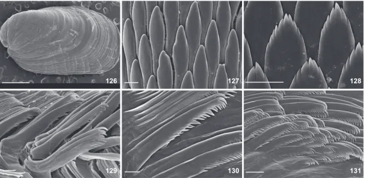
CLARK (1984) suggested that B. agassizii is similar to Pleurobranchopsis aurantiaca Verrill, 1900 due to similarities in their descriptions, except for the absence of a shell in P. aurantiaca. CLARK (1984) noted that the shell is highly trans-parent and easily overlooked in living animals, which could explain why VE R R I L L (1900) established the shell-less Pleurobranchopsis. However, CLARK (1984) did not synonymize Figures 19-24. Berthella agassizii, SEM: (19-20) shell, MNRJ 14983; (20) detail near protoconch; (21) elements of jaw, MNRJ 14982; (22-24) radula; (22) innermost lateral teeth, MNRJ 30346; (23) lateral teeth, MNRJ 11062; ((22-24) outermost lateral teeth, MNRJ 14992. Scale bars: 19 = 1mm, 20 = 500 µm, 21-22, 24 = 10 µm, 23 = 5 µm.
19 20 21
24 23
these two species because Verrill’s description lacked sufficient diagnostic characteristics to allow synonymy in the absence of the holotype of P. aurantica; thus, he preferred to consider P. aurantica as nomen dubium.
Besides that, P. aurantiaca has a problematic nomencla-tural and taxonomic history. THIELE (1931) inferred that Pleurobranchopsis was a subgenus of Pleurobranchus, thus the new name combination Pleurobranchus aurantiaca (Verrill, 1900) became a homonymy of Pleurobranchus aurantiacus Risso, 1818, species described from the Mediterranean Sea. To solve this nomenclatural problem, THIELE (1931) created a new name for species from the Bermudas, Pleurobranchus verrilli Thiele, 1931. Berthella agassizii and P. verrilli are well established as
dis-tinct species, however we would like to emphasize the differ-ences between these two species. In spite of Verrill’s brief de-scription, there are some characteristics that clearly distinguish these two species: in P. verrilli the gill is attached laterally along its entire length, while in B. agassizii we find 5-6 pinnae that are free from the body wall (Fig. 12); the mantle, gill and foot are orange in P. verrilli, whereas in B. agassizii the mantle is pink, the foot is white, and the gill with the upper pinnae are rosy (Figs. 1-2); P. verrilli is relatively larger, between 32 mm to 36 mm, whereas B. agassizii measures 7 mm to 23 mm; and the possible egg mass find by VERRILL (1900) had numerous bright orange eggs in many rows, while the egg masses of B. agassizii are white (Fig. 3) (BANDEL 1976, present study).
Figures 25-28. Berthella agassizii, digestive system. (25) dorsal view, MNRJ 31140; (26) ventral view,MNRJ 31140; (27) foregut sec-tioned longitudinally from ventral side,MNRJ 14983; (28) odontophoral cartilages, MNRJ 14983. Scale bars: 1.0 mm.
25
26
Berthella punctata
sp. nov.
Figs. 4-6, 29-52Berthella sp.: Valdés et al., 2006: 110; PADULA et al., 2012: 3.
Diagnosis. Living specimens translucent white to brown, passing through rosy (Figs. 4-6); one dark pink spot, almost brown, in middle of mantle, surrounded by white patches (Figs. 4-6). Anal opening lying approximately above 5° pinnae (Fig. 31). Elements of jaw without denticles (Figs. 45-46). Presence of jugal muscle m1vb, originating into dorsal portion of mouth, inserting into ventral portion of m5 (Figs. 49-51). Voluminous stomach covering entire dorsal portion of pharyngeal bulb (Figs. 49-50). Ampulla very thick and elongated, 8-17 times thicker than deferent duct (Figs. 34-35); deferent duct curved, but not convoluted (Figs. 34-35).
Description. External morphology (Figs. 4-6, 29-31). Liv-ing specimens translucent white to brown, passLiv-ing through rosy (Figs. 4-6); one dark pink spot, almost brown, in middle of mantle, which are surrounded by white patches (Figs. 4-6); rhinophores, oral veil, gill and foot of same pattern color as mantle. Living specimens up to 13 mm in length; length of mature preserved specimens 6-9 mm; width 4 mm; length of foot 5-6 mm; width of foot 2 mm. Body oval and oblong. Mantle covered foot entirely. Mantle surface smooth in its middle; honeycomb appearance in mantle edge, larger honey-comb appearance in the posterior portion than anterior por-tion. Oral veil thin and trapezoidal connected with the head region (Fig. 30); laterally, oral tentacles with deep notch, cor-responding almost its length. Rhinophores rolled joined at their bases, up to two-thirds of its length. Gill exposed laterally (Fig. 31); 2/5 length of body; main rachis smooth, without tubercles; alternate pinnae; simple pinnae; 17-22 pinnae; 7-13 pinnae free from body wall, attached by branchial membrane. Anal opening lying approximately above 5° pinnae (Fig. 31). Pre-branchial pore opening beside main rachis. Genital aperture surrounded by collar (Fig. 31). Penis retractable. Foot slightly pointed at posterior end with elongated metapodial gland; metapodial gland 0.3 times foot length; anteriorly bilabiated, upper lip notched, smaller than lower one. Eyes localized just behind rhinophores.
Mantle (Figs. 39-42). Mantle, rhinophores and oral veil covered by stellate spicules (Fig. 42). Two types of spicules in mantle: linear (length: 90-180 µm (Fig. 39), few spicules with 440-450 µm (Fig. 40); thickness: 10 µm) and stellate with four-five rays irregular and in different plans (ray length: 30 µm; ray thickness: 10 µm) (Figs. 41-42). Rod-like spicules calcare-ous; stellate spicules partially calcareous, but not entirely formed by calcium carbonate because in sodium hypochlorite they partially dissolve, it is also composed of an organic ma-trix not identified.
Shell (Figs. 43-44). Translucent white, with light golden tones in some parts of shell, visible according to incidence of light; subquadrangular in outline; slightly convex profile;
ap-proximately two times longer than wide. Length 5.9 mm, width 2.9 mm (in preserved specimen with 8 mm in length); and, length: 6.8 mm, width 2.9 mm (in preserved specimen with 9 mm in length). Spire with 1.5 whorls. Protoconch smooth (Fig. 44). Lines of growth distinct; immediately after protoconch, with longitudinal sculptures transverse to lines of growth (Fig. 44); shell smooth after the last line of growth. Shell above heart, on right side of blood gland. Shell covering almost all visceral mass.
Circulatory system (Fig. 32). Circulatory system of B. punctata sp. nov. identical to the B. agassizii as described above. Reproductive system (Figs. 33-37). Ampulla very thick and elongated, eight to 17 times thicker than deferent duct (Figs. 33-35); curved, but not convoluted (Figs. 34-35). Spermoviduct branching into two ducts, shorter and wider oviduct leading to female gland mass, other duct leading to prostate (Figs. 34-35). Deferent duct glandular in 63% to 79% its length (Figs. 34-35). Prostatic portion of deferent duct folds once to twice; near its distal end it joins with elongated and curved penial gland and narrows near into elongated penis. Penial gland four to five times thicker than deferent duct (Figs. 34-35). Penis (Fig. 39) (in preserved specimen with 8 mm length: length 0.94 mm; width 0.3 mm); (in preserved specimen with 9 mm length: length 0.95 mm; width 0.4 mm); completely contractible. Bursa copulatrix and seminal receptacle join vagi-nal duct in its proximal portion (Figs. 34-35, 37). Elongated to rounded bursa copulatrix (Figs. 34-35, 37). Seminal receptacle stalked and bean-shaped; twice or same volume than bursa copulatrix (Fig. 37). Vagina not convoluted; about same diam-eter of deferent duct (Figs. 34-35). Vaginal opening immedi-ately ventral to penis. Genital aperture surrounded by fold.
Nervous system (Fig. 38). Nervous system of B. punctata
sp. nov. very similar to B. agassizii as described above, with the following exceptions: cp7 absent in cerebro-pleural gan-glia. Nerves leaving buccal ganglia: nb1 inserting into esopha-gus; nb2 inserting into salivary ducts; nb3 leading to m5; connective cerebro-pleural-buccal shortly after nb3. Connec-tive cerebro-pleural-pedal (ccpp) extremely short and it can only be seen after carefully dissecting.
por-Figures 29-38. Berthella punctata sp. nov. (29) whole dorsal view, dotted line indicates position of shell internally, MZSP 97088; (30) ventral view, MZSP 97088; (31) detail of lateral view, MZSP 97088; (32) Dorsal view, organization of internal organs, MZSP 97088; (33-37) reproductive system, dorsal view; (33-34) MZSP 115665; (34) deflected; (35) deflected MZSP 97088; (36) penial papilla, MZSP 115665; (37) detail of the connection between bursa copulatrix and seminal receptacle, MZSP 115665; (38) central nervous system, dorsal view, MZSP 97088. Scale bars: 1.0 mm.
29
33
32
30
34
35
38 36
tion of digestive gland. Radula rectangular; formula 72 × 38.0.38 (from preserved specimen 8 mm length); 65 × 47.0.47 (from preserved specimen 9 mm length). Radula lacking a rachidian tooth; innermost lateral tooth hook-shaped; subsequent lat-eral plates hook-shaped, larger and more developed in center of rows, base of tooth not enlarged (Fig. 47); outermost later-als teeth less developed (Fig. 48). Esophagus internally with longitudinal folds (Fig. 52). Salivary gland small and in front of digestive gland (Fig. 49). Ducts of salivary glands entering pharynx musculature laterally to esophagus, opening into base of pharyngeal cavity between radula and jaw plates (Fig. 50); convoluted; without visible ampulla. Voluminous stomach covering all dorsal portion of pharyngeal bulb (Figs. 49-50); thin lines internally (Fig. 52); stomach passing dorsally into the digestive gland and surrounds the digestive gland (Figs. 49-50). Posteriorly, stomach passes into intestine (Fig. 49); in-testine long and thin and embedded in dorsal portion of the digestive and hermaphrodite glands and, opening laterally in body (Fig. 49); internally, intestine with longitudinal folds (Fig. 52).
Type material. Holotype MNRJ 34013, 12/x/2014, tide pool, J. Alvim coll. Paratypes: MZSP 115665, off Espírito Santo, Ilha da Trindade, Praia do Príncipe-Pedra da Garoupa (20°31’35,58”S/29°18’94,38”W), 16/vii/2013, 8m depth, D. Abbate & P. Lima colls. [1 dissected]; MZSP 97088, Alagoas, Saco da Pedra 11/i/2008, V. Padula coll. [1 dissected].
Type locality. Guarapari (20°39’35”S/40°28’31”W), Espírito Santo, Brazil.
Specimen records. Puerto Rico (VALDÉS et al. 2006); Bra-zil: Alagoas, Saco de Pedra (PADULA et al. 2012); Espírito Santo: Ilha da Trindade and Guarapari (present study).
Distribution. Caribbean Sea to Brazil.
Etymology. punctata, from Latin ”punctatus” means dot-ted. The name refers to the dark pink spot or almost brown in middle of mantle.
Remarks. Berthella punctata sp. nov. fits perfectly in Berthella, presenting all general characteristics of the genus, including the ovate and large shell (Figs. 43-44); elliptical or convex body (Figs. 4-6); generally smooth gill rachis; hook-shaped radular teeth (Figs. 47-48); smooth jaw elements (Figs. 45-46).
Berthella punctata sp. nov. is clearly distinguished from most species of the genus by its external morphology and color, including the two species that occur in Brazil, B. stellata and B. agassizi. The most similar species to B. punctata sp. nov. are Berthella africana (Pruvot-Fol, 1953) and B. tamiu, mainly due to the pigmented spot in the middle of the mantle.
Berthella punctata sp. nov. has some similarities with B. africana from Morocco, mainly due to the beige to brown col-oration with a dark spot in the middle of the mantle. PRUVOT -FOL (1953) made a mistake in the description of B. africana that was clarified some years later by GANTÈS (1956): the plates that Gantès sent to Pruvot-Fol represented B. africana (Pruvot-Fol, Figures 39-42. Berthella punctata sp. nov., spicules, MZSP 115665.
(39-40) linear spicules into the mantle, black arrow indicates spi-cule; (41-42) stellate; (41) spicules into the mantle, white arrow indicates spicule; (42) spicules into the rhinophores. Scale bar: 100 µm.
39
40
41
1953: pl. 49-51), but the description made by PRUVOT-FOL (1953) for B. africana actually corresponds to Berthella aurantiaca (Risso, 1818). Berthella africana was poorly described and the type is probably lost, but based on the redescription of this species by GANTÈS (1956), it can be distinguished from B. punctata sp. nov. by the absence of denticles on the elements of the jaw in B. punctata sp. nov. (Fig. 45-46), once B. africana was described with two or three denticles. Recently, ORTEA et al. (2012) con-cluded that Berthella canariensis Cervera, Gosliner, Garcia-Gomez & Ortea, 2000 is a junior synonym of B. africana. Considering that, assuming that B. canariensis was adequately described, B. punctata sp. nov. differs from it in the proportion of the bursa copulatrix and the seminal receptacle, and the position of the junction in relation to the vaginal duct, the shape of the penial gland, the proportion of the ampulla, the prostatic portion of the deferent duct, not as convoluted as in B. africana/B. canariensis, and the proportion of the shell in relation to the body. Thus, we conclude that B. africana and B. punctata sp. nov. are distinct species.
Berthella punctata sp. nov. closely resembles B. tamiu from Mexico. These similarities are: brownish mantle, number of pinnae in the gill (B. punctata sp. nov.: 21; B. tamiu: 26), uni-cuspidate elements of jaw (without denticles in relation to the main cuspid), radula formulae (B. punctata sp. nov.: 72 × 38.0.38; B. tamiu: 72 × 50.0.50), hook-shaped teeth without any denticles, genital opening with a ring-fold without flaps and slender penis. The unique difference according to the
de-scription made by EV. MARCUS (1984) is that B. tamiu has a small reflective circlet in the middle of the jaw elements (Fig. 57), which was not observed in B. punctata sp. nov. (Figs. 45-46). Recently, four microscope slides of the syntypes of B. tamiu were found (MZSP 121400, Figs. 53-60), including the cerebro-pleural complex (Fig. 53), transversal cuts of the mantle (Figs. 54-55), radula and jaw platelets (Figs. 56-57) and penis (Figs. 58-60), which make the comparison between these two spe-cies possible. There are sparse linear spicules in the mantle of the syntypes as well as in the mantle B. punctata sp. nov. (Figs. 39-40, 55); the small reflective circlet in the middle of the jaw elements mentioned by EV. MARCUS (1984) is not a preserva-tion artifact (Fig. 57), since it can be observed all over the jaw. In contrast, even when we examined the elements of B. punctata
sp. nov. in profile (Fig. 46) the small reflective circlet was never observed, the elements are always superficially smooth; the penis of B. tamiu is relatively larger (1533 µm in length) (Figs. 59-60) than that of B. punctata sp. nov. (940-950 µm in length) (Fig. 36); the deferent duct of B. tamiu is highly convoluted (Fig. 59), while in B. punctata sp. nov. it is not (Figs. 34-35). Slides with the complete reproductive system of the syntypes of B. tamiu were not found. B. tamiu is undoubtedly the most similar species to B. punctata sp. nov., however based on the differences in the jaw elements, penis, deferent duct and bathy-metric distribution, B. tamiu occurs at great depths (146-265 m), while B. punctata sp. nov. is found in tide pool to a 8 m depth, we prefer to maintain these two species as distinct. Figures 43-48. Berthella punctata sp. nov., SEM. (43-44) shell, MZSP 97088; (44) detail near protoconch; (45) elements of jaw, MZSP 97088; (46) lateral view of the elements of jaw, MZSP 115665; (47-48) radula, MNRJ 115665; (47) lateral teeth; (48) outermost lateral teeth. Scale bars: 43 = 1.0 mm, 44 = 200 µm, 45, 47-48 = 20 µm, 46 = 50 µm.
43
46
44 45
Figures 49-52. Berthella punctata sp. nov., digestive system, MZSP 97088. (49-50) dorsal view; (50) stomach deflected; (51) ventral view; (52) foregut sectioned longitudinally from ventral side. Scale bars: 1.0 mm.
49
50
The color pattern of B. punctata sp. nov. is very similar to Pleurobranchus caledonicus Risbec, 1928 from New Caledonia, which probably belongs to Berthella; both species have a brown-ish mantle, with a dark spot in the middle of the mantle sur-rounded by a light ring (Figs. 4-6). However, this dark spot in the middle of the dorsum is only a dark blotch in B. punctata
sp. nov., while RISBEC (1928) described it as a hole in P. caledonicus. Gary Cobb (pers. comm., 16 January 2014) found a specimen from Queensland, Australia, with the same features of P. caledonicus, except for the mark on the dorsum, which
stands out for its coloration but is not a hole. It is possible that RISBEC (1928) misinterpreted this dark spot in the middle of the dorsum as being a hole. Some differences between B. punctata sp. nov. and P. caledonicus are as follows: first, B. punctata sp. nov. (up to 24 mm in length) is smaller than P. caledonicus (up to 50 mm in length); second, there is a differ-ence in the size of shell. The shell of B. punctata sp. nov. is 5.9 mm in length in a 8 mm long preserved specimen, while the shell of P. caledonicus alone is 18 mm in length (RISBEC (1928) did not mention the size of the he specimen analyzed); third, Figures 53-60. Berthella tamiu, syntypes microscopic slides, MZSP 121400. (53) cerebro-pleural complex; (54-55) transverse cut of mantle; (55) linear spicule, black arrow indicates spicule; (56) radula and jaw plates; (57) detail of jaw elements, white arrow indicates small reflective circlet; (58-60) region near penis; (60) detail of penial papillae. Scale bars: 55 = 200 µm, 57 = 100 µm, 59 = 2.0 mm, 60 = 1.0 mm.
55 56
59
58
57
the jaw elements of B. punctata sp. nov. lack denticles (one cusp) (Figs. 45-46), while RISBEC (1928: fig. 68) described the jaw elements of P. caledonicus as having one or two cusps; fourth, the radular formulae differ in both species (B. punctata sp. nov.
72 × 38.0.38, in a 8 mm long preserved specimen; P. caledonicus 130 × 150.0.150). Additionally, RISBEC (1928) mentioned the presence of a mucus gland on the left side of the pharyngeal bulb near the mouth of P. caledonicus, which was not observed in B. punctata sp. nov. A future study of specimens of P. caledonicus from New Caledonia is necessary to elucidate the correct generic allocation of P. caledonicus.
VALDÉS et al. (2006) illustrated a morphotype named as Berthella sp. from Puerto Rico that closely resembles B. punctata
sp. nov. in the external morphology and coloration. A detailed comparison between specimens from Brazil and Puerto Rico is not possible because there is no data available on the internal anatomy in VALDÉS et al. (2006).
Berthella stellata
(Risso, 1826)
Figs. 7-10, 61-85
Pleurobranchus stellatus Risso, 1826: 41; Mazzarelli, 1891: 73, figs. 1-5. Pleurobranchus pellucidus Pease, 1860: 24; Risbec, 1928: 63.
Syn-onymized by GOSLINER & BERTSCH (1988). Bouvieria stellata: Vayssière, 1898: 302.
Berthella stellata: Pruvot-Fol, 1954: 223; Thompson, 1981: 74, fig. 4; 1985: 225, fig. 2; Gosliner & Bertsch, 1988: 50, figs. 7-12; Redfern, 2001: 168, pl. 117, fig. 293A-293B; pl. 72: fig. 693C; 2013: 296, figs. 814A-E; García et al., 2002: 50; Valdés et al., 2006: 110; García et al., 2008: 88; Rios, 2009: 417. Berthella pellucidus: Thompson, 1970: 188, fig. 8.
Berthella pellucida: Kay, 1979: 443; Willan, 1984: 40, figs. 6, 7, 17, 18, 29, 37-39, 44.
Berthella tupala Er. Marcus, 1957: 416, figs, 58-69; Er. Marcus, 1958: 57; Ev. Marcus & Er. Marcus, 1964: 198; 1967: 43, fig. 52; Er. Marcus & Ev. Marcus, 1970: 54, fig. 102; Bertsch, 1975: 124, figs. 1-7; Ev. Marcus, 1984: 53, figs. 17-19; Rios, 1994: 206. Synonymized by GOSLINER & BERTSCH (1988). Berthella postrema Burn, 1962: 140, figs. 1b, 2b, 4; pl. 1: fig. 2;
pl. 2: figs. 3-4. Synonymized by GOSLINER & BERTSCH (1988). Berthinellops serenitas Burn, 1962: 143, figs. 1d, 2d, 5; figs. 1d, 2d, 5; pl. 1: fig. 4; pl. 2: figs. 5-6. Synonymized by SABELLI et al. (1990). Berthella stellata albocrossata Heller & Thompson, 1983: 328,
figs. 5A-5C. Synonymized by GOSLINER & BERTSCH (1988).
Type material. Type presumed lost (fide ARNAUD 1978). Type locality. Nice, Mediterranean Sea.
Description. External morphology (Figs. 7-8, 61-64). Liv-ing specimens translucent white with opaque white marks, more concentrated in middle of mantle, sometimes forming one cruces (Figs. 7-8); rhinophores, oral veil, gill and foot translucent white (Figs. 7-8). Living specimens up to 18 mm in length; length of preserved specimens 3-9 mm; width 2-7 mm; length of foot 3-6 mm; width of foot 1-3 mm. Body oval and oblong. Mantle
cov-ered foot entirely. Mantle surface ranges from smooth, in juve-niles, to a rough appearance. Oral veil broad and trapezoidal connected with head region (Figs. 62-63); laterally, oral tentacles with deep notch, corresponding almost its length (Fig. 63). Rhinophores rolled joined at their bases, up to 1/4 of its length. Gill exposed laterally (Figs. 63-64); 1/2 to 1/3 length of body; main rachis smooth, without tubercles; alternate pinnae; simple pinnae; 7-11 pinnae; 3-6 pinnae free from body wall, attached by branchial membrane. Anal opening lying approximately above 3º-4° pinnae (Fig. 64). Pre-branchial pore opening beside main rachis, slightly above genital pore (Fig. 64). Genital aper-ture surrounded by collar (Fig. 64). Penis conical and retractable (Fig. 66). Foot slightly pointed at posterior end with metapodial gland (Fig. 62); metapodial gland 0.2 times foot length; anteri-orly bilabiated, upper lip notched, smaller than lower one. Eyes localized just behind rhinophores.
Mantle (Figs. 68-70). Two types of spicules in mantle: lin-ear, rod-like (length: 28 µm; thickness: 2.47 µm) (Fig. 68) and stellate with four-six rays irregular and in same plane or direct towards different plans (ray length: 2.88-20.18 µm; ray thick-ness: 1.44-4.32 µm) (Figs. 69-70). Rod-like spicules calcareous; stellate spicules partially calcareous, but not entirely formed by calcium carbonate because in sodium hypochlorite they partially dissolve, it is also composed of an organic matrix not identified. Shell (Figs. 9-10, 71-72). Two color types of shells were observed. First type, in small specimens up to 9 mm long alive, white and very fragile (Fig. 9); subquadrangular; covers all over dorsum; approximately two times longer than wide; length 3.2 mm, width 1.6 mm (in preserved specimen with 4 mm in length); and, length 2 mm, width 1.7 mm (in preserved speci-men with 3.5 mm in length). Second type brown with first whorl translucent white (Fig. 10), not fragile as first type; con-vex, subquadrangular; covers approximately 70% of mantle; approximately two times longer than wide; length 5.7 mm, width 3.6 mm (with 16 mm long alive); and, length 8.8 mm, width 5.3 mm (in with 18 mm long alive). Spire with 1.5-1.7 whorls (Fig. 71). Protoconch smooth (Fig. 72). Lines of growth distinct; immediately after protoconch, with longitudinal sculp-tures transverse to lines of growth (Fig. 72); anterior portion of last whorl only lines of growth are recognizable (Fig. 71).
Circulatory system (Fig. 65). Circulatory system of B. stellata identical to the B. agassizii as described above.
in its ¾ distal portion. Rounded bursa copulatrix. Seminal re-ceptacle stalked and elongated; representing 70% of length of bursa copulatrix. Vagina not convoluted; about same diameter or two times wider than deferent duct. Vaginal opening imme-diately ventral to penis. Genital aperture surrounded by fold. Nervous system (Fig. 67). Nervous system of B. stellata very similar to B. agassizii as described above, with the follow-ing exceptions: cp2 bifurcatfollow-ing near base, both cp2a and cp2b
inserting dorso-laterally into mantle. Nerves leaving buccal ganglia: nb1 inserting into esophagus; nb2 inserting into sali-vary ducts; nb3 leading to m5; connective cerebro-pleural-buc-cal shortly after nb3.
Digestive system (Figs. 73-85). Digestive system of B. stellata very similar to B. agassizii as described above, with the following exceptions: Muscle surrounding jaws (mj) strong, well-developed (Fig. 79). Jaws light yellow, lighter posteriorly. Each Figures 61-67. Berthella stellata. (61) whole dorsal view, dotted line indicates position of shell internally, MNRJ 31246; (62) ventral view, MNRJ 31246; (63-64) lateral view; (63) MNRJ 31246; (64) detail near gill, MNRJ 30345; (65) dorsal view, organization of internal organs, MNRJ 31333; (66) reproductive system deflected, dorsal view, MNRJ 30345; (67) central nervous system, dorsal view, MNRJ 30345. Scale bars: 1.0 mm.
61 62
65
63
64
jaw plate showing alternate rows formed by elongated and den-ticulate elements with cruciform lateral expansion (29 longitu-dinal and 20 transversal elements, in specimen of 9 mm long alive; 47 longitudinal and 22 transversal elements, in specimen of 18 mm long alive); elements consist on a main cusp with 3-5 denticles in each side (Fig. 73), which could be of different sizes and not symmetric; some specimens presents posterior elements flattened in its middle (Fig. 74). Pair of m1va originating in pos-terior portion of oral canal, running in middle of buccal mass and inserting near radular sac (Figs. 81-82). Single auxiliary muscle m10a absent. Pair of strong retractor muscles originates in most posterior portion of m5 (Fig. 81); separated approxi-mately the anterior 2/3 of length and jointed in its posterior portion, laying above anterior portion of digestive gland.
Odontophore cartilage resembles a rhombus in outline (Fig. 85). Radula rectangular, two times longer than wide; formula 59 × 53.0.53 (in specimen of 18 mm long alive); 49 × 45.0.45 (in specimen of 16 mm long alive); 46 × 56.0.56 (in specimen of 9 mm long alive). Radula lacks rachidian tooth (Fig. 75); lateral plates smooth hook shaped, without denticles; base of tooth enlarged (square) and concave; innermost lateral tooth hook-shaped and could be smooth (Fig. 75) or with one denticle in its base (Fig. 76); subsequent lateral plates hook-shaped, larger and more developed in center of rows; outermost lateral teeth bifid (Fig. 78), unusually smooth (Fig. 77). Esophagus sac-like tube passing into tube-like stomach (Fig. 84); thin walled. Stomach internally with longitudinal folds (Fig. 84).
Material examined. Puerto Rico: MZSP 121398, 18/xi/1964, G. Warmke coll. [one microscope slides: radula and jaw]. Brazil: Rio Grande do Norte: Baia Formosa: MZSP 97065, 05/vii/2009, V. Padula coll. [1]. Espírito Santo: Guarapari (20°39’28”S/ 40°28’29”W): MNRJ 34011, 12/x/2014, J. Alvim coll. [1]. Rio de Janeiro: Cabo Frio: Praia das Conchas: MZSP 97539, 16/x/2009, V. Padula coll. [1]; Final do Canal de Itajurú: MZSP 97540, 07/ viii/2009, V. Padula coll. [1]; MZSP 97557, 23/iv/2009, V. Padula coll. [1]; MNRJ 31333, 15/iii/2013, J. Alvim coll. [2 dissected]; MZSP 97529, 01/ix/2010, V. Padula coll. [1]; Ilha do Papagaio: Figures 68-70. Berthella stellata, spicules, MNRJ 31330. (68)
lin-ear; (69-70) stellate. Scale bar: 15 µm.
Figures 71-72. Berthella stellata, SEM of shell, MNRJ 31246; (72) detail near protoconch. Scale bars: 71 = 1.0 mm, 72 = 200 µm.
72
68
69
70
MZSP 97514, 30/vii/2008, V. Padula coll. [3]; Arraial do Cabo: Prainha: MNRJ 11020, 13/ii/2007, J. Alvim coll. [1 dissected]; MNRJ 31246, 10/iii/2012, J. Alvim & P. Romano colls. [3; 2 dis-sected]; MNRJ 31330, 16/iii/2013, J. Alvim coll. [2; 1 disdis-sected]; Praia do forno: MZSP 25923, vii/1957, Er. Marcus coll. [1]; MNRJ 12784, 19/i/2008, J. Alvim coll. [1 dissected]. São Paulo: Ubatuba, MZSP 121399, part of the holotype of B. tupala, ix/1955 [one microscope slide: 121399]. Santa Catarina: Itapema: Praia do canto: MNRJ 30345, 04/i/2012, J. Alvim coll. [1 dissected].
Specimen records. Indo-Pacific tropics from South Af-rica (Gosliner 1987 apud GOSLINER & BERTSCH 1988); Australia (BURN 1962, THOMPSON 1970); Marshall Islands (WILLAN 1984); New Caledonia (RISBEC 1928); Pacific Ocean: Hawaii (PEASE 1860, KAY 1979); Mexico: Baja California, Gulf of the California (GOSLINER & BERTSCH 1988); Mediterranean Sea: Nice (RISSO 1826); Naples (MAZARRELLI 1891); Coast of Sicily (VAYSSIÈRE 1898); Rovinj (PRUVOT-FOL 1954, THOMPSON 1981); Greece (GOSLINER & BERTSCH 1988); Sudanese Red Sea (HELLER & THOMPSON 1983); Atlantic Ocean: Florida (EV. MARCUS & ER. MARCUS 1967, ER. MARCUS & EV. MARCUS 1970); Puerto Rico (ER. MARCUS & EV. MARCUS 1970); Bahamas (REDFERN 2013): Abaco (REDFERN 2001); Panama (BERTSCH 1975); Belize, Curaçao, Bermuda, Honduras, Cayman Islands, Virgin Islands, St. Lucia, Martinique; Venezuela (VALDÉS et al. 2006); Colombia (EV. MARCUS 1984); Grenada (GOSLINER & BERTSCH 1988); Brazil (ER. MARCUS 1957): Fernando de Noronha (GARCÍA et al. 2002); Rio Grande do Norte(present study); Rio de Janeiro(EV. MARCUS & ER. MARCUS 1967): Cabo Frio (ER. MARCUS
1958), Arraial do Cabo (present study); São Paulo(ER. MARCUS & EV. MARCUS 1970): Ubatuba, Ilhabela (ER. MARCUS 1957); Santa Catarina (present study).
Distribution. Indo-pacific tropics from South Africa; South Pacific Ocean; North Pacific Ocean; Mediterranean Sea; Sudanese Red Sea; Atlantic Ocean: Florida to Brazil (Fernando de Noronha to Santa Catarina).
Remarks. Berthella stellata was originally described from the Mediterranean Sea and is a well-known species with a wide-spread worldwide distribution. B. stellata is easily recognized by its white-yellowish translucent body surface with scattered or medially situated opaque white markings, sometimes form-ing a cross. Several species of Berthella were described as hav-ing a translucent body with white markhav-ings, and for many years they were considered distinct due to their disjunct geo-graphic distributions (e.g., B. tupala, from Brazil, Caribbean Sea and South Africa; B. stellata albocrossata, from Sudanese Red Sea; B. postrema, from Australia; B. pellucida, from Hawaii, Australia and New Caledonia). GOSLINER & BERTSCH (1988), in a review of Berthella with white markings, concluded that they are all the same species because the variation exhibited through-out the world is expressed in individuals within a single lim-ited geographical area.
In fact, the general color is highly variable in specimens of B. stellata, from translucent whitish to yellowish brown, as well as the opaque white marks on the dorsum, which could have different patterns or be absent (e.g., RISSO 1826, PEASE 1860, Figures 73-78. Berthella stellata, SEM of jaw and radula. (73-74) elements of jaw; (73) MNRJ 31246; (74) MNRJ 30345; (75-78) radula; (75-76) innermost lateral teeth, MNRJ 31246; (75) smooth; (76) with one denticle in its base; (77-78) outermost lateral teeth; (77) smooth, MNRJ 30345; (78) bifid, MNRJ 31246. Scale bars: 73, 77 = 20 µm, 74 = 50 µm, 75-76, 78 = 5 µm.
75
78 74
77 73
Figures 79-85. Berthella stellata, digestive system, MNRJ 31333. (79-80) dorsal view; (80) pharyngeal bulb with esophagus deflected; (81-82) ventral view; (82) detail of the pharyngeal bulb; (83) pharyngeal bulb partly sectioned longitudinally from ventral side; (84) foregut sectioned longitudinally from ventral side; (85) odontophoral cartilages. Scale bars: 79-84 = 1.0 mm, 85 = 0.5 mm.
79
81
82
83
85
by many authors (RISSO 1826, PEASE 1860, RISBEC 1928), while only ER. MARCUS (1957) described it as brown for B. tupala and BURN (1962) as pale fawn for B. postrema. In the specimens of the present study, there are two color types: in small live specimens, up to 9 mm long, the shell is white and very fragile (Fig. 9); in larger specimens the shell is brown, with the first whorl translucent white (Fig. 10) and not fragile. Also, there is a correlation between the length of the shell and its color: white shells are smaller than brown shells. Some intermediate shell color, as light gold, are rare. The innermost lateral teeth of the radula vary consider-ably, sometimes in the same specimen; they can be smooth or bear one denticle at the base. GOSLINER & BERTSCH (1988) found all outermost lateral teeth with secondary denticles, as many other authors did (ER. MARCUS 1957, WILLAN 1984, Gosliner 1987 apud GOSLINER & BERTSCH 1988). In most specimens studied here, the outermost lateral tooth has a secondary basal denticle. In only one specimen (MNRJ 30345, from Santa Catarina) the outermost tooth is smooth (Fig. 77). This fact was already re-ported by VAYSSIÈRE (1898) and RISBEC (1928). Therefore, the radula is also a really variable character within this species.
Despite the wide geographic distribution of B. stellata, there are only a few publications describing its reproductive sys-tem, fully or partially (e.g., ER. MARCUS 1957, BURN 1962, WILLAN 1984, EV. MARCUS 1984, GOSLINER & BERTSCH 1988). GOSLINER & BERTSCH (1988) studied the reproductive system of specimens from different localities and concluded that the duct of the seminal receptacle may join the vagina at the base of the bursa copulatrix or near the middle of the vaginal duct. In specimens from Bra-zil, the bursa copulatrix and the seminal receptacle join the vagi-nal duct at its ¾ distal portion. ER. MARCUS (1957) described the reproductive system of B. tupala without mentioning the semi-nal receptacle, which led GOSLINER & BERTSCH (1988) to suggest that the description of the arrangement of the reproductive or-gans was wrong. We agree with this assumption, since B. tupala was originally described based on specimens from Brazil, as the material studied here, and we found the seminal receptacle in all specimens dissected, as expected. BURN (1962) also described only one allosperm receptacle and that is most likely an error.
Berthella tupala is the only species originally described from Brazil among the synonymies of B. stellata. The original description of B. tupala, as well as the description in the present work, agrees with the descriptions of B. stellata (e.g., BURN 1962, WILLAN 1984, EV. MARCUS 1984, GOSLINER & BETSCH 1988). The wide geographical distribution of B. stellata is quite question-able. The characters traditionally used to distinguish species
with further descriptions of specimens of different localities.
Berthellina
Gardiner, 1936
Berthella auct. non Blainville, 1824: VAYSSIÈRE, 1896: 115. Berthellina GARDINER, 1936: 198. Type species by original
desig-nation: Berthellina engeli Gardiner, 1936.
Description (adapted from WILLAN 1983, 1987, THOMPSON 1970, EV. MARCUS 1984). Body elliptical or convex; internal shell between one-quarter and one fifth the body length, or absent; pedal gland never present; gill rachis smooth; anus at poste-rior end of gill membrane; radula teeth elongate, lamelliform, with denticles on distal section of posterior edge; jaw elements smooth or denticulate with forward denticles. Pair m4, main dorsal tensor muscle of radula, well developed, originating in lateral region of cartilages, surrounding them ventrally, insert-ing into subradular membrane. Pair m5, secondary dorsal ten-sor muscle of radula, covering median portions of cartilage, extending up to dorsal region; originating in posterior surface of cartilages; inserting laterally in mj. Pair m7 absent. Pair m10d (dorsal) absent. Pair m10v (ventral), protractor muscle of odontophore, connected posterior portion of canal oral with ventral portion of m5. Single auxiliary muscle m10a, ventral tensor muscle of radula, originating in anterior portion of oral canal, running in middle of buccal mass and inserting into radular sac. Pair of strong retractor muscles originates in most posterior portion of m5 and radular sac.
Remarks. GARDINER (1936) proposed the genus Berthellina, based on B. engeli, to include species with lamellate radular teeth. VAYSSIÈRE (1896) presented his classification scheme for Pleurobranchoidea and described each genus and subgenus of it. However, according to GARDINER (1936), the species called Berthella by VAYSSIÈRE (1896) in fact represents Berthellina. The general features of Berthellina that VAYSSIÈRE (1896) pointed out were: polygonal elements of the jaw, usually without lateral denticles, and lamellar radular teeth with serrate edges at the upper half of the teeth.
Berthellina circularis
(Mörch, 1863)
Figs. 86-87, 91-112
Berthella circularis Mörch, 1863: 31; Vayssière, 1898: 277. Pleurobranchus circularis: Pilsbry, 1896: 200; Bergh, 1897: 134,
figs. 31-37, pl. 10.
Berthellina amarillia: Ev. Marcus & Er. Marcus, 1957: 21; 1962: 465; 1967: 163; 1969: 12.
Berthellina circularis: Ev. Marcus & Er. Marcus, 1962: 465; Ev. Marcus, 1984: 58, figs. 28-33; García et al., 2008: 208; Rios, 2009: 418.
Berthellina quadridens auct. non Mörch, 1863: Ev. Marcus & Er. Marcus, 1962: 463, figs. 9-13, in part; 1969: 12, fig. 4; Ev. Marcus, 1972: 78; 1979: 132; Rios, 1994: 206, pl. 69, in part; 2009: 417, in part; Valdés et al., 2006: 109, in part; García et al., 2008: 208; Padula et al., 2012: 8; fig. 5C.
Gminotoplax amarillus: Abbott, 1974: 347.
Type material. Not located at the Natural History Mu-seum of Denmark (ZMUC).
Type locality. St. Thomas.
Description. External morphology (Figs. 86-87, 91-93). Living specimens semi-translucent rosy in all parts of body, on laterals color more intense than center and mantle edge (Figs.
86-87). Living specimens up to 31 mm in length; length of mature preserved specimens 12-26 mm; width 7-14 mm; length of foot 6-21 mm; width of foot 5-12 mm. Body oblong. Mantle covered foot partially (Fig. 91). Dorsum seems smooth, but covered by very small papillae, which were retracted into tiny dorsal perforations. Oral veil thin and trapezoidal, that con-nects with head region (Fig. 92); laterally, oral tentacles with deep notch, corresponding to almost its length (Fig. 92). Rhinophores rolled joined at their bases, up to 2/3 of the length (Fig. 93). Gill exposed laterally; 1/3 to 1/2 length of body; main rachis smooth with alternated pinnae (Fig. 92); bi-tripinnate pinnae; 13-15 pinnae; 7 pinnae free from body wall, attached by branchial membrane. Anal opening lying over the end of gill membrane. Pre-branchial pore opening approximately be-side main rachis (Fig. 92). Nephropore under first pinnae. Geni-tal aperture surrounded by collar (Figs. 92, 94-95). Penis semi-internal, never completely internal when retracted (Figs. Figures 86-90. Living species of Berthellina from Brazil. (86-87) Berthellina circularis, MNRJ 12935, 21 mm length of preserved specimen, photo: V. Padula; (86) dorsal view; (87) ventral view; (88-90) Berthellinaignissp. nov.; (88) dorsal view, MNRJ 95965, 18 mm length of preserved specimen, photo: F. Santos; (89) dorsal view, MNRJ 97042, 9 mm length of preserved specimen, photo: V. Padula; (90) ventral view, MNRJ 97042, 9 mm length of preserved specimen, photo: V. Padula.
86
87
88
89
Figures 91-97. Berthellina circularis. (91) dorsal view, dotted line indicates position of shell internally, MNRJ 12935; (92) lateral view, MNRJ 18770; (93) Dorsal view, organization of internal organs, MNRJ 12935; (94) detail of genital apertures, MNRJ 12935; (95) reproductive system deflected, dorsal view, MNRJ 12935; (96) detail of the connection between bursa copulatrix and seminal recep-tacle, dorsal view, MNRJ 12935; (97) central nervous system, dorsal view, MNRJ 12935. Scale bars: 91-93 = 5.0 mm, 94-97 = 1.0 mm.
92
95
97 96
91
93
92, 94-95). Foot rounded at posterior end and could projected beyond the notum (Figs. 86, 91); metapodial gland absent; an-teriorly bilabiated, upper lip notched, smaller than lower one. Mantle (Figs. 98-100). Mantle and some parts of foot covered by spicules. Spicules stellate (ray length: 350-490 µm; ray thickness: 20-30 µm) (Figs. 98-99); difficult to see how many rays it have, many broken (Fig. 100), some with three-four rays in the same plane. Spicules partially calcareous, but not en-tirely formed by calcium carbonate because in sodium hy-pochlorite they partially dissolve, it is also composed of an organic matrix not identified (Fig. 100).
Shell (Fig. 101). White, decalcified and fragile; two times longer than wide; yellowish periostracum. Convex; subqua-drangular in outline (Fig. 101). Length 4.3 mm, width 2.7 mm (in preserved specimen with 22 mm in length). Spire with 1.5 whorls. Protoconch smooth. Lines of growth distinct; imme-diately after protoconch, with longitudinal sculptures trans-verse to lines of growth; anterior portion of last whorl only lines of growth are recognizable. Shell above heart, on right side of blood gland (Fig. 91).
Circulatory system (Fig. 93). Pericardium well developed in anterior portion of body (near cerebro-pleural ganglia). Blood flowing into auricle from gills, kidney and venous sinuses. Ef-ferent branchial vessel connecting gill with the auricle. Au-ricle on right side, ventAu-ricle on left; auAu-ricle with thin wall; ventricle muscular. Blood gland small, creamy covering left part of pericardium. Blood gland close or joined to aorta; it covers partially ventricle.
Reproductive system (Figs. 94-96). Ampulla elongated, two times wider than deferent duct; curved, sometimes slightly con-voluted (Fig. 95). Spermoviduct branching into two ducts, long oviduct leading to female gland mass, other duct leading to pros-tate (Fig. 95). Prospros-tate globular and granular; five-seven times wider than deferent duct. Deferent duct not convoluted, nar-rowing into not cuticular penis and penial gland (Fig. 95). Penis semi-internal, never completely internal when retracted (Fig. 94). Penial gland elongated, not-convoluted. Penis half-moon shape (in preserved specimen with 21 mm length: length 1.7 mm; in preserved specimen with 25 mm length: length 1.8 mm). Seminal receptacle stalked (Figs. 95-96). Rounded bursa copulatrix; seminal receptacle two times more volume than semi-nal receptacle (Fig. 96). Vagina not convoluted; two times wider than deferent duct. Vagina opening immediately ventral to pe-nis. Genital aperture surrounded by wide flap (Fig. 94).
Nervous system (Fig. 97). Nerve ring above oral canal. Cerebral and pleural ganglia fused. Eyes located latero-centrally of cerebro-pleural complex; eyes borne upon very short opti-cal nerves. Rhinophoral ganglia placed at bases of rhinophores, near cerebro-pleural ganglia; two main nerves leaving from rhinophoral nerves, runing until distal portion of rhinophores; rhinophoral nerves with many secondary nerves, perpendicu-lar in relation to main nerves. Nerves leaving cerebro-pleural ganglia: cp2 bifurcating near origin, both cp2a and cp2b
in-serting dorsally into mantle; cp6 inin-serting dorsally into mantle; cp3 runing until median portion of body wall, near anterior portion of digestive gland in left side, inserting into mantle laterally. Connective between visceral and cerebro-pleural gan-glia well developed, which has two main nerves. Connective between buccal and cerebro-pleural ganglia leading from ven-tral view of cerebro-pleural ganglia, in the most anterior por-tion of ganglion. Nerves leaving buccal ganglia: nb1 bifurcating near origin, both nb1a and nb1b inserts into esophagus; nb2 inserting into salivary ducts; connective cerebro-pleural-buc-cal shortly after nb1; nb3 inserting into m4; nb4 inserting into Figures 98-100. Berthellina circularis, stellate spicules, MNRJ 12935. (98) into the foot, black arrow indicates spicules; (99) into the mantle, black arrow indicates spicules; (100) some broken spi-cules due sodium hypochlorite. Scale bar: 200 µm.
81
100
radula sac; nb5 inserting into retractor muscles of buccal bulb.Connective cerebro-pleural-pedal (ccpp) extremely short and can only be seen after carefully dissecting. Pedal commis-sure short. Pedal ganglia smaller than cerebro-pleural complex: np1 inserting ventrally into oral veil; np2 inserting anteriorly into foot; np3 innervating foot; np4 inserting ventrally into foot and runs until the most posterior portion of body. In the right side of pedal ganglion, just after the np2, there is a con-nective that leads to genital ganglion.
Digestive system (Figs. 102-112). Mouth transversal in middle of snout tip. Oral canal muscular just posterior to mouth, representing approximately 1/3 of pharyngeal bulb length; in-ternally rough (Figs. 111-112). Muscle surrounding jaws (mj) strong, pair of large jaws located in its inner surface (Figs. 111-112), mj originating in lateral and dorsal surfaces of oral canal, inserting into lateral and dorsal regions of buccal mass. Jaws amber, lighter posteriorly; jaw of two plates surrounding radula inside buccal cavity; elongated, reaching level of radula. Each jaw plate showing alternate rows formed by elongated elements with a cruciform lateral expansion (104 longitudinal and 70 transversal, in preserved specimen of 12 mm long and 109 lon-gitudinal and 64 transversal, in preserved specimen of 21 mm long) (Figs. 102-103);each element with 1-5 small denticles, but some elements could be not symmetric; anterior elements worn. Pair of dorsal jugal muscles m1d (Fig. 107), inserting into m4, connecting to dorsal-anterior end of oral canal. Pair m4, main dorsal tensor muscle of radula, well developed, originating in
lateral region of cartilages, surrounding them ventrally, insert-ing into subradular membrane (Figs. 107-109). Pair m5, second-ary dorsal tensor muscle of radula, covering median portions of cartilage, extending up to dorsal region; originating in poste-rior surface of cartilages; inserting laterally in mj (Figs. 107-109). Pair m7 absent. Pair m10d (dorsal) absent. Pair m10v (ventral), protractor muscle of odontophore, connecting posterior por-tion of oral canal with ventral porpor-tion of m5 (Fig. 108). Single auxiliary muscle m10a, ventral tensor muscle of radula, origi-nating in anterior portion of oral canal, running in middle of buccal mass and inserting into radular sac (Fig. 108). Pair of strong retractor muscles originates in most posterior portion of m5 and radular sac (Figs. 107-109, 111-112); separated in 1/2 its total length and jointed in its posterior portion, laying above anterior portion of digestive gland (Fig. 108). Odontophoral cartilages subquadrangular in outline (Fig. 110). Radula amber; formula 108 × 144.0.144 (from a preserved specimen with 21 mm length) and 56 × 107.0107 (from a preserved specimen with 12 mm length). Rachidian tooth absent. Lateral teeth long and slender with subtermial denticles (Figs. 104-106). Innermost eral tooth with 4-8 irregular denticles (Fig. 104). Subsequent lat-eral plates larger and more developed in center of rows with 10-17 irregular denticles (Fig. 105). Outermost laterals teeth less developed up to 10 denticles (Fig. 106). Aperture of acid gland located between jaw plates (Figs. 111-112). Duct of acid gland thin (same width as salivary duct) (Fig. 107); runs between stom-ach (until median portion of stomstom-ach) and hermaphrodite gland Figures 101-106. Berthellina circularis, SEM. (101) shell, MNRJ 12935; (102-103) elements of jaw, MNRJ 18770; (104-106) radula, MNRJ 18770; (104) innermost lateral teeth; (105) lateral teeth; (106) outermost lateral teeth. Scale bars: 101 = 200 µm, 102-105 = 10 µm, 106 = 20 µm.
101 103
106 105
102
Figures 107-112. Berthellina circularis, digestive system. (107) dorsal view, MNRJ 12935; (108) ventral view, MNRJ 12935; (109) dorsal view, detail of posterior portion with esophagus deflected, MNRJ 12935; (110) odontophoral cartilages, MNRJ 18770; (111-112) foregut sectioned longitudinally from ventral side, MNRJ 12935. Scale bars: 107-108 = 5.0 mm, 109, 111-112 = 2.5 mm, 110 = 1.0 mm.
108
111
112 109
107