JOANA PALHARES CAMPOLINA
Plan of nutrition in dairy goats diets on mammary
gland development
Dissertação apresentada ao Programa de Pós-Graduação em Zootecnia da Escola de Veterinária da Universidade Federal de Minas Gerais como requisito parcial para obtenção do grau de Mestre em Zootecnia
Área de concentração: Produção Animal Orientador: Prof. Dr. Iran Borges
Ostra feliz não faz pérola
“… Pois havia num fundo do mar uma colônia de ostras. Eram ostras felizes. Sabia-se que eram felizes porque dentro de suas conchas saía uma delicada melodia, música aquática, como se fosse um canto gregoriano, todas cantando a mesma música. Com um exceção: de uma ostra solitária que fazia um solo solitário. Diferente da alegre música aquática ela cantava um canto muito triste. As ostras felizes riam dela e diziam: Ela não sai da sua depressão… Não era depressão. Era dor. Pois um grão de areia havia entrado dentro de sua carne e doía, doía, doía. E ela não tinha jeito de se livrar dele, do grão de areia. Mas era possível livrar-se da dor. O seu corpo sabia que, para se livrar da dor que o grão de areia lhe provocava, em virtude de suas asperezas, arestas e pontas, bastava envolvê-lo com uma substância lisa, brilhante e redonda. Assim, enquanto cantava seu canto tristem o seu corpo fazia o trabalho – por causa da dor que o grão de areia lhe causava. Um dia, passou ali um pescador com seu barco. Lançou a rede e toda a colônia de ostras, inclusive a sofredora, foi pescada. O pescador se alegrou, levou-as para casa e sua mulher fez uma deliciosa sopa de ostras. Deliciando-se com as ostras, de repente seus dentes batem num objeto duro que estava dentro de uma ostra. Ele o tomou nos dedos e sorriu de felicidade: era uma pérola, uma linda pérola. Apenas a ostra sofredora fizera uma pérola. Ele a tomou e deu-a de presente para sua esposa. Isso é verdade para as ostras. E é verdade para os seres humanos. No seu ensaio sobre
O nascimento da tragédia grega a partir do espírito da música, Nietzche observou que os gregos, por oposição aos cristãos, levavam a tragédia a sério. Tragédia era tragédia. Não existia para eles, como existia para os cristãos, um céu onde a tragédia seria transformada em comédia. Ele se perguntou então das razões por que os gregos, sendo dominados por esse sentimento trágico da vida, não sucumbiram ao pessimismo. A resposta que encontrou foi a mesma da ostra que faz uma pérola: eles não se entregaram ao pessimismo porque foram capazes de transformar a tragédia em beleza. A beleza não elimina a tragédia, mas a torna suportável. A feliciade é um dom que deve ser simplesmente gozado. Ela se basta. Mas ela não cria. Não produz pérolas. São os que sofrem que produzem beleza, para parar de sofrer. Esses são os artistas. Bethoven – como é possível que um homem completamente surdo, no fim da vida, tenha produzido uma obra que canta a alegria? Van Gogh, Cecília Meireles, Fernando Pessoa…”
Acknowledgments
A Deus pelo dom da vida, por ser meu grande guia e proporcioanr muito luz em meu caminho.
Aos meus pais, Clélio e Alda, pelo incentivo, apoio, ensinamentos e amor incondicional. Vocês são meus grandes mestres e tem minha profunda admiração!
Aos meus irmãos, Bernardo e Gabriela, pelo companheirismo e amizade. Por me acolherem em suas casas e proverem o rango que tanto me fez falta. Por me guiarem e ajudarem a passar por esta difícil etapa. Muito obrigada!
Às minhas lindas sobrinhas, Duda e Bebela, por tornerem a jornada mais leve, promoverem horas seguidas de Peppa pig, por trazerem doçura à minha vida.
Aos meus cunhados, tios, primos e demais familiares que sempre me apoiaram. Obrigada!
Ao meu amor, meu melhor amigo, meu porto seguro, meu companheiro. Gui, com certeza não chegaria até o final sem seu apoio e incentivo, seu amor, sugestões e conversas. Cedeu aos fim de semana de descanso para me ajudar, fazer companhia, e me fazer ainda mais feliz. Xu, muito obrigada!
Ao professor Iran, grande mestre, grande pessoa, no qual vejo meu espelho. Obrigada por confiar um experimento tão grande e tão importante! Obrigada por acreditar, obrigada por me orientar. Sua escolha foi um acerto, pois nunca fui tão desafiada e aprendi tanto! Meus sinceros agradecimentos!
Ao professor Rômulo pelo grande apoio na graduação e na pós, pelo empréstimo do ultrassom e pelo carinho!
Aos professores Elias Facury e Antônio Último pelo apoio, pelo socorro clínico e ajuda durante o experiemnto!
À professora Fabiola pelo apoio e orientação nas coletas de sangue.
Ao professor Marc Henry pelos ensinamentos de ultrassom, pelo apoio e ajuda no experimento.
Ao professor Monteiro, pelas constantes conversas. Minha profunda admiração, grande pesquisador e professor.
Ao Nepper, grande grupo de pesquisa e trabalho! Aprendi muito com vocês! A todos presentes no grupo durante minha jornada no mestrado!
Ao Luiz e Gabi, meus parceiros de experimento. Me ensinaram a respeito de paciência e calma. Luiz, você tornou nossos dias mais leves com suas piadas e jeito simples de ser. Gabi, mostrou q atrás de uma pequena menina vive uma grande mulher, com muita garra e perseverança.
Ao Dodô, Zê, Lu, Cimara, Flávio (Dona Maria), Pedro, Léo e Tassinha. Meus companheiros de pós, meus companheiros de grupo, de aprendizado, de farra!
À Ju, Jizenio, Celso e Joelma, meus queridos ics que me ensinaram mto sobre trabalho em equipe. Ju com suas frases de efeito, Jizênio com seu entusiasmo, Celso descomplicando o que eu tornava complicado e Joelma com sua garra e força de vontade. Muito obrigada!
Às estagiarias, Helen, Isabella, Elen, Melina e Jéssica.
À Pamela e Camila por me ajudarem com o ultrassom e as intermináveis análises. Muito obrigada!
À Hemilly, minha eterna chefa, grande amiga, grande mentora. Um dos melhores corações que já conheci!
Ao Luigi, grande amigo, mentor e pessoa. Sem sua ajuda não teria ido tão longe. Obrigada pelos ensinamentos, pelos “toques”, pelo apoio e ajuda na estatística! Um grande orientador!
Aos amigos da Vet, Dri, Ra, Carol, Gabi, Paulinha, Ju, Mamma, Paulão, Vitim e Rafa pelo apoio sempre! Obrigada.
Aos amigos da vida, Ana, Rê, MC, Mari, Tam, Ci e Renatinha, por compreenderem minha distância, mas sempre estarem ao meu lado!
Aos funcionário da fazenda, especialmente Douglas, Carlinhos, Seu Luiz, Marciano e Marquinhos! Sem vocês estaria perdida! Obrigada pela amizade sincera, pela ajuda e pelas farras!
Aos companheiros de CT, Helmut, Márcio, Ju, Mosquito e Otoni! Obrigada!
À todos funcionários da Escola de Veterinária, que ajudaram tanto durante o período!
À todas as pessoas que me ajudaram em qualquer parte do trabalho, principalmente aos que contribuiram em algum momento do experimento.
Ao capril Triqueda, pelo empréstimo dos animais .
Às cabras, especialmente Julieta, por nos ensiarem tanto, e serem tão pacientes e amorosas. Foi ótimo trabalhar com vocês.
À CAPES, pela bolsa de estudos.
SUMMARY
CHAPTER 1. INTRODUCTION ... 17
CHAPTER 2. LITERATURE REVISION ... 20
2.1ANATOMY OF THE MAMMARY GLAND ... 20
2.1.1 Connective tissues ... 21
2.1.2 Blood supply ... 22
2.1.3 Lymphatic system ... 23
2.1.4 Nervous system ... 24
2.1.5 Secretory tissue ... 24
2.2ONTOGENY OF MAMMARY GLAND ... 26
2.3HORMONAL REGULATION OF MAMMARY DEVELOPMENT ... 31
2.3.1 Estrogen ... 32
2.3.3 Prolactin ... 33
2.3.4 Growth Hormone ... 34
2.3.5 Insulin ... 34
2.3.6 IGF-1 ... 35
2.3.7 Placental Lactogen ... 35
2.3.8 Glucocorticoids ... 35
2.3.9 Thyroid Hormones ... 36
2.3.10 Leptin ... 36
2.4NUTRITION IMPACT ON MAMMARY DEVELOPMENT ... 37
2.5TECHNIQUES FOR STUDY MAMMARY GROWTH ... 46
2.5.1 Biopsy ... 46
2.5.2 Comparative slaughter ... 47
2.5.3 Water displacement and plaster cast ... 47
2.5.4 Palpation and udder measures ... 48
2.5.5 Magnetic resonance imaging ... 48
2.5.6 Ultrasound ... 48
2.6HYPOSTASIS ... 50
2.7MAIN OBJECTIVE ... 50
2.8SPECIFIC OBJECTIVES ... 50
2.9REFERENCES ... 51
CHAPTER 3. EFFECT OF AVERAGE DAILY GAIN, DRY MATTER, CRUDE PROTEIN AND METABOLIZABLE ENERGY INTAKE ON MAMMARY GLAND DEVELOPMENT AND BODY GROWTH IN SAANEN KIDS ... 58
3.1ABSTRACT ... 58
3.2INTRODUCTION ... 59
3.3MATERIAL AND METHODS ... 60
3.3.1 Animals, design and management ... 60
3.3.2 Reproduction ... 62
3.3.3 Body measurements ... 62
3.3.4 Mammary gland ultrasound and measurements ... 63
3.3.5 Digital Image Processing ... 64
3.3.6 Statistical Analyses ... 65
3.4.RESULTS ... 66
3.4.1Body Growth ... 66
3.4.2 Mammary biometry ... 67
3.4.3 Mammary ultrasound ... 68
3.4.4 Allometric Growth ... 70
3.5.DISCUSSION ... 71
3.6.CONCLUSION ... 76
8.REFERENCES ... 77 CHAPTER 4. IMPLICATIONS ... 79
LIST OF TABLES
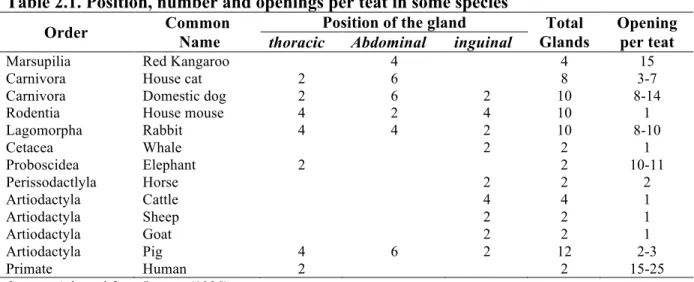
TABLE 2.1.POSITION, NUMBER AND OPENINGS PER TEAT IN SOME SPECIES ... 21
TABLE 2.2.EMBRYONIC AND FETAL MAMOGENESIS IN CATTLE, PIGS AND HUMANS ... 28
TABLE 2.3.TOP FINDINGS IN DOMESTIC ANIMALS MAMMARY GLAND NUTRITION IMPACT ... 38
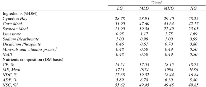
TABLE 3.1.FORMULATION AND DIET NUTRIENT COMPOSITION ... 61
TABLE 3.2.AVERAGE DAILY GAIN,TOTAL DRY MATTER INTAKE,TOTAL METABOLIZABLE ENERGY INTAKE AND TOTAL CRUDE PROTEIN INTAKE OF SAANEN GOATS ... 66
TABLE 3.3.DEPENDENT VARIABLES AND STANDARD DEVIATION VALUES OF BODY MORPHOMETRIC MEASURES OF SAANEN GOATS ... 67
TABLE 3.4.DEPENDENT VARIABLES AND STANDARD DEVIATION VALUES OF MAMMARY GLAND MORPHOMETRIC MEASURES OF SAANEN GOATS ... 67
TABLE 3.5.DEPENDENT VARIABLES AND STANDARD DEVIATION VALUES OF MAMMARY GLAND ULTRASOUND ANALYZES OF SAANEN GOATS ... 69
TABLE 3.6.DAILY GROWTH RATES OF BODY AND MAMMARY GLAND MORPHOMETRY AND MAMMARY PARENCHYMA OF 37SAANEN GOATS ... 70
LIST OF FIGURES
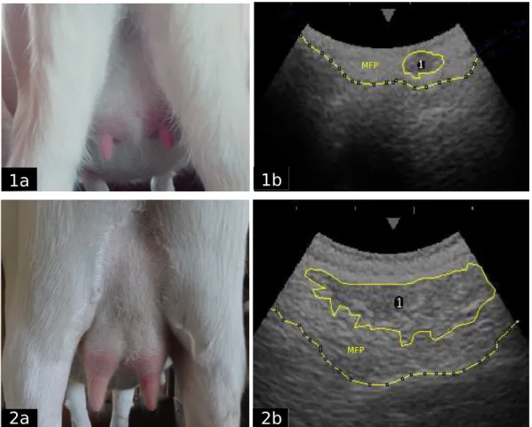
FIGURE 3.1.APPEARANCE OF UDDERS AT THE BEGINNING AND END OF THE TRIAL (1A-2A), ULTRASOUND IMAGES (1B-2B) SHOWING THE PARENCHYMA AREA (1), THE MAMMARY FAT PAD (MPF) SEPARATED FROM THE BODY WALL WITH A DASHED LINE.GREY TICK MARKS ON THE TOP OF THE ULTRASOUND SCREEN ARE SPACED 1 CM APART. ... 64 FIGURE 3.2.ALLOMETRY COEFFICIENTS OF BODY PARTS AND PAR AREA THROUGH THE
LIST OF ABBREVIATIONS
2D: Two Dimensional ADF: Acid Detergent Fiber ADG: Average Daily Gain BG: Body girth
BL: Body length BMP: Bitmap
BST: Bovine somatotropin BW: Body Weight
CP: Crude Protein
CPI: Crude Protein intake CW: Chest width
d: day
DFFT: Dried-fat-free tissue DMI: Dry matter intake DNA: Deoxyribonucleic acid
E2: Estradiol
g: gram
GH: Growth Hormone h: hour
HG: Heart girth HG: High Gain Hz: Hertz
IGF-1: Insulin-like growth factor 1
Kg: Kilogram LG: Low Gain LL: Leg length LP: Leg perimeter M: Million
ME: Metabolizable Energy
MEI: Metabolizable Energy intake MFP: Mammary fat pad
MG: Mammary Gland mg: Milligrams
MHG: Medium-high Gain MHz: Megahertz
mL: Milliliters
MLG: Medium-low Gain
MRI: Magnetic Resonance Imaging mRNA: messenger Ribonuclec acid NDF: Neutral Detergent Fiber NEFA: Non esterified fatty acids NSC: Nonstructural Carbohydrates PCA: Principal Component Analyses P4: Progesterone
PAR: Parenchyma Prl: Prolactin
PUF: Polyunsaturated Fats
RH: Rump height RL: Rump length
RNA: Ribonucleic acid
RW: Rump width
SH: Sternum height (SH), SL: Shoulder length SW: Shoulder width T3: Tiiodothyronine
T4: Thyroxine
ABSTRACT
Rearing replacement dairy animals is one of the most costly phases on a dairy farm, foe being it an unproductive period. For that reason, high plane of nutrition is given to provide high gains and decrease age at sexual maturity. However, this high energy and protein consumption, during the prepubertal period, may affect body growth and mammary secretory tissue development. The aim of this experiment was to evaluate the effect of different average daily gains (ADG) and dry matter (DMI), crude protein (CPI) and metabolizable energy intake (MEI) on body and mammary development. Forty Saanen kids, 109.1±21.4 days old and 12.9±2.9 kg initial body weight, had their body and gland morphometry, and mammary parenchyma (PAR) measured until 30 days of pregnancy. They were separated in four diets with ten animals each, where each group received a different protein-rate diet – 15, 17, 18 and 19% –, allowing different ADG (90, 130, 170 and 210 g/day) and nutrient consumption. Mammary morphometry was obtained weekly, whereas body morphometry and mammary PAR at every fourteen days. For PAR access, a B-mode ultrasound with a 5.0 MHz convex probe was used to examine in a 2-dimension PAR area, perimeter and grey scale, of both glands, throughout the trial. Data was analyzed with a multiple regression using RStudio software. Throughout the trial, all variables augmented. ADG had the major impact, influencing positively all body measurements (p<0.01) and mammary teat growth (p<0.01), PAR area (p<0.01), PAR perimeter (p<0.001). MEI influenced only rump length and chest width (p<0.05) on body growth, right PAR area and perimeter (p<0.05). CPI and DMI did not influence body growth; however, protein affected considerably PAR development, increasing area, perimeter, and maximum grey value. Allometry coefficients of PAR area were low when compared to other body coefficients. However, it was possible to visualize a change of proportion in fifteen times for this variable, which means that PAR area increased more proportionally with time. Increasing levels of CPI, conversely from MEI, had an important and positive impact on body and mammary growth. Mammary growth could not be associated with any body growth pattern.
RESUMO
A recria é uma das fases mais caras em uma fazenda leiteira, já que é um período improdutivo. Por essa razão, dietas com altos planos nutricionais são dadas para possibilitar altos ganhos e diminuir a idade de maturidade sexual. No entanto, este alto consumo de energia e proteína, durante o período de pré-púbere, pode afetar o crescimento do corpo e desenvolvimento do tecido secretor mamário. O objetivo deste trabalho foi avaliar o efeito de diferentes ganhos de peso diário (GPD) e matéria seca (CMS), proteína bruta (CPB) e consumo de energia metabolizável (CEM) no corpo e desenvolvimento mamário. Quarenta cabritas Saanen, com 109,1 ± 21,4 dias de idade e 12,9 ± 2,9 kg de peso corporal inicial, tiveram seu corpo e morfometria da glândula, e parênquima mamário (PAR) medidos atingirem 30 dias de gestação. Elas foram separadas em quatro grupos com dez animais cada, onde cada grupo recebeu uma dieta com diferentes taxas de proteína - 15, 17, 18 e 19% -, permitindo diferentes GPD (90, 130, 170 e 210 g/dia) e consumo de nutrientes. A morfometria mamária foi obtida semanalmente, e a morfometria corporal e PAR mamário, a cada quatorze dias. Para a mensuração interna da glândula, um ultra-som modo B com uma probe convexa de 5,0 MHz foi usado para examinar, em duas dimensões, a área de PAR, o perímetro e a escala de cinza, de ambas as glândulas, por todo o periodo experimental. Os dados foram analisados com regressões múltiplas usando software Rstudio. Durante o experimento, todas as variáveis aumentaram. GPD teve o maior impacto, influenciando positivamente em todas as medidas do corpo (p <0,01) e no crescimento das tetas mamárias (p <0,01), área de PAR (p <0,01), o perímetro de PAR (p <0,001). CEM influenciou apenas no comprimento garupa e largura no peito (p <0,05), na área da glândula direita do PAR e perímetro (p <0,05). CPB e CMS não influenciaram no crescimento do corpo, no entanto, CPB afetarou o desenvolvimento consideravelmente do PAR, aumentando área, perímetro e valor máximo de cinza. Os coeficientes alométricos de área do PAR foram baixos em comparação com outros coeficientes corpo. No entanto, é possível visualizar uma mudança da proporção em quinze vezes para esta variável, o que significa que a área de PAR cresceu mais proporcionalmente ao longo do tempo. Níveis crescents de CPB tem um importante e positivo impacto no crescimento corporal e da glândula mamária. O crescimento mamário não pode ser associado a qualquer padrão de crescimento do corpo.
CHAPTER 1. Introduction
Goats have been domesticated since ancient times, to provide milk, meat, skin and hair. They are highly adaptable to harsh conditions such as different climates, regions and altitudes, have the ability to graze bad quality forages and still produce milk and meat, and have high body resistance to draughts and long distance walks.
According to FAOSTAT (2015), there are one billion goats worldwide. Asia and Africa are the continents with the biggest flocks, with respectively 586 and 364 M goats, contributing to associating goat production with underdevelopment and poverty, The latter is not entirely true. It could more likely be justified by historical connections, easy management, food for rural populations and as well as providing work to women and children, low investments, and consequently low risks. On the other hand, about 150 years ago, more exactly after the two world wars, Mediterranean countries like France, Spain, Greece and Italy, as well as the USA, invested in dairy goat to produce milk and top quality cheese. These countries started to make genetic improvements, allied with superior nutritional management, thus, increasing milk production. They also developed a highly organized chain with the creation of social bonds of goat breeders and other classes (Boyazoglu et al., 2005).
Brazil, currently with 8,9 M goats, occupies the 22nd position in number of goats and the 18th position in goat milk with production of 153 thousand tons of milk a year (FAOSTAT, 2015). Most of the herd is concentrated in the Northeast of the country, spread in small native breed flocks and raised in extensive management. Nevertheless, it is in the southeast of Brazil where most of the specialized dairy herd is situated, with approximately 25% of national goat milk production (IBGE, 2013). Constituted mostly by European dairy breeds – Saanen, Alpine and Toggenburg – milk production is now driven by gastronomy and powdered milk.
on most Brazilian farms, it is nutrition that is not always adequate and the ratio formulation is mostly unbalanced.
Between birth and the beginning of milk production, raising female kids is extremely arduous because the nanny goat does not provide any benefits until giving birth and the onset of milk production; therefore, it is a long term breeder investment. That is the reason why a rapid development from neonate to puberty, together with fast growth and elevated daily body gain is desirable. To achieve high gains, dairymen give high protein and energy concentration ratio, to make the animals develop fast. However, this rush could compromise body growth, in particular mammary gland growth, and consequently future milk production.
The mammary gland is one of the organs that suffers most alterations during all animal life, contemplating size, structure, composition and activity (Akers and Denbow, 2013). Its growth takes over complexity factors involving animal, environment and nutrition. Anyway, despite a large number of studies on mammary gland development in mice, rats, sows, cows and sheep, few works were found on goat mammary gland peculiarities.
Many techniques have been developed to accompany organ growth such as comparative slaughter, biopsy, metric measurements, nuclear magnetic resonance imaging (MRI) and ultrasound. This last technique permits to see in real time, in two dimensions and in 256 gray scale, the complexity of the analyzed body part with little invasion. There is no need to slaughter or remove part of the organ, which allows following the changes in the evaluated period.
The scarcity of research on dairy goat mammary gland development is a motivation for investigating the implication of different nutritional planes, leading to divergent average daily gains, in female kids from weaning to pregnancy, and their consequences on body and mammary gland growth.
were measured every two weeks to compare the different growth of body parts with mammary gland parenchyma.
CHAPTER 2. Literature Revision
The mammary gland produces “nature’s most perfect food”. Understanding its anatomy, histology, ontology, physiology and biochemistry allows manipulation of animal production and helps preventing maladies that affect not just milk production but human and animal health (Schmidt, 1971). A better knowledge of these characteristics drives researchers to work with the mammary gland in all its aspects, helping increasing efficiency not only for dairy cows, but also for sheep, goats, water buffalos and camels.
For this study, goats were chosen not only because of their socio and economic importance, but mainly due to the lack of works on nutrition and mammary gland development in this domestic ruminant. Since goats are not taken as an experimental model, literature revision will be made comparing different mammals, but preferably does used on dairy farms.
2.1 Anatomy of the mammary gland
The mammary gland is an anatomical structure characteristic of mammals, whose primary function is transferring food from parent to offspring (Larson, 1985; Ferreira et al., 2013). It also has thermoregulatory, immunological, antibiotic and behavioral functions (Akers, 2002), important to seal bonds between mother and progeny, and helping offspring survival under harsh conditions. Because soft tissues are not well preserved as fossils, evolutionary origins of the mammary gland are unclear (Akers and Denbow, 2013). It is suggested that this organ arose from the combination of sweat and sebaceous glands, since it follows apocrine and meocrine modes of secretion (Kon and Cowie, 1961; Akers, 2002; Capuco and Akers, 2010).
from two for sheep and goats, to four for cows and buffalos, with only one opening per teat. Sows have mammary glands distributed all over the ventral part of the body, with two openings per teat. Primates, like humans, have only two mammary glands, located in thoracic position, but a substantial number of openings per teat (Larson, 1985) (Table 2.1).
Table 2.1. Position, number and openings per teat in some species
Order Common
Name
Position of the gland Total
Glands
Opening per teat
thoracic Abdominal inguinal
Marsupilia Red Kangaroo 4 4 15
Carnivora House cat 2 6 8 3-7
Carnivora Domestic dog 2 6 2 10 8-14
Rodentia House mouse 4 2 4 10 1
Lagomorpha Rabbit 4 4 2 10 8-10
Cetacea Whale 2 2 1
Proboscidea Elephant 2 2 10-11
Perissodactlyla Horse 2 2 2
Artiodactyla Cattle 4 4 1
Artiodactyla Sheep 2 2 1
Artiodactyla Goat 2 2 1
Artiodactyla Pig 4 6 2 12 2-3
Primate Human 2 2 15-25
Source: Adapted from Larson (1985).
In rodents and lagomorphs, the mammary glands do not develop very much beyond the primary ducts, having a flat aspect form, thus permitting a more accurate study of the whole organ. On the contrary, in ungulates, the highly branched ducts show a more developed mammary gland (Larson, 1985).
2.1.1 Connective tissues
Patterns of mammogenesis in bovine, ovine and caprine are similar, even when ruminants have a different number of glands with size variation among them. Located in the udder, the mammary gland is protected by skin and a thin layer of connective tissue below , uniting the four or two mammary quarters. The skin and the connective tissue provide protection but little sustentation and stabilization, although the main support is provided by a thick, elastic and prominent ligament attached to the pelvic bone, known as the median suspensory ligament. Composed of mesordermic elastic and fibrous connective tissues, especially elastin, this ligament divides the udder in two halves, allowing the animal to absorb shocks when walking and lying down, due to its great strength (Turner, 1952; Schmidt, 1971; Larson, 1985).
tissue that bonds the dorsal surface of the udder with the abdominal wall, the lateral suspensory ligaments, the lamellar plates that extend from the ligaments to the mammary parenchyma and the subpubic and prepubic tendons (Turner, 1952). The strong tendons of the abdominal muscles also help udder support. Weight and udder capacity increase until a determined time, with their maximum enlargement between the first and second lactation (Schmidt, 1971). With aging, these connective tissues lose their support capacity and the udder becomes pendulous. Because of this reason, for dairy animal longevity, it is recommended that the udder should extend well forward, be strongly attached, be balanced and symmetrical, with its floor above the animal’s knuckle and have a soft, pliable and elastic texture (Turner, 1952). Unfortunately, despite their importance, connective tissues are not among the main objectives of studies and, instead, neglected and left aside.
2.1.2 Blood supply
In the mammary gland, blood supply it is extremely important since it carries essential components needed for milk production. In virtue of that, studies on the concentration of blood components in mammary vessels, and its influence on milk composition are common, although animal mammary angiogenesis does not receive much attention. In humans, breast vascularization and factors that stimulate it, are largely investigated because of their relation with breast cancer (Akers, 2002).
For rabbits, rats, mice, sows, bitches and queens, since their glands are located on each side of the midline extending from the thoracic to the abdominal region, blood supply comes from several vessels of the external pudendal arteries and their branches, and from vessels that supply the body and abdominal and thoracic walls (Schmidt, 1971).
Blood draining, different from the arterial route, leaves the udder by two divergent paths in most animals with inguinal glands. The first path is a combination of the cranial and caudal branches of the mammary vein, forming the venous circle. Then, this venous circle becomes the external pudendal vein, going all the way back paralleled to the arteries with the same name, until it reaches the vena cava and the right atrium of the heart. The second route, despite its massive size, is less important and physiologically minor. It is composed by the subcutaneous abdominal vein, or simply milk vein, and unites with the internal thoracic vein in the xiphoid process to enter the anterior vena cava (Turner, 1952; Schmidt, 1971; Larson, 1985, Akers, 2002). In virgin ruminants, the first path drains practically all mammary blood back to the heart. That is the reason why, as well as the fact that the blood from the second route mixes with that coming from the reproductive tract, the milk vein is not recommended for mammary gland metabolite measurements (Akers, 2002). For animals with glands spread all through the thoracic and abdominal area, the veins follow the opposite way back to the parallel arteries (Schmidt, 1971).
2.1.3 Lymphatic system
allowing flows from alveolar to interstitial spaces, altering osmolarity and hydrostatic pressure (Schmidt, 1971; Akers, 2002). Udder massaging towards the lymph node and onset of frequent milking helps the draining system (Akers, 2002).
2.1.4 Nervous system
Mammary gland development and milk synthesis and secretion have small influence on the nervous system. Denervated animals continue milk production for a period of time, as isolated cells are capable of continued producing (Akers, 2002). However, interactions between the nervous and endocrine system on milk ejection and removal are essential. Signals from the external environment such as sounds, eye contact with the offspring and tactile contact on the mammary gland stimulate oxytocin release and, consequently, milk release. However, since sheep and goats have large gland cisterns, milk ejection is not as essential for milk removal, as is for the cow (Marnet et. al., 1998; Lérias et. al., 2014). Denervated ewes had only 20% retention of the total milk volume (Marnet and McKusick, 2001).
The inguinal mammary gland is innervated by four pairs of lumbar nerves that communicate directly with the spinal cord. The first lumbar nerve does not reach the parenchyma, only the cranial part of the udder and abdominal wall. The second lumbar never innervates the anterior parts of the udder. Branches from the second, third and fourth nerves, anastomose forming the inguinal nerve composed by afferent and efferent fibers. This nerve enters the inguinal ring and branches in the anterior and posterior inguinal nerve, innervating the gland parenchyma, udder skin and teat. The perineal nerves that arise from the sacral nerves innervate the posterior part of the udder (Turner, 1952; Larson, 1985). Animals with thoracic and abdominal glands have their innervation coming from the subcutaneous nerve arising from the fourth cervical nerve (only for rabbits), the lateral cutaneous nerves from the third lumbar nerve to the third thoracic nerve, the external spermatic nerve, and the ventral cutaneous branches of intercostal nerves (Schmidt, 1971).
2.1.5 Secretory tissue
change milk composition and osmolarity. The single circular cuboidal thick cell layer arrangement of the epithelial cells forms the alveolus, or acinus. The alveolus is surrounded by myoepithelial cells and intertwined blood vessels that provide nutrition, support and contraction. A group of alveoli joined by a common duct and surrounded by connective tissue forms a lobule, and many lobules bound also by connective tissue forms a unit called lobo (Turner, 1952; Schmidt, 1971; Larson, 1985; Akers, 2002).
An udder considered hard at palpation, indicates large amount of connective tissue probably due to mastitis, although it also can be an inherited characteristic (Schmidt, 1971). All these units are connected by two cell layer ducts, followed by bigger ducts, that run towards the gland cistern, forming a dense and compact tissue. This cistern does not produce milk, but works as a storage compartment where the tortuous system of ducts empty. There is a continuous connection with the gland cistern, only separated by an annular fold, with the teat cistern and canal, where the milk is secreted through. The milk is stored inside alveoli, luminal spaces of ducts, and in the gland cistern, even though storage quantities will vary among animal species (Akers, 2002). It is known that small ruminants have proportionally bigger gland cisterns than cows (40 to 80% of the total volume), providing bigger space for outside alveoli storage (Marnet and McKusick, 2001).
2.2Ontogeny of the mammary gland
Ontogeny refers to the development of an individual organism, anatomic structure or behavioral feature from its earliest stage, until maturity, and in this case, mammary gland growth. In the female, although the mammary gland changes through all animal life, it can be divided into five distinct phases: prenatal, prepubertal, postpubertal, pregnancy and lactation (Schmidt, 1971). This classification could also be given as mammogenesis, lactogenesis,
galactopoiesis and involution.
The prenatal phase goes from the early stages of embryo development until parturition. Hormones are not required for normal development, although it is because of sexual hormones that males have undeveloped mammary glands (Akers, 2002). It is in this phase that all basic structures are formed. Prepubertal development occurs between birth and puberty, and it is then when large gland growth and development occurs (Sinha and Tucker, 1969). After reaching puberty, the gland continues developing, almost entirely under hormonal influence, but not as fast as in the prepubertal period. It is during pregnancy, that a resumption of gland development takes place, especially during the last third of gestation, when major growth occurs. With the advent of lactation, the gland is all developed and producing milk. Through all this period it will suffer transformations, restructuration, and will start its regression after the lactation peak (Schmidt, 1971; Akers, 2002). It will be fully-grown after the second lactation, inasmuch the cycle of growing and involution will be repeated at each pregnancy and lactation, reaching its maximum weight and production around the sixth lactation (Turner, 1952)
Mammary development of the placental mammal fetus is comparable in all species (Schmidt, 1971; Akers, 2002). Mammary growth starts in an ectoderm area on both sides of the midline on inguinal, thoracic or the entire abdominal surface depending on the specie. It is referred to as mammary band and occurs on day 32 of embryo development in the cow, 21 in the sow and 35 in humans (Akers, 2002). One or two days after this development begins, the mammary band narrows to form a mammary streak, preceded by a mammary line (Larson, 1985).
1961). Complete cell proliferation, turning into a hemispherical or convex shape, will form the primitive mammary bud, symbolizing an important differentiation and transition stage of mammary development, since it is from the mammary bud that all individual epithelial gland structures arise. Until then, male and female embryos show the same evolution in mammary growth. After, differences between sexes will occur and the embryo will become a fetus (Schmidt, 1971; Akers, 2002).
The mammary buds turn to a spherical shape and sink entirely into the mesenchyme leaving a small opening causing a depression called mammary pit in bovines, or just a depression in rodents (Akers, 2002). The tissue around the mammary bud forces it to rise above the surrounding epithelium, and at the same time, an opening on the distal ends of the bud occurs leading to teat formation (Larson, 1985). In humans, the first sign of nipple prominence appears only with four months, but occurs earlier in fetal life in other species (Kon and Cowie, 1961). Inasmuch, in some animals, males do not present teats, so it can be concluded that male gland development, at this point, is slower that in females (Schmidt, 1971). In male mice, rats, horses and beavers, the mammary apparatus grows only to the primary sprout stage and, therefore, do not develop teats or nipples (Akers, 2002).
The invagination of the mammary bud, pushing the mesenchyme cells aside and arranging them give rise to the primary sprout, starting the formation of the teat and the gland cisterns, and the major ducts of the udder. The center cells of the primary sprout are separated and the teat and cistern lumen are formed. The teat opening is known as galactophore, and the number of openings will vary between animals. Ruminants and rodents have just one opening per teat, but humans and carnivores have multiples (Larson, 1985).
The cistern lumen continues growing and enlarging, and on its distal end, it narrows to form the keratinized streak canal. Secondary sprouts, or secondary buds in rodents, arise from the end of the first sprout in various angles to give form to the duct system. In ruminants, only primary ducts are seen on male mammary apparatus (Larson, 1985). Tertiary sprouts develop from the secondary ones, and form other sprouts until all mammary duct system is arranged, even if this growth is small in the fetus (Schmidt, 1971). This duct system grows in coordination and organization, not allowing duct overlapping.
until puberty, so the mammary ducts do not grow over an adipose tissue like in other mammals, but over a fibrous stromal tissue (Sheffield, 1988; Akers, 2002). In females, the mammary fat pad is considerably bigger than in males and in case of major mammals is extremely necessary for mammogenesis. This fat pad is very important for duct network formation in puberty and maintenance of ductal and alveolar architecture in the adult mammary gland as a structural support. It is also responsible for setting the hormonal milieu, helping mammary morphogenesis regulation (Landskroner-Eiger et. al., 2010). Connective tissue cells form whorls will be further replaced by lipids and secretory tissue when the alveoli appear.
The development aforementioned occurs in the first half of fetal growth and non-secretory tissues are well formed at time of birth, even if the mammary gland is configured by rudimentary ducts, and the udder volume is mainly due to the fat pad(Table 2.2).
Table 2.2. Embryonic and fetal mamogenesis in cattle, pigs and humans
Source: Adapted from Turner (1952), Kon and Cowie (1961) and Larson (1985).
From birth to conception, despite small alveolar development, the duct system proliferates through the adipose and connective tissues. In rodents and ruminants, this happens in a more extensive way than in carnivores and rabbits (Akers, 2002). However,
Stages of development
Cattle Pigs Human Goat
Age of embryo days Crown-rump of embryo, mm Age of embryo days Crown-rump of embryo, mm Age of embryo days Crown-rump of embryo, mm Age of embryo days Crown-rump of embryo, mm Mammary band
32 14 21 10 35 6
Mammary streak
34 16 22 12 36 8 25 4-5
Mammary line 35 17 23 15 37 10
Mammary crest
37 19 25 18 40 13
Mammary hillock
40 21 26 20 42 15 28
Mammary bud 43 25 28 22 49 20 37 30
Early teat formation
65 80 40 50 120
Primary sprout 80 120 50-70 75-120 150 120-150 113
Secondary sprouts
90 160 77 130
Canalization of primary
sprout
100 190 180-200 88 226
Development of gland and teat cisterns
rodents have limited and sparse collagenous tissues with a unilocular adipose tissue. It is from these tissue that mammary parenchyma arises in an actively duct growth characterized by the presence of bulbous expansions, called terminal end buds (Capuco and Ellis, 2013). Before puberty, there is no ductal branching, for that and other reasons, mice and rats are not very good models for peripubertal mammary growth (Knight and Sorensen 2001). Differently from rodents, ruminants, humans and other animals provided with an udder, have a more inter and intralobular parenchyma with an arborescent shape, called terminal duct unit, that penetrates deeper into the mammary fat pad while the parenchyma grows (Capuco and Ellis, 2013).
Variables such as mammary gland weight or area, DNA content and parenchyma mass are used to compare mammary development with body grow. To know if the gland fills in allometric law 𝑦 =𝑎𝓍!, where 𝑦 can be referred as mammary size, 𝑎 as the intercept, 𝑥 as the
body weight and 𝑏 as the power function on this equation. When 𝑏= 1, it means that the gland is growing at the same speed as the body, i.e., it has an isometric growth. When𝑏> 1, the gland is growing faster than the body with a positive allometric growth. When 𝑏< 1, it is an enantiometric growth, the organ growth is slower when compared with the body, having a negative allometric growth (Anderson, 1976; Akers, 2002).
The length of the prepubertal period varies from specie to specie. In dairy cattle, onset of puberty occurs between six and eight months, whereas in beef and zebu cattle, this happens later. For small animals like mice, rats and dogs, it takes place after 28, 40 days and five to eight months, respectively, and for humans, it occurs after several years. An allometric growth occurs soon after birth (Capuco and Ellis, 2013), preceded by an isometric growth, returning to allometric before and through the first estrous cycles. In dairy heifers, the second allometric period appears after three months (Akers, 2002), and, in mice, at 22 days (Schmidt, 1971). The mammary gland increases in the connective tissue and fat deposition, but especially in the mazy ducts. According to many authors, this is a highly critical phase during which alterations in mammary growth could dramatically affect milk production (Schmidt, 1971; Larson, 1985; Akers, 2002).
After reaching puberty, the period that animals reach maturity and are capable of reproducing, some mammary development occurs, especially under hormone influence. Alveolar development begins, especially controlled by progesterone. Estrogen and Insulin- as growth factor I -help ductular growth, elongation branching and thickening. The influence of these three hormones leads to an expansion and establishment of the milk secretory tissue (Knight and Sorensen 2001; Capuco and Ellis, 2013), since in every cycle a hormonal surge stimulates parenchyma proliferation to prepare the gland for future milk production. Prolactin and somatotropin are also important to mammary development. However, as some species have short cycles without a luteal phase, such as rodents, estrogen works in synergism with prolactin and somatotropin, and duct development begins only at conception (Larson, 1985).
Although mammary development begins in the uterus and an expressive growth occurs before puberty, it is only during pregnancy that its major maturation eventuates, when more than 60 percent of development happens, reaching up to 90 in some species (Akers, 2002). This growth can be described by the equation 𝑌=𝑎!", where 𝑌= mammary size, 𝑡 = days of gestation, 𝑎 and 𝑏 are the constants (Sheffield, 1988).
Duct length and cistern size increase, vascular and lymphatic systems take a definitive form, as the amount of secretory tissue, which replaces the fat pad, form the alveoli and lobules. This lobule-alveolar tissue starts to be formed, as the alveoli start to increase remarkably in size and number during the second half of gestation. All this happens in a more languid way in the beginning of pregnancy but increases absurdly in the last third, when the alveoli also start to show secretory activity (Schmidt, 1971).
With the progression of lactation, cells that are destroyed and eliminated are not replaced in the same amount, thus, beginning tissue remodeling and giving start to the involution phase. If milk removal is stopped, intramammary pressure increases, preventing precursors to move towards the secretory cells and blood to flow adequately, accelerating the involution phase (Akers, 2002).
Dairy animals such as cows and goats lose their secretory capacity with the lactation progress (Elsayed et. al. 2009), but maintain the alveolar structure and renew progenitor cells and epithelial cells during the period that they are not producing milk, known as the dry period. Differences in milk production between beef and dairy animals are due to increased parenchymal mass and enhanced activity of the secretory cells in dairy animals (Akers, Capuco and Keys, 2006). Rodents, on the contrary, lose their alveolar structure but have their progenitor cells also renewed (Akers, 2002). Stress factors, mastitis, decreased milk frequency, as well a new pregnancy are also responsible for a negative milk production yield since they are related to apoptotic cell death and consequently, epithelium reduction. In absence of gestation, secretion also continues diminishing with the gradual loss of epithelial cells and cell activity (Capuco and Ellis, 2013). This loss has close relation with lactation persistency, varying between species and breeds, milking frequency, nutritional requirements coping, number of parturitions, hormonal influences, photoperiod, bovine somatotropin supplementation, prolactin levels, antioxidants and local mechanisms (Capuco et al., 2003; Svennersten-Sjaunja and Olsson, 2005, Capuco and Ellis, 2013). At remodeling, cell increase and mammary gland growth will take place in the subsequent lactations.
2.3 Hormonal regulation of mammary development
Hormones are regulatory organic chemical products, synthetized in small
concentrations by endocrine glands and secreted into the blood in response to internal or
external stimuli. They are transported to their target cells where they bind specifically with
receptor molecules. Some hormones bind with specific protein receptors on the cell surface
membrane, while others enter the target cells and bind to specific cytoplasmic receptors.
Thus, each particular hormone-target cell interaction results in a variety of biological
responses (Larson, 1985; Rodwell, 2015) that play a central role in mammogenesis,
lactogenesis and galactopoiesis. Interaction between systemic hormones and local growth
The interaction between cell and hormone can occur in three different ways: endocrine
action, where the hormone is secreted by specialized cells into the blood to travel to the target
cells; paracrine action, which acts locally by diffusing from its source to the neighboring
target cells; and autocrine action where the hormone acts in the same cell that produced it. All
these three interactions may act agonistically or antagonistically with target cell enzymes and
molecules, or gene expression modulation (Rodwell, 2015).
2.3.1 Estrogen
Estrogen is a steroid hormone, synthesized mainly in the ovaries, that has a direct
effect on mammogenesis and induces secretions of growth factors from the pituitary, kidney
and mammary gland (Rodwell, 2015) – IGF-1, epidermal growth factor (not in cattle),
fibroblast growth factor, transforming growth factor-α, hepatocyte growth factor and
macrophage colony stimulating – help to mediate its effects (Tucker, 2000). Estrogen
receptors appear initially with onset of puberty, and the number of receptors increases with
increasing tissue weight and estrogen concentrations (Larson, 1985).
Because of its mitogenic effect, in rodents, estrogen induces stroma proliferation,
followed by ductular epithelium proliferation. In cattle, neither adipocytes nor fibroblasts of
the mammary stroma proliferate in estrogen response (Tucker, 2000), but there is an intense
duct growth (Schmidt, 1971). Estrogen increases number of progesterone receptors and,
during pregnancy, plus progesterone to induce lobule-alveoli formation (Larson, 1985).
Estrogen is involved in the beginning of lactation in the periparturient period causing
prolactin release and increase of prolactin receptors on the mammary gland. In humans, its
administration may suppress lactation by interference on milk-ejection reflex (Tucker, 2000).
During late gestation, estrogen and progesterone concentrations are elevated, what
may lead to believe that both are responsible for mammary allometric growth (Larson, 1985;
Akers, 2002). They accelerate cell division rate, especially in the terminal end buds, since
high affinity binding receptors are located in this epithelial cell, as well as in the adipose and
connective tissues of the mammary gland. In prepubertal ovariectomized animals, mammary
development is impaired. However, ovary transplantation to other parts, as well as hormone
supplementation, restores normal mammary growth (Larson, 1985).
Like estrogen, progesterone is a steroid hormone, produced by the corpus luteum, ovaries, adrenal glands and, during pregnancy, by the placenta. It induces DNA synthesis and cell division at the end buds and mammary duct walls where its receptors are located (Tucker, 2000). Estrogen- progesterone synergism induces lobule-alveolar growth and, consequently,
mammary development. It s an important hormone to suppress lactation during pregnancy because it prevents prolactin from increasing its receptors in the mammary gland, blocks glucocorticoid receptors on the mammary tissue, reduces estrogen-prolactin synergism, and inhibits synthesis initiation of milk components such as lactose, α-lactalbumin and casein. However, after prepartum, the concentration decreases and at the onset of lactation,
progesterone has no more effect on milk yield, possibly due to its higher affinity for milk fat
than for its own receptor (Tucker, 2000).
2.3.3 Prolactin
Prolactin (Prl) is a peptide hormone, produced by the pituitary gland, known as the primary lactogen hormone. It has the ability to regulate its own receptors, in an “up-down regulation” system, where a gradual increase of the hormone concentration augments its number of receptors (Larson, 1985). Besides inducing mammary gland lobule-alveolar growth, it also regulates gonadal function and influences maternal behavior and the immune system (Hennighausen et al., 1997). Prolactin is essential for lactation onset, due to its indirect effect on ductal side branching and terminal end bud regression, and as its direct effect increasing lobule-alveolar development during pregnancy by epithelial cell metabolism stimulation (Tucker, 2000). In sheep, pigs, rodents and primates, Prl depletion during lactation can limit milk secretion and lactation persistency, conversely what happens in goatsand cows, where its concentration does not interfer after lactation onset. Nevertheless, it is possible that dairy ruminant mammary tissue developed a high avidity for blood circulating Prl, permitting adequate responses even with very low levels of the hormone (Knight, 2001).
Prl is responsible for maintaining mRNA concentrations for the synthesis of milk proteins (Svennersten-Sjaunja and Olsson, 2005). There is also some evidence that Prl itself, in the absence of steroid hormones, has a modest induction on mammary growth (Knight, 2001). Furthermore, without Prl, steroid hormones fail to stimulate mammogenesis. However, P4 antagonizes its effect on the prolactin receptor and that is the reason why pre-partum P4
concentration decrease is needed to unlock Prl receptors and onset lactation. Prl is also inhibited in cold weather and during short photoperiods, but stimulated by milking and
persistency (Knight, 2001). Prl increases intestinal calcium absortion and facilitates uptake of long-chain fatty acids for milk fat syntesis (Svennersten-Sjaunja and Olsson, 2005).
2.3.4 Growth Hormone
The growth hormone (GH), or somatotropin, is a peptide hormone produced by the pituitary gland and responsible for growth stimulation, cell reproduction and regeneration (Rodwell, 2015). In ruminants, GH, replacing Prl, binds to receptors on hepatocytes and
stimulates IGF-1 production, causing a mitogenic effect on the mammary gland, helping milk
secretion persistence. Inasmuch, it influences fat synthesis and, together with Prl, has an
impact on milk production (Svennersten-Sjaunja and Olsson, 2005), despite little change in
blood concentrating during all lactation period. Apparently, it is the primary galactopoietic
hormone in most mammals, with exception of rodents (Akers, 2002).
Bovine somatotropin (bst) is an identical synthetic growth hormone, widely used on
dairy farms to increase milk production and lactation persistency without gross changes in
milk composition. Bst increases the parenchymal mass and mammary cell numbers, without
health implications (Tucker, 2000). It causes corporal store mobilization and leads to
increasing mammary gland ability to syntetize milk components by nutrient and energy
partitioning from the body and blood flow. It has a dose dependent response and lasts just a
few days. Therefore, constant injections are needed to increases lactation efficiency, causing
insulin action inhibition, lypolisis, hepatic glucose increase, mammary gland glucose uptake
and lactose syntesis, NEFA utlilization for fat syntesis and amino acids for milk protein
syntesis (Akers, 2002).
2.3.5 Insulin
Insulin is a peptide hormone produced in the pancreas and is responsible for regulating the metabolism of carbohydrates and fats by the absorption of glucose, and is involved in the nutrient portioning to the mammary gland during lactation. Despite its little effect on
mammogenesis in vivo, in in vitro experiments it has demonstrated to be essential because of the IGF-1 mimic effect. However, in cattle, insulin serum concentrations decrease during
pregnancy, as well as the mammary gland uptake of glucose, acetate, β-hydroxybutyrate,
triglycerides and amino acids, independent of insulin, transpiring that insulin concentration
does not limit mammogenesis in this specie (Tucker, 2000), despite exogenous insulin
2.3.6 IGF-1
Insulin-like growth factor 1 (IGF-1) is a peptide hormone with similar molecular structure of insulin, produced by a wide spectrum of tissues, but synthesized primarily in the liver and locally in the stromal mammary gland cells under GH control (Zhou et at., 2008). It is essential for normal mammary development due to its ability to stimulate epithelial cell development and, consequently, rapid mammary growth (Cannata et al., 2010).It mediates, in association with GH and estrogen, cell growth, differentiation, development, maintenance and apoptosis prevention (Akers, 2002; Akers et al., 2005). Long-day photoperiod can increase IGF-1 secretion, positively influencing lactation persistency (Svennersten-Sjaunja and Olsson, 2005). Additionally, the feeding level can also affect IGF-1, since it is deeply related
with mammary growth and feed nutrition level. In heifers on high feeding level, IGF-1 has a reduced sensitivity due to an increasing production of local binding proteins that inhibit its effects (Sejrsen et al., 2000). Inasmuch, in ovariectomized heifers, there is less local IGF-1 and also fewer receptors and IGF-1 mRNA, reducing epithelial cell proliferation rates (Akers et al., 2005).
2.3.7 Placental Lactogen
Placental lactogen is a placental peptide hormone, with similar structure and function to GH or PRL in most species, binding in a common receptor site of these two hormones in the epithelial membrane. Its secretion increases at mid pregnancy until parturition. In rodents, its concentration is correlated with the number of fetuses, and consequently, with mammary
growth stimulation. Similarly, in small ruminants, the greatest secretion of placental lactogen
coincides with the most intense growth of the lobule-alveolar system (Tucker, 2000). It is also
involved in the preparation of the mammary gland for lactation, fetal growth and maternal
metabolism alterations. However, despite all its influence in many species, the role of
placental lactogen in cattle physiology is still unclear (Akers, 2002).
2.3.8 Glucocorticoids
In general, its concentrations remain low throughout gestation, increasing in the last portion, just before delivery of the offspring, when it competes with progesterone for the binding receptor in the epithelial cell, reducing progesterone block for milk production, regulating α-lactalbumin and β-casein secretions. Glucocorticoids binding is also positively correlated with uptake into mammary tissue, amplification of prolactin effects on lactogenesis
(Tucker, 2000), and it is important for the maintenance of milk production in dairy species
(Akers, 2002). Cortisol has an effect on the lactating animal metabolism, helping maintain the
epithelial secretory activity (Svennersten-Sjaunja and Olsson, 2005), as well as, together with
Prl, regulate secretory cell differentiation (Akers, 2002).
2.3.9 Thyroid Hormones
Thyroxine (T4) and tiiodothyronine (T3) are thyroid hormones, produced by the
thyroid gland, partially composed by iodine. T4 stimulates oxygen consumption, protein
synthesis, milk yield and T3, Ca and P metabolism. Although thyroid hormones are not
essential for mammogenesis, hypothyroidism retards ductal and lobule-alveolar development. T4 given in low doses enhances estrogen and progesterone effects on mammary growth during
pregnancy, as well as mammary growth itself, and increases milk production. T3 enhances Prl
ability to stimulate lactose, as well as GH synthesis regulator and alteration of GH hepatic receptor enhancing IGF-1 synthesis. It is also known that surgical removal of the thyroid gland reduces milk yield (Larson, 1985; Akers, 2002), noting that parathyroid removal also occurs in this type of surgery. Thus, thyroid hormones are not essential for mammary development, since thyroidectomized animals conceive and lactate (Akers, 2002); however they are needed for maximal mammary growth and strongly influence milk synthesis (Larson, 1985).
2.3.10 Leptin
Discovered just in 1994, leptin is a protein hormone, synthesized primarily by adipose
cells, but also by a variety of tissues like stomach, skeletal muscle, pituitary, mammary tissue
and placenta. Its secretion follows the circadian rhythm, nutritional and physiological status
(Zieba et al., 2005), with potential effects on many parts of the body, activity behavior,
thermoregulation and stress (Friedman, 2014), presenting a link between metabolic status and
neuroendocrine axis (Barb And Kraeling, 2004; Svennersten-Sjaunja and Olsson, 2005). It is
also associated to reproduction and reproductive behaviors acting as a metabolic gate,
inducing luteinizing hormone release to promote puberty onset, if there is no nutritional
On the mammary gland it is a powerful Prl release stimulator, possibly interacting
with this hormone to regulate metabolism, growth, osmotic homeostasis or cell maturation
(Tipsmark et al., 2008). Its presence was found in mammary adipocytes, epithelial and
myoepithelial cells during pregnancy and lactation, since its gene is expressed in mammary
tissue of dairy ruminants. Leptin concentrations increase over pubertal development and
postpartum (Barb and Kraeling, 2004). As discussed in a review (Chilliard et al., 2001),
leptin synthesis by the mammary adipocytes may be due to a paracrine role in epithelial cell
growth by the hormone itself, or indirectly, by mediated growth factors. It has also been also
suggested that leptin has a role in duct and alveoli formation, and that elevated nutritional
planes increase leptin production by the mammary stroma, nevertheless, no direct action on
epithelial cells was found (Thorn et al., 2006).
2.4 Nutrition impact on mammary development
On dairy farms, nutrition is responsible for a large amount of the total costs and, although cows, goats and ewes pay these expenses with milk production, the young dairy replacement animals do not provide any revenue during the rearing period, considered one of the most costly phases in the dairy business (Whitlock et al., 2002).
The rearing period can be divided in four phases: from birth to weaning (lactating), from weaning to puberty (prepubertal), from puberty to conception (pospubertal) and pregnancy period (Sejrsen et al., 2000). For that reason, attempts to provide a decrease of this rearing phase length are made by providing high plane nutrition with the purpose of achieving high gains, decrease sexual maturity age and, consequently, breeding and conceiving earlier leading to advanced milk production. This high feeding level has been studied in sheep and bovines for the past 50 years in an attempt to suggest the best rearing management without affecting mammary secretory tissue development and, consequently, changes on total milk amount, as well as development of techniques that allow selecting good replacement animals at young age.
and higher body weight gain due to genetic potential for growth is positively correlated to milk yield.
The negative impact of high-gain diets in the development of the ductal epithelium would influence secretory epithelium (alveoli) development and, consequently, milk production (Sejrsen et al., 2000). Since duct developments occur at early stages of the animal’s life, the peripubertal period has often been characterized as the most critical and coincides with one of the allometric mammary growth phases.
However, despite the large number of studies in bovines, the consequences of high-gain diet on mammary gland development and reduction of lifetime milk production still remains controversial (Capuco et al., 2003; Meyer et al.; 2006a, b), mainly because of interacting factors (Daniels et al., 2009). Some studies compare mammary growth of animals at equivalent body weight, but disregard their age. So, individuals reared with greater rate gains will achieve target body weight at a younger age, ignoring their physiological development for further comparisons. Conversely, other studies compare equivalent ages, but not the animal’s body weight, and few studies make interactions between weight and age. Additionally, most analyses use parenchyma and fat pad weight or size, DNA and RNA amount, and protein and ether extract mass only in the studied phase, but only a small number verified mammary subsequent lactation performances to prove negative consequences of high-gain diets.
Despite the consistent data that high-gain induces fattening and impairs mammary growth, information relating these prepubertal impacts and ensuing lactations are needed (Capuco et al., 2003). The mammary gland high plasticity might allow a compensatory growth during other development stages, providing suitable development of the parenchyma that was impaired previously (Capuco et al. 1995).
To better illustrate, a summary of the top studies on the influence of nutrition on the domestic animal mammary gland through the past years is represented in Table 2.3.
Table 2.3. Top findings on the domestic animals mammary gland nutrition impact
Year Authors Studied animal
Findings
1966 Srivastava and Turner
Rats - MG DNA decrease on the restricted feeding group
- Feed restriction up to 50% during 19 days, under E2 and P4 stimuli,
has a less depressive effect on MG growth, than endocrine gland removal
Year Authors Studied animal
Findings
1982 Serjrsen et al.
Heifers - Ad libitum feeding during prepuberty lowered mammary secretory tissue and DNA content
- No difference in pospubertal growth between restricted and ad libitum feeding.
- Mammary parenchyma was not affected by plane nutrition in pre and pospubertal heifers.
1990 Stelwagen and Grieve
Heifers - Increasing plane of nutrition resulted in fatter mammary gland, but did not affect total mammary DNA.
1995 Bowden et al.
Goats - Kids fed with ad libitum intake had greater ADG, larger mammary glands, increased adipose deposition and connective tissue.
- Kids on the restricted diet had more DNA and protein per gram of mammary gland, indicating greater secretory development.
1995 Capuco et al.
Heifers - Heifers reared at silage corn high rate gain had reduced parenchymal DNA and RNA, greater volume of adipocytes and lower volume of epithelial cells.
- Milk production did not differ among treatments.
1997 Kim et al. Rats - Compensatory nutritional regimen during peripubertal growth phase stimulated mammary epithelial cell proliferation and improved lactation performance.
1999 Lammers, Heinrichs
and Kensinger
Heifers -Accelerated prepubertal growth regimen decreased first lactation fat-corrected milk yield in 7,1%, but increased udder size, probably because of increased fat deposition.
2001 Lyvers-Peffer and Rozeboom
Gilts - Restricted energy regimen during prepubertal growth may limit mammary development but enhance lactation performance.
- Restricted regimen had less parenchymal tissue, but during lactation consumed more feed and tended to wean heavier litters.
2002 Whitlock et al.
Heifers - Dietary protein did not affect puberty age, body weight and withers height gain, average mammary parenchyma, teat length, mammary development.
- Feeding low-protein diets increases risk of impaired mammary development when heifers are fed for rapid growth
2005 Guerreiro et al.
Ewes - Nutritional planes based on grazing with constant supplementation did no influence mammary development.
- Group with high ADG had more fat on the mammary gland. - Milk production did not differ among diets.
2006 Meyer et al.
Heifers - Mammary fat pad weight, lipid and DNA content were greater in heifers with elevated gains, but their mammary parenchyma weight and DNA were lower when compared to restricted heifers.
- However, when age was analyzed as a covariant term, level of nutrient intake had no effect on parenchyma composition.
- Dynamics of allometric and isometric mammary growth were also unaffected by the level of nutrient intake.
- Treatment did not negatively influence epithelial cell proliferation or the parenchyma DNA accretion rate.
Year Authors Studied animal
Findings
2006 Sørensen et al.
Gilts - High feeding level from 3 months of age to puberty has a stimulatory effect on mammary development in prepubertal gilts. - Ad libitum group had higher mammary tissue, RNA and DNA content compared to restricted feeding.
2015 Albino et al.
Heifers - Mammary lipid content was influenced by metabolizable protein: metabolizable energy rates in diets of heifers subjected to accelerated growth rates.
- Diets with MP:ME ratios less than 38g/Mcal might induce fat accumulation in mammary parenchyma.
2015 Esselburn et al.
Heifers - Diet did not affect mass, protein or DNA content of mammary parenchyma in young heifers.
A classical study carried out by Sejrsen et al. (1982), investigating the influence of plane of nutrition on mammary development in heifers, found that ad libitum feeding during prepuberty lowered mammary secretory tissue in 23%, connective tissue in 32%, increased the amount of adipose tissue and mammary total weight when compared to restricted feeding. However, no differences were found in the pospubertal phase, concluding that heifers raised with high-gains during gland allometric grow might have less secretory tissue.
Using the same animals, Sejrsen et al. (1983) measured GH, Prl, insulin and glucocorticoids hormone concentration to correlate their levels with mammary development. GH was elevated in restricted heifers, whereas prolactin, insulin and glucocorticoids were lower when compared with ad libitum animals, leading to the conclusion that mammary secretory tissue was positively correlated with GH concentration and extra-parenchymal adipose tissue was negatively correlated. However, when adipose tissue, parenchymal DNA and hydroxyproline were compared pospuberty, no differences were found. The variations obtained earlier in the experiment, when the animals did not onset puberty, were probably due to age differences at slaughter time and not because of feeding level, since age and weight interaction was not statically made, mammary development might be underestimated.