S H O R T R E P O R T
Open Access
Multi-antigen print immunoassay (MAPIA)-based
evaluation of novel recombinant
Leishmania
infantum
antigens for the serodiagnosis of canine
visceral leishmaniasis
Isaac Queiroz de Oliveira
1, Rodrigo Araujo Silva
1, Michel Vergne Sucupira
2, Edmilson Domingos da Silva
2,
Alexandre Barbosa Reis
3,4, Gabriel Grimaldi Jr
1, Deborah Bittencourt Mothé Fraga
1,3,5and Patrícia Sampaio Tavares Veras
1,3*Abstract
Background:Domestic dogs are the principal reservoir hosts ofLeishmania infantumin regions where visceral leishmaniasis (VL) is endemic. Although serologic methods are frequently used for the screening of infected dogs, antibody-based tests require further assessment, due to lack of sensitivity and specificity. In this study, we employed a multi-antigen printing immunoassay (MAPIA) to compare the antibody responses to novel recombinant proteins ofL. infantumwith the potential for the detection of canine VL.
Findings:MAPIA strips were prepared employing 12 recombinant proteins. Antibody reactivity to these antigens was compared using a panel of sera collected from clinically asymptomatic (n = 16) and symptomatic (n = 41) culture-positive animals. Our findings showed that the canine immune response to antigen differs between dogs and depends on infection status. Using this screening assay, when five out of the 12 antigens were combined, an overall 81% detection rate ofL. infantum-infected dogs was achieved.
Conclusions:We conclude that MAPIA is an effective screening tool to rapidly select multiple antigens of diagnostic utility to be used in a more sensitive point of care diagnostic test such as the Dual-Path Platform (DPP) multiplex test for the rapid detection of infected dogs.
Keywords:MAPIA, Recombinant antigens, Leishmaniasis
Background
Zoonotic visceral leishmaniasis (VL) caused by L. infan-tumis an important emerging parasitic disease in many regions [1]. In the neotropics, L. infantumtransmission to humans occurs as a result of Lutzomyia longipalpis
bites [2]. Accordingly, dogs are the major source of L. infantumfor humans; thus, early and accurate detection of infected dogs is critical to successfully controling the spread of leishmaniasis [3,4]. Additionally, it is also im-portant to highlight the value of a reliable test to screen
seronegative dogs before vaccination and to confirm in-fection before culling of seropositive dogs.
Current parasitological diagnostic tests, including microscopic examination and in vitro culturing, offer limited sensitivity with respect to the direct detection of
Leishmania. In addition, parasite-specific antibody tests, such as the immunofluorescent-antibody test [IFAT], direct agglutination test [DAT], enzyme-linked immuno-sorbent assay [ELISA], although widely used to diagnose infection, employ crude antigens derived from whole-parasite extracts and lack the appropriate sensitivity and specificity required for accurate serodiagnosis [5-11].
Recently, the use of recombinant protein-based immu-nochromatographic testing, such as lateral-flow and Dual-Path Platform (DPP®) technologies, has overcome
* Correspondence:pveras@bahia.fiocruz.br 1
Laboratório de Patologia e Biointervenção, Centro de Pesquisas Gonçalo Moniz, FIOCRUZ, Rua Waldemar Falcão, 121 (Candeal), Salvador, BA, Brazil 3Instituto de Ciência e Tecnologia de Doenças Tropicais, INCT-DT, Salvador, BA, Brazil
Full list of author information is available at the end of the article
the practical limitations of other serological-based methods in the field [6,11-13]. Previous studies have indicated the promising potential of antigen-based serodiagnostic assays for VL, employing a cocktail of antigens [14] or chimeric proteins [15] that cover a broad spectrum of immunoreac-tivities [16-19]. Although these tests perform well, they do have limitations; for example, DPP® shows high sensitivity (98%) and specificity (96%) towards sera from symptom-atic dogs, but shows a low sensitivity of only 47% towards sera from asymptomatic dogs [13].
With this in mind, we used the screening test, multi-antigen print immunoassay (MAPIA), to further char-acterize antibody responses in order to select those L. infantumrecombinant proteins with a greater capacity to be utilized for the serodiagnosis of canine visceral leish-maniasis (CVL). MAPIA is more efficient, cost-effective, and reproducible than other screening techniques. In addition, as MAPIA is a membrane-based assay, it can easily be developed into a rapid test that utilizes thin-layer immunochromatography, similar to rapid diagnostic tests for other infectious diseases [20]. This advantage is im-portant because our future goal is to generate a more reli-able DPP® assay [13], using MAPIA to carefully select multiple antigens for the effective serodiagnosis of L. infantum-infected dogs.
Methods
Leishmania infantumantigens
A set of 12 recombinant L. infantum antigens (rLci1A, rLci2B, rLci3, rLci4, rLci5, rLci6, rLci7, rLci8, rLci10, rLci11, rLci12, rLci13) was previously selected from DNA libraries based on antibody reactivity using sera from cultupositive dogs [21,22]. Histidine-tagged re-combinant proteins were produced after sub-cloning DNA fragments as described previously [21]. The anti-gens were then purified by affinity chromatography using PD-10 Desalting Workmate nickel-sepharose col-umns (Amersham Pharmacia Biotech AB, Sweden), in accordance with the manufacturer’s instructions.
Dog sera and infection status
A panel of 138 canine sera was used. Negative control sera were obtained from 40 kennel dogs from Pelotas, Rio Grande do Sul (a VL-free area of Brazil). These dogs tested negative for L. infantum via serology, culturing, and qPCR of splenic aspirate [23]. To test for cross-reactivity of the 12 recombinant antigens with other pathogens, we also screened sera from dogs infected with Leishmania braziliensis (n = 10), Trypanosoma cruzi(n = 10),Babesiaspp. (n = 10), andEhrlichia canis
(n = 11). To determine sensitivity, the antibody reactivity was assessed using a panel of 57 sera from symptomatic (n = 41) and asymptomatic (n = 16) culture-positive dogs. All infected dogs enrolled in the study were selected
during epidemiological surveys of CVL carried out in four endemic areas in Brazil: Camaçari, Bahia; Dias D’Àvila, Bahia; Jequié, Bahia; and Pancas, Espírito Santo. At the time of sampling, dogs were clinically examined for seven typical signs of CVL and were scored clinically as asymp-tomatic if they had total scores of 0 to 4 and as symptom-atic if they had scores greater than 4 [8].
MAPIA strip preparation
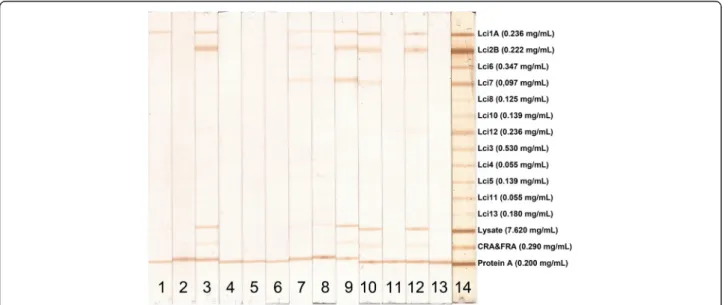
Antigens were sprayed onto a 0.45-μm, pore-size nitrocel-lulose membrane (HiFlow Plus HFB24004, Millipore, MA) in parallel bands via use of a semi-automatic air-brush printing device (CAMAG automatic TLC sample 4, CAMAG, Muttenz, Switzerland) with a volume of 5 μL/ mm. As described by Lyashchenko and collaborators [20], each antigen solution was printed in 15 cm length lines using the concentration of antigen according to solubility in phosphate-buffered saline (PBS): Lci1 = 0.236 mg/mL, Lci2 = 0.222 mg/mL, Lci3 = 0.530 mg/mL, Lci4 = 0.055 mg/mL, Lci5 = 0.139 mg/mL, Lci6 = 0.347 mg/mL, Lci7 = 0.097 mg/mL, Lci8 = 0.125 mg/mL, Lci10 = 0.139 mg/mL, Lci11 = 0.055 mg/mL, Lci12 = 0.236 mg/mL, Lci13 = 0.180 mg/mL. Three additional lines were saturated with
L. majorlysate = 0.7620 mg/mL, recombinant CRA&FRA
T. cruziproteins = 0.290 mg/mL, and a protein A solution = 0.200 mg/mL. The printed nitrocellulose membranes were dried in ambient air and cut into 5-mm strips.
Serum incubation and antibody detection
Before incubation with test sera, strips were blocked for 1 h in 800 μl of PBS with 0.3% Tween 20 (Calbiochem, La Jolla, CA) and 5% instant nonfat dry milk at room temperature while rocking. Then, the strips were incu-bated with 1:100 dilution of each serum for 30 min at room temperature while rocking. After being triple-washed with PBS-Tween under agitation for 5 min at 37°C, the strips were incubated with 1 mL of goat anti-dog IgG antibodies conjugated with peroxidase (1:150 dilution), at room temperature for 1 h, and then washed twice. Enzyme activity was visualized by incubating the strips for 5 min with 1 mL of substrate-chromogenic so-lution (5 mg of DAB in 19,995 μL of PBS, with 5μL of hydrogen peroxidase). To stop the reaction, the strips were rinsed extensively in distilled water at room temperature. The strips were then dried and immedi-ately stored in dark conditions until the results were visually read and digitalized.
Data analysis
Ethical approval for animal use
All experiments involving animals were performed in compliance with Brazilian federal law for animal experi-mentation (Law 11794). In conformity with the Oswaldo Cruz Foundation (FIOCRUZ) animal experimentation guidelines, and according to instructions outlined in the Brazilian Ministry of Health’s manual for the surveillance and control of VL. The present study was approved by the Institutional Review Board (CEUA protocol no. 015/ 2009) of the Gonçalo Moniz Research Center in Bahia, Brazil (CPqGM-FIOCRUZ/BA).
Finding
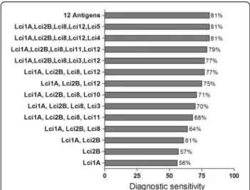
All MAPIA procedures were optimized with regard to antigen concentrations and serum dilution (data not shown). A total of 138 sera from clinically symptomatic (n = 41) and asymptomatic (n = 16)L. infantum-infected dogs, healthy controls (n = 40) and animals harboring other infections (n = 40) were tested against the selected panel of 12 antigens. The results showed variable antigen recognition patterns among the evaluated serum sam-ples, as indicated in Figure 1. As shown in Table 1, the individual sensitivities of the 12 recombinant proteins coated onto nitrocellulose membranes ranged from 4 to 58% for identifying parasite-positive dogs. Nonetheless, each of the antigens detected some positive sera that others missed. When the individual recombinant pro-teins were combined, the total sensitivity increased to
81% (Figure 2), revealing that the antigens complemen-ted each other. The well-known heterogeneous humoral immune response that develops in L. infantum-infected dogs [24] likely involves multiple antigens that are differ-entially recognized by the serum of each animal depend-ing on the state of disease [8]. Therefore, further research into the development of a more reliable rapid test based on the combination of multiple antigens in a DPP format should be pursued.
Efforts to develop a diagnostic test based on antibody-detection for VL or CVL have been underway for de-cades although few of such tests are chromatographic immunoassays currently used in endemic countries [12,13,25]. Prior studies have demonstrated that anti-bodies against these antigens are mainly detectable in cases with advanced stages of disease, while they are much less detectable in sera from asymptomatic cases [12,13]. Variable multi-antigen recognition by canine serum provides an explanation for the variable perform-ance of commercial tests for detecting infected dogs. Here, we found the recombinant antigens evaluated were more sensitive in symptomatic cases (89%) in compari-son to dogs without signs of VL (56%), thus confirming that these antigens are differentially recognized at differ-ent stages of infection. Using MAPIA, the Lci2B and Lci1A antigens gave the best results in terms of sensitiv-ity (76% and 71%, respectively) for the detection of dogs with active disease. Although individually, none of these
antigens performed well in detecting asymptomatic dogs (sensitivity ranging from 6 to 38%), 46 out of 57 serum samples recognized two groups of combined antigens (namely, rLci1A, rLci2B, rLci8, rLci12 and rLci4 or rLci1A, rLci2B, rLci8, rLci12 and rLci5), thus improving the overall sensitivity of the test to 81%. This result is in agreement with previously published results suggest-ing that more than one recombinant antigen may be useful to maximize the sensitivity of diagnostic tests for CVL [22,26].
As shown in Table 1, the individual antigens displayed variable specificities (ranging from 86% to 99%). The rLci1A antigen had a specificity of 90% and the rLci2B
antigen had a specificity of 91%. These results are in ac-cordance with previous study, which reported specificity values of 92% for rLci1A and 95% for rLci2B [27]. In addition, rLci8, which composes the groups of combined antigens, also showed a very high specificity value (99%). By contrast, the other three antigens, rLci4, rLci12 and rLci5, included in at least one of the two groups of com-bined antigens, showed lower specificity values of, re-spectively, 86, 88 and 89%, reducing overall specificity value to 74% (Table 1).
MAPIA has previously proven to facilitate rapid screen-ing of antigens, since in comparison to ELISA [20], it is an easy and rapid test that simultaneously detects the re-sponse of sera to multiple antigens coated onto a single nitrocellulose strip. Indeed, the serological performance of antigens in MAPIA shown to be a good predictor of their performance in several point-of-care assays [14,15,28]. Here, we reveal MAPIA to be a useful tool forL. infantum
antigen selection for the future development of an immu-nodiagnostic test for CVL. This screening test allowed the selection of two sets of antigens (rLci1A, rLci2B, rLci8, rLci12 and rLci4 or rLci1A, rLci2B, rLci8, rLci12 and rLci5) offering both high sensitivity and specificity; our re-sults illustrate the benefit of utilizing a more effective multi-antigen point-of-care test in DPP format for applica-tion in mass assessment surveys of L. infantum-exposed dogs.
Competing interests
The authors declare that they have no competing interests.
Authors’contributions
Conceived and designed the experiments: EDS, GG Jr, DBMF, PSTV. Performed the experiments: IQO, RAS, MVS. Analyzed the data: GG Jr, DBMF, PSTV. Contributed reagents/materials/analysis tools: ABR. All authors read and approved the final version of the manuscript.
Table 1 Sensitivity and specificity of MAPIA with recombinant antigens ofLeishmania infantumfor the serodiagnosis
of canine visceral leishmaniasis
Study groups (n) Reactivity with individualL. infantumantigens (number of positive sera)
Lci1A Lci2B Lci3 Lci4 Lci5 Lci6 Lci7 Lci8 Lci10 Lci11 Lci12 Lci13 Any antigen
Dogs without signs of VL (16) 3 2 4 6 4 1 2 1 4 2 6 1 9
Dogs with signs of VL (41) 29 31 8 15 15 1 9 4 3 8 13 3 37
Total (57) 32 33 12 21 19 2 11 5 7 10 19 4 46
% Sensitivity 56 58 21 37 33 4 19 9 12 18 33 7 81
Normal control (40) 0 1 2 2 2 1 3 1 2 2 4 3 6
Dermal leishmaniasis* (10) 6 3 0 2 2 0 1 0 0 0 0 0 6
Trypanosomiasis* (10) 1 1 1 1 1 0 1 0 1 0 3 0 3
Babesiosis* (10) 1 2 3 3 3 0 1 0 3 1 2 2 4
Ehrlichiosis* (11) 0 0 0 1 1 0 1 0 1 0 1 1 2
Total (81) 8 7 7 11 9 1 7 1 7 3 10 6 21
% Specificity 90 91 91 86 89 99 91 99 91 96 88 93 74
*Sera of dogs affected with other infections.
Figure 2Increased diagnostic sensitivity by combining the individual recombinant antigens for detecting clinically symptomatic and asymptomaticLeishmania infantum
Acknowledgments
This work was supported by INCT-DT, FAPESB, and FIOCRUZ. The authors would like to thank Dr. Flávia W. Cruz McBride for support to obtain negative control samples in Pelotas. We are also grateful to Andris K. Walter for providing English revision and consulting services. In addition, this manuscript has been edited by native English-speaking experts fromBioMed Proofreading LLC.
Author details
1Laboratório de Patologia e Biointervenção, Centro de Pesquisas Gonçalo Moniz, FIOCRUZ, Rua Waldemar Falcão, 121 (Candeal), Salvador, BA, Brazil. 2
Laboratório de Tecnologia Diagnóstica, Instituto de Tecnologia em Imunobiológicos, Bio-Manguinhos, FIOCRUZ, Rio de Janeiro, RJ, Brazil. 3Instituto de Ciência e Tecnologia de Doenças Tropicais, INCT-DT, Salvador, BA, Brazil.4Laboratório de Imunopatologia, Núcleo de Pesquisas em Ciências Biológicas, Universidade Federal de Ouro Preto, Ouro Preto, MG, Brazil. 5Departamento de Medicina Veterinária Preventiva e Produção Animal, Escola de Medicina Veterinária e Zootecnia, Universidade Federal da Bahia, Salvador, BA, Brazil.
Received: 8 September 2014 Accepted: 11 January 2015
References
1. Alvar J, Velez ID, Bern C, Herrero M, Desjeux P, Cano J, et al. Leishmaniasis worldwide and global estimates of its incidence. PLoS One. 2012;7(5):e35671. 2. Quinnell RJ, Courtenay O. Transmission, reservoir hosts and control of
zoonotic visceral leishmaniasis. Parasitology. 2009;136(14):1915–34. 3. Ashford DA, David JR, Freire M, David R, Sherlock I, Eulalio MC, et al. Studies
on control of visceral leishmaniasis: impact of dog control on canine and human visceral leishmaniasis in Jacobina, Bahia. Brazil Am J Trop Med Hyg. 1998;59(1):53–7.
4. Moreira Jr ED, Mendes de Souza VM, Sreenivasan M, Nascimento EG, Pontes de Carvalho L. Assessment of an optimized dog-culling program in the dynamics of canineLeishmaniatransmission. Vet Parasitol. 2004;122(4):245–52. 5. Aisa MJ, Castillejo S, Gallego M, Fisa R, Riera MC, de Colmenares M, et al.
Diagnostic potential of Western blot analysis of sera from dogs with leishmaniasis in endemic areas and significance of the pattern. Am J Trop Med Hyg. 1998;58(2):154–9.
6. Alvar J, Molina R, San Andres M, Tesouro M, Nieto J, Vitutia M, et al. Canine leishmaniasis: clinical, parasitological and entomological follow-up after chemotherapy. Ann Trop Med Parasitol. 1994;88(4):371–8.
7. Falqueto A, Ferreira AL, dos Santos CB, Porrozzi R, da Costa MV, Teva A, et al. Cross-sectional and longitudinal epidemiologic surveys of human and canineLeishmania infantumvisceral infections in an endemic rural area of southeast Brazil (Pancas, Espirito Santo). Am J Trop Med Hyg. 2009;80(4):559–65. 8. Quinnell RJ, Courtenay O, Davidson S, Garcez L, Lambson B, Ramos P, et al.
Detection ofLeishmania infantumby PCR, serology and cellular immune response in a cohort study of Brazilian dogs. Parasitology. 2001;122(Pt 3):253–61. 9. Reithinger R, Quinnell RJ, Alexander B, Davies CR. Rapid detection of
Leishmania infantuminfection in dogs: comparative study using an immunochromatographic dipstick test, enzyme-linked immunosorbent assay, and PCR. J Clin Microbiol. 2002;40(7):2352–6.
10. Dye C, Vidor E, Dereure J. Serological diagnosis of leishmaniasis: on detecting infection as well as disease. Epidemiol Infect. 1993;110(3):647–56. 11. Lombardo G, Pennisi MG, Lupo T, Chicharro C, Solano-Gallego L. Papular
dermatitis due toLeishmania infantuminfection in seventeen dogs: diagnostic features, extent of the infection and treatment outcome. Parasit Vectors. 2014;7:120.
12. da Costa RT, Franca JC, Mayrink W, Nascimento E, Genaro O, Campos-Neto A. Standardization of a rapid immunochromatographic test with the recombinant antigens K39 and K26 for the diagnosis of canine visceral leishmaniasis. Trans R Soc Trop Med Hyg. 2003;97(6):678–82.
13. Grimaldi Jr G, Teva A, Ferreira AL, dos Santos CB, Pinto I, De-Azevedo CT, et al. Evaluation of a novel chromatographic immunoassay based on Dual-Path Platform technology (DPP(R) CVL rapid test) for the serodiagnosis of canine visceral leishmaniasis. Trans R Soc Trop Med Hyg. 2012;106(1):54–9. 14. Lyashchenko KP, Greenwald R, Esfandiari J, Chambers MA, Vicente J,
Gortazar C, et al. Animal-side serologic assay for rapid detection of
Mycobacterium bovisinfection in multiple species of free-ranging wildlife. Vet Microbiol. 2008;132(3–4):283–92.
15. Lyashchenko KP, Greenwald R, Esfandiari J, Meylan M, Burri IH, Zanolari P. Antibody responses in New World camelids with tuberculosis caused by
Mycobacterium microti. Vet Microbiol. 2007;125(3–4):265–73. 16. Boarino A, Scalone A, Gradoni L, Ferroglio E, Vitale F, Zanatta R, et al.
Development of recombinant chimeric antigen expressing
immunodominant B epitopes ofLeishmania infantumfor serodiagnosis of visceral leishmaniasis. Clin Diagn Lab Immunol. 2005;12(5):647–53. 17. Costa MM, Penido M, dos Santos MS, Doro D, de Freitas E, Michalick MS,
et al. Improved canine and human visceral leishmaniasis immunodiagnosis using combinations of synthetic peptides in enzyme-linked immunosorbent assay. PLoS Negl Trop Dis. 2012;6(5):e1622.
18. Passos S, Carvalho LP, Orge G, Jeronimo SM, Bezerra G, Soto M, et al. RecombinantLeishmaniaantigens for serodiagnosis of visceral leishmaniasis. Clin Diagn Lab Immunol. 2005;12(10):1164–7.
19. Rosati S, Ortoffi M, Profiti M, Mannelli A, Mignone W, Bollo E, et al. Prokaryotic expression and antigenic characterization of three recombinant
Leishmaniaantigens for serological diagnosis of canine leishmaniasis. Clin Diagn Lab Immunol. 2003;10(6):1153–6.
20. Lyashchenko KP, Singh M, Colangeli R, Gennaro ML. A multi-antigen print immunoassay for the development of serological diagnosis of infectious diseases. J Immunol Methods. 2000;242(1–2):91–100.
21. Oliveira GG, Magalhaes FB, Teixeira MC, Pereira AM, Pinheiro CG, Santos LR, et al. Characterization of novelLeishmania infantumrecombinant proteins encoded by genes from five families with distinct capacities for serodiagnosis of canine and human visceral leishmaniasis. Am J Trop Med Hyg. 2011;85(6):1025–34.
22. Teixeira MC, Oliveira GG, Silvany MA, Alcantara-Neves NM, Soares MB, Ribeiro-Dos-Santos R, et al. A strategy for identifying serodiagnostically relevant antigens ofLeishmaniaor other pathogens in genetic libraries. Biologicals. 2007;35(1):51–4.
23. Solca Mda S, Guedes CE, Nascimento EG, Oliveira GG, dos Santos WL, Fraga DB, et al. Qualitative and quantitative polymerase chain reaction (PCR) for detection ofLeishmaniain spleen samples from naturally infected dogs. Vet Parasitol. 2012;184(2–4):133–40.
24. Solano-Gallego L, Riera C, Roura X, Iniesta L, Gallego M, Valladares JE, et al.
Leishmania infantum-specific IgG, IgG1 and IgG2 antibody responses in healthy and ill dogs from endemic areas. Evolution in the course of infection and after treatment. Vet Parasitol. 2001;96(4):265–76.
25. Mettler M, Grimm F, Capelli G, Camp H, Deplazes P. Evaluation of enzyme-linked immunosorbent assays, an immunofluorescent-antibody test, and two rapid tests (immunochromatographic-dipstick and gel tests) for serological diagnosis of symptomatic and asymptomaticLeishmaniainfections in dogs. J Clin Microbiol. 2005;43(11):5515–9.
26. Lyashchenko KP, Greenwald R, Esfandiari J, Greenwald D, Nacy CA, Gibson S, et al. PrimaTB STAT-PAK assay, a novel, rapid lateral-flow test for tuberculosis in nonhuman primates. Clin Vaccine Immunol. 2007;14(9):1158–64. 27. de Souza CM, Silva ED, Ano Bom AP, Bastos RC, Nascimento HJ, da Silva
Junior JG. Evaluation of an ELISA for canine leishmaniasis immunodiagnostic using recombinant proteins. Parasite Immunol. 2012;34(1):1–7.
28. Buddle BM, Wilson T, Denis M, Greenwald R, Esfandiari J, Lyashchenko KP, et al. Sensitivity, specificity, and confounding factors of novel serological tests used for the rapid diagnosis of bovine tuberculosis in farmed red deer (Cervus elaphus). Clin Vaccine Immunol. 2010;17(4):626–30.
Submit your next manuscript to BioMed Central and take full advantage of:
• Convenient online submission
• Thorough peer review
• No space constraints or color figure charges
• Immediate publication on acceptance
• Inclusion in PubMed, CAS, Scopus and Google Scholar
• Research which is freely available for redistribution