O r i g i n a l A r t i c l e
Acute kidney injury after snakebite accident treated in a Brazilian
tertiary care centre
POLIANNA L. M. M. ALBUQUERQUE,1,2GERALDO B. SILVA JUNIOR,3CAMILLA N. JACINTO,2JULIANNA B. LIMA,2 CAROLINE B. LIMA,2YAGO S. AMARAL,2MARIA DO SOCORRO B. VERAS,1ROSA M. S. MOTA4and
ELIZABETH F. DAHER2
1Toxicological Assistance Center, Instituto Dr. José Frota,2Post-Graduation Program in Medical Sciences, Department of Internal Medicine, School of Medicine, 4Department of Statistics, Sciences Center, Federal University of Ceará, and3Post-Graduation Program in Collective Health, Health Sciences Center, School of Medicine, University of Fortaleza, Fortaleza, Ceará, Brazil
KEY WORDS:
acute kidney injury, complications, ophidic accident, risk factors, snakebite.
Correspondence:
Dr Elizabeth F. Daher, Rua Vicente Linhares, 1198, CEP: 60135-270, Fortaleza, Ceará, Brazil. Email: ef.daher@uol.com.br
Accepted for publication 11 August 2014. Accepted manuscript online 14 August 2014.
doi:10.1111/nep.12327
SUMMARY AT A GLANCE
The manuscript describes the
epidemiology and clinical features of AKI among victims of snake bite in Brazil. Importantly, a significant proportion did not show full renal functional recovery.
ABSTRACT:
Aim: Acute kidney injury (AKI) is one of the main causes of morbidity and
mortality in cases of envenomation by venomous snakes. The present study was carried out to investigate the clinical and laboratory manifestations in accidents with venomous snakes and the risk factors associated with AKI in these accidents.
Methods: A retrospective study was carried out with patients victims of
snakebite admitted to a reference centre. AKI was defined according to the RIFLE and AKIN criteria.
Results: A total of 276 patients were included, of which 230 (83.7%) were
males. AKI was observed in 42 cases (15.2%). The mean genus involved in the accidents was Bothrops (82.2%). Mean age of patients with AKI was higher than in patients without AKI (43±20 vs. 34±21 years,P= 0.015). The time elapsed between the accident and medical care was higher in the AKI group (25±28 vs. 14±16h,P= 0.034), as well as the time elapsed between the accident and the administration of antivenom (30.7±27 vs. 15±16 h, P= 0.01). Haemodialysis was required in 30% of cases and complete renal function recovery was observed in 54.8% of cases at hospital discharge. There were four deaths, none of which had AKI. Factors associated with AKI were haemorrhagic abnormalities (P= 0.036, OR = 6.718, 95% CI: 1.067– 25.661) and longer length of hospital stay (P= 0.004, OR = 1.69, 95% CI 1.165– 2.088).
Conclusion: Acute kidney injury is an important complication of snakebite accidents, showing low mortality, but high morbidity, which can lead to partial renal function recovery.
Envenomation by venomous snakes belongs to the group of main neglected tropical diseases1and constitutes an
impor-tant public health problem. It has a worldwide distribution, affecting mainly rural populations in Asia, Africa, Latin America and Oceania.1 In Latin America, there are four
groups of clinically relevant poisonous snakes: Bothrops,
Crotalus,LachesisandMicrurus.2
In Brazil, there has been an increase in the number of envenomation accidents caused by poisonous animals reported from 1986 to mid-2012, according to data from the Ministry of Health (http://portal.saude.gov.br/portal/ arquivos/pdf/clipping30072010.pdf). The main venomous
snakes involved in this type of accident in Brazil are similar to those observed in Latin America. Acute kidney injury (AKI) is a result of the accidents with the genusBothropsand
Crotalus.2
Snakebite-associated AKI in tropical countries and its impact on morbidity and mortality has been the subject of some studies.2,3The clinical picture resulting from snakebites
varies according to the involved species and the amount of inoculated venom. Systemic manifestations consist mainly of renal,3haemorrhagic (haematuria, haematemesis, brain and
cavity haemorrhages) and neurological alterations, common in Crotalus accidents (paresthesia, paralysis with craniocaudal
progression, starting with ptosis and ophthalmoplegia, which may develop into paralysis of the respiratory muscles resulting in acute respiratory failure).4
The present study was carried out to investigate the char-acteristics of AKI associated with venomous snakebites and the involved risk factors.
METHODS
Patients
This is a retrospective study carried out in a tertiary hospital in the city of Fortaleza, state of Ceará, Brazil.
We included all the patients treated at Instituto Dr. José Frota from January 2003 to December 2012, with a history of poisonous snakebite. All the patients included in the study were hospitalized due to the accident.
The victims of non-poisonous (n=243) or non-identified snake-bites (n=628) were excluded. One patient that was diagnosed with heart failure (clinical syndrome that result in any structural or func-tional impairment of ventricular filling or ejection of blood – ejection fraction≤40%, according to the Guidelines of the American Heart Association),5 was excluded. This patient, victim of a venomous
snakebite, had heart failure, but not AKI.
The snakes were identified by the patients themselves. In some cases, the patients brought the snake to the hospital. The physician assessed the site of the bite and classified the snake through anam-nesis and laboratory exams were requested.
A semistructured form was used for data collection from each patient, containing epidemiological, clinical and laboratory information.
The study protocol was reviewed and approved by the Committee of Ethics from Instituto Jose Frota, in Fortaleza, Brazil (protocol 119/12).
Definitions
Acute kidney injury was defined according to the RIFLE and AKIN classifications, according to creatinine levels within the first 48 h after hospital admission.6,7Baseline creatinine was the one
meas-ured 48 h before its elevation. Three or more serum creatinine measurements were obtained from each patient studied during this period. A time constraint of 48 h for the diagnosis of AKI was proposed. Creatinine measurements were performed using the modified kinetic Jaffé method, at 500 nm, without deproteinization. Patients were classified according to the worst criteria (creatinine or urine output). Oliguria was considered as urinary volume<400 mL/ day after 24 h of adequate volume replacement.
Renal function recovery was based on the creatinine levels at the time of hospital discharge. Partial recovery of renal function at the time of discharge was considered as creatinine above the basal level (i.e., patients still classified as one of the RIFLE or AKIN stages at the time of hospital discharge).
Study groups
Patients were classified into two groups: patients with normal renal function (Non-AKI) and patients with AKI. When comparing the
two groups, differences in clinical manifestations, laboratory findings and risk factors for AKI were assessed.
Clinical and laboratorial parameters
At physical examination, signs and symptoms were evaluated and the following aspects were recorded: age, gender, snake species, severity of accident, the median time between the accident and the administration of the antivenom, dose of antivenom, length of hos-pital stay and haemorrhagic manifestations. The use of herbal rem-edies was investigated, and their use constituted an exclusion criterion, as some herbs are nephrotoxic and could be responsible for AKI development.
The following laboratory parameters were evaluated: serum creatinine, urea, sodium, potassium, haematocrit, haemoglobin, leu-kocytes, platelets, prothrombin time (PT) and partially activated thromboplastin time (aPTT).
Treatment protocol
Patients were classified according to the severity of the snakebite accident using the Brazilian Ministry of Health criteria, based on local and systemic clinical manifestations established in specific tables for each accident (see appendix). The administration of spe-cific antivenom was performed according to those criteria.
Statistical analysis
The results are expressed in tables and means (mean±SD) for quan-titative variables. Univariate and multivariate analyses of clinical and laboratory data were performed using the SPSS program, release 21.0, 2012 for MacBook (SPSS Inc. Chicago, IL, USA), to investigate the possible risk factors associated with acute kidney injury.
The Kolmogorov-Smirnov test was used to verify the normal distribution of continuous variables. The Levene test was used to compare the variability of the means. In the of normal data distri-bution, the comparison between two means was made by Student’s
t-test. In case of non-normal data, the Mann–Whitney test was applied and the variables were expressed as medians. Analysis of variance (ANOVA) was used to compare means between three or more independent groups, with a post-hoc analysis through Bonferroni’s method. Pearson’sχ2test, likelihood ratio and Fisher’s
exact test were used for association and homogeneity tests in the distribution of categorical data.
Odds ratio was estimated when the association was significant. Confidence intervals of 95% were calculated. The factors included in the multivariate model (logistic regression) through the Backward method, were those factors that showed statistical significance level <20% in the univariate analysis (Mann–Whitney andχ2test).
Sta-tistical significance was set at 5% (P<0.05).
RESULTS
Demographic and clinical characteristics at admission
The AKI group (n=42) had a mean age of 43±20 years (median 48), of which 32 (76.2%) were males and 38 (90.5%) were from the rural area. Of the snake species involved in the accident, 30 (71.4%) were Bothrops, 10 (23.8%)Crotalusand two (4.8%)Micrurus. As for the severity of the accident, nine (21.4%) were mild, 19 (45.2%) mod-erate and 14 (33.3%) severe. The median time between the accident and antivenom administration was 24 h (3–96 h). The median time of hospital stay was 8 days, (2–38 days). The median dose of administered antivenom was 8 vials (0–33 vials) (Table 1).
The non – AKI group (n=198) had a mean age of 34±21 years (median 33), of which 198 (84.6%) were males and 198 (85.3%) were from the rural area. Of the snake species involved in the accident, 197 (84.2%) were Bothrops, 18
(7.7%)Crotalusand 17 (7.3%)Micrurus. As for the severity of the accident, 30 (12.8%) were mild, 154 (65.8%) moderate and 50 (21.4%) severe. The median time between the acci-dent and antivenom administration was 9 h (1–72 h). The median time of hospital stay was 3 days, (1–8 days). The median dose of administered antivenom was 8 vials (0–25 vials) (Table 1).
Distribution of patients according to the RIFLE and AKIN classifications
Acute kidney injury was observed in 42 (15.2%) patients according to the AKIN and 41 (18.9%) patients according to the RIFLE criteria. Patients were classified as AKIN 1 (15; 35.7%), AKIN 2 (3; 7.1%), AKIN 3 (24; 57.1%), R (12; 29.2%), I (5; 12.2%) and F (24; 58.5%). Due to the small number of patients in each group, only two groups were used in the analysis: AKI and non-AKI, determined by serum creatinine, according to the AKIN criteria.
Comparison of laboratory tests and clinical findings
A comparison of clinical characteristics between patients classified as AKI and non-AKI is summarized in Table 2. The groups differed regarding the presence of haemorrhagic alterations (OR 3.016,P=0.001) and lesion site (OR 0.47,
P=0.022). As for PT (OR 0.383,P=0.005) and aPTT (OR 0.478,P=0.042) alterations in the groups, there was statis-tical difference between them. The presence of myalgia showed a tendency to significance, but did not differ between the groups (P=0.053).
Among the patients with haemorrhagic alterations
(n=274), 19 developed concomitant AKI.
The mean values of creatinine and urea at admission and at discharge were higher in the AKI group (Table 3). Haemo-globin (9.4±2.5 vs. 12.7±2.5,P<0.0001) and haematocrit (28.9±9.0 vs. 39.1±11.0; P=0.002) levels and platelet count (95 690±79 200.8 vs. 212 227±90 913;P<0.0001)
were lower in the AKI group. Leukocyte count
(13 568.3±5178.4 vs. 11 135±3011.5; P=0.044) was lower in the non-AKI group (Table 3).
Table 1Demographic characteristics of patients who developed or not acute kidney injury (AKI), victims of accidents with venomous snakes
AKI (n= 42) Non-AKI (n= 234) P
Gender
Male 32 (76.2%) 198 (84.6%) 0.182
Female 10 (23.8%) 36 (15.4%)
Age (years)† 43±20 34±21 0.015
Area
Rural 38 (90.5%) 198 (85.3%) 0.47
Urban 4 (9.5%) 34 (14.7%)
Snake species
Bothrops 30 (71.4%) 197 (84.2%) 0.015
Crotalus 10 (23.8%) 18 (7.7%)
Micrurus 2 (4.8%) 17 (7.3%)
Lachesis – 2 (0.9%)
Severity of accident
Mild 9 (21.4%) 30 (12.8%)
Moderate 19 (45.2%) 154 (65.8%) 0.741
Severe 14 (33.3%) 50 (21.4%)
Time between the accident and antivenom administration (hours)‡
24.0 (3–96) 9.0 (1–72) 0.01
Length of hospital stay (days)‡ 8.0 (2–38) 3.0 (1–8) <0.001
Dose of antivenom (vials)‡ 8 (0–33) 8 (0–25) 0.196
†Values expressed as mean±standard deviation. ‡Values expressed as median (minimum – maximum). Significant Mann–Whitney test:P<0.05.
Table 2Clinical aspects of acute kidney injury (AKI) and non-AKI groups in victims of accidents with venomous snakes
AKI (n= 42) Non-AKI (n= 234) P OR 95%CI
LL UL
Nervous system alteration 12 (28.6%) 38 (16.4%) 0.06 2.042 0.960 4.34
Haemorrhagic manifestation 22 (52.4%) 62 (26.7%) 0.001 3.016 1.541 5.905
Local lesion 24 (57.1%) 172 (74.5%) 0.022 0.457 0.232 0.902
Palpebral ptosis or diplopia 7 (16.7%) 18 (7.8%) 0.065 2.378 0.926 6.107
Myalgia 8 (19%) 21 (9.1%) 0.053 2.364 0.970 5.764
Altered PT 9 (21.4%) 88 (37.9%) 0.005 0.383 0.187 0.784
Altered aPTT 8 (19%) 71 (30.3%) 0.042 0.478 0.220 1.037
Clinical outcome
Among the patients who developed AKI, 25 (64.1%) achieved renal function recovery, 12 (30.8%) had partial recovery, no patient died and two (5.1%) were transferred to another reference hospital. Of the patients who did not have AKI (non-AKI) four (1.7%) died, and two (0.9%) were transferred to another hospital.
The causes of death were gastrointestinal bleeding (1), CNS haemorrhage (1), anaphylaxis to the antivenom (1) and haemorrhagic diastasis (1). Of the patients who developed AKI, 13 (30.6%) underwent haemodialysis.
Risk factors for AKI
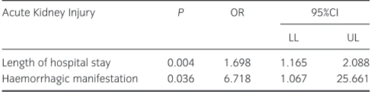
Independent factors associated with AKI were the presence of haemorrhagic alterations on admission (OR=6.718, 95% CI=1.067 to 25.661,P=0.036) and a longer hospital length of stay (OR=1.698, 95% CI=1.165–2.088, P=0.004), as shown in Table 4.
DISCUSSION
The present study showed important clinical and laboratory aspects of snakebite-associated AKI. As the kidney is a highly vascularized organ, it is very susceptible to toxins.8AKI is an
important complication of envenomation accidents, being one of the main causes of mortality.9
The predominance of the male gender was observed in the group that developed AKI (76.2%), as well as in the group who did not develop it (84.6%), with no difference between these groups regarding gender. Many studies in the literature have demonstrated the predominance of males in snakebite
accidents.10–14However, studies have shown that the
distribu-tion of the male gender was not different in the groups that did or did not develop AKI.15–17Feitosaet al.18has described the
predominance of males in the state of Ceará, Brazil (over 70%). Borgeset al.,19 in a study carried out in the Amazon
region, also showed a predominance of males (81.3%). Patients who developed AKI in the study were older, with a significant statistical difference between the groups. Some studies have shown a prevalence of AKI in older patients.16,17
Pinho et al.15 showed a predominance of AKI in younger
patients, as they concentrate larger amounts of venom in a lower body surface area. On the other hand, older patients would have less viable glomerular mass, which could make them more susceptible to toxins.
The predominant area of origin in both AKI (90.5%) and non-AKI (85.3%) groups was the rural area, with no statis-tical difference between the groups, corroborated by the fact that the accident is associated with people’s activities in the countryside. Akaniet al.,20in their study, highlighted a strong
correlation between human activity type (rural or urban) and the occurrence of snakebite accidents.
In our study, the snake species distribution in the AKI and
non-AKI groups showed a predominance of the Bothrops
genus in both groups, consistent with the fact that this is the most prevalent snake species in Brazil, with more than 30 species being found throughout the national territory.8Other
studies have obtained the same finding.13,14,21The second most
prevalent species involved in snakebite accidents in this study is theCrotalus. It is important to note the strong association between theCrotalusspecies and the development of AKI. AKI is more commonly seen in Crotalus than inBothropsaccidents, as the crotalic venom is more nephrotoxic.22
As for the severity of the accidents, most were classified as being of moderate severity followed by severe, as observed in both groups (AKI and non-AKI), with no statistical difference. According to Amaralet al.4most snakebite accidents reported
are mild ones, which corroborates the findings of Limaet al.,12
Oliveiraet al.,14and Lemoset al.21Miseet al.11described the
prevalence of moderate cases followed by severe cases, data that are consistent with the findings in our study.
Many studies have shown a tendency to the development of AKI in patients that waited longer between the snakebite
Table 3Comparison of laboratory findings between the groups of patients that developed acute kidney injury (AKI) and those who did not develop it (Non-AKI)
AKI (n= 42) Non-AKI (n= 274) P
Sodium (mEq/L)† 137.4 (±5.2) 140 (±5.7) 0.06
Potassium (mEq/L)† 4.5 (±0.9) 4.0 (±0.4) 0.17 Creatinine at AKI
diagnosis (mg/dL)†
3.07 (±2.7) 0.94 (±0.38) <0.0001
Creatinine at discharge (mg/dL)†
3.0 (±2.9) 0.9 (±0.35) <0.0001
Urea at AKI diagnosis (mg/dL)†
107.1 (±74.1) 37.6 (±27.5) <0.0001
Urea at discharge (mg/dL)†
72.9 (±50.72) 32.4 (±16.3) <0.0001
Haemoglobin (g/dL)‡ 9.42 (±2.46) 12.7 (±2.52) <0.0001 Haematocrit (%)‡ 28.86 (±9.0) 39.1 (±11.0) 0.002 Leukocyte count
(/mm3)‡
13568.3 (±5178.4) 11135 (±3011.45) 0.044
Platelet count (/mm3)‡
95690.0 (±79200.8) 212227 (±90913) <0.0001
†Mann–Whitney. ‡Student’st-test. Significant:P<0.05.
Table 4 Factors associated with acute kidney injury (AKI) in 276 patients, victims of accidents with venomous snakes
Acute Kidney Injury P OR 95%CI
LL UL
Length of hospital stay 0.004 1.698 1.165 2.088
Haemorrhagic manifestation 0.036 6.718 1.067 25.661
and antivenom administration.15–17 In our study, the
multi-variate analysis showed no significant difference between AKI and non-AKI groups regarding the time elapsed between the bite and antivenom administration, which is in conflict with data from some studies that defined longer time between the bite and antivenom administration as an inde-pendent risk factor for the development of AKI.15,16As this is
a retrospective study, there may have been variables that were not informed, which could have influenced the analysis of this parameter (time elapsed between the snakebite and antivenom administration). The small sample size may have interfered with the results.
Regarding the length of hospital stay, AKI and non-AKI groups differed significantly, which was consistent with the literature.15–17 There was an association between length of
stay and the development of AKI, which may represent major clinical complications for the patient, leading to longer hospital stays.
The presence of haemorrhagic disorders, even without coagulation alterations, was an important finding in this study. Bleeding is one of the most important effects induced by snakebites. Damage to the microvasculature is a conse-quence of metalloproteinase action, which leads to disten-sion, oedema and rupture of capillary walls.23Santoroet al.24
showed the presence of a platelet-inhibitory factor found in the plasma of rabbits injected with Bothrops venom and described the presence of fibrin thrombi in the histological analysis of lungs and kidneys of these rabbits, suggesting that renal abnormalities can occur even without coagulation alterations (PT and aPTT).
The association between haemorrhagic manifestations and AKI development may represent a typical feature of the venom of poisonous snakes found in north-eastern Brazil, mainly represented in the study by the Bothrops
species. However, the association between haemorrhagic manifestations and AKI development should be interpreted with caution, considering the confidence interval was too long.
Fonsekaet al.25demonstrated that haemorrhagic
manifes-tations secondary to snakebites can be severe. Silva et al.26
reported the presence of petechiae and severe congestion in the lungs, kidneys and gastrointestinal tract in victims of snakebite accidents. Maduwage et al.27 reported cases of
fatal snakebite accidents that developed consumptive coagulopathy and AKI. Moriarity et al.,28 based on a
retro-spective study, concluded that coagulation tests should be requested for all patients, as it would be impossible to iden-tify which groups would be more susceptible to bleeding. The importance of haemorrhagic manifestations as a risk factor for the development of AKI can indicate a particular physiopathological aspect of the snakes found in north-eastern Brazil.
In the present study, laboratory parameters showed higher levels of urea and creatinine in patients with AKI, when compared to the non-AKI group. These results were
expected, according to the use of AKIN and RIFLE classifica-tions. The haemoglobin and haematocrit levels in the group that developed AKI were lower, with a statistically significant difference (P<0.0001, P=0.002). This fact may represent haemorrhagic alterations secondary to Bothrops poisoning, resulting from the action of zinc-containing metallo-proteinases, which produce lesions in the basal capillary membrane, associated with thrombocytopenia and coagula-tion disorders.4 The haemoglobin and haematocrit levels
were not included in the multivariate analysis, as several cases’ records lacked this information.
Athappan et al.16 described the presence of intravascular
haemolysis as an independent risk factor for the
develop-ment of AKI (OR=3.2, P=0.01), which justifies the
decrease in platelet and haemoglobin levels in our sample. Platelets can also be reduced due to the coagulation system activation and intravascular fibrin formation after factor X activation by the bothropic venom.29
The study mortality was 1.4% in snakebite accidents with poisonous snakes, which was considered low, consistent with data in the national literature,4,12,14,21showing the low
lethal-ity of snakebites. Most patients were cured; however, 13 patients persisted with renal function deficit until hospital discharge.
In summary, AKI was an important complication of snake-bite accidents in our study. Morbidity can be high, leading to persistent kidney dysfunction. A longer duration of hospital stay and the presence of haemorrhagic alterations were inde-pendent risk factors for the development of AKI.
As the present is a retrospective study, one of its limita-tions was the lack of information in the medical records, which prevented some statistical inferences. Prospective studies may further elucidate the factors that lead to snakebite-associated AKI. At admission, some patients showed higher levels of creatinine with no variation during hospitalization. This fact should be seen as a limitation/bias in the diagnosis of AKI (these cases were excluded from the analysis, as they did not meet the RIFLE or AKIN criteria). This is a single-centre study and it is necessary to perform similar investigations in other centres to better analyze the results and to assess the impact of these accidents in our country.
REFERENCES
1. Gutiérrez JM, Theakston DG, Warrell DA. Confronting the neglected problem of snakebite envenoming: The need for a global partnership.PLoS Med.2006;3: e150.
2. Pinho FMO, Yu L, Burdmann EA. Snakebite-induced kidney injure in Latin America.Semin. Nephrol.2008;28: 354–62.
3. Amaral CFS, Rezende NA, Silva OAet al. Insuficiência renal aguda secundária a acidentes ofídicos botrópico e crotálico: Análise de 63 casos.Rev Inst Med Trop São Paulo1986;28: 220–7.
4. Amaral CFS, Bucaretchi F, Araújo FAAet al.Manual de diagnóstico e tratamento de acidentes por animais peçonhentos, 2aedição. Brasília:
5. Yancy CW, Jessup M, Bozkurt Bet al. Guideline for the
management of heart failure: Executive summary.Circulation2013; 128: 1810–52.
6. Bellomo R, Ronco C, Kellum JA, Mehta RL, Palevsky P. Acute renal failure – Definition, outcome measures, animal models, fluid therapy and information technology needs: The Second
International Consensus Conference of Acute Dialysis Quality Initiative (ADQI) Group.Crit. Care2004;8: R204–R212.
7. Mehta RL, Kellum JA, Shah SVet al. Acute kidney injury network (AKIN): Report of an initiative to improve outcomes in acute kidney injury.Crit. Care2007;11: R31.
8. Sitprija V, Sitprija S. Renal effects and injury induced by animal toxins.Toxicon2012;60: 943–53.
9. Chugh KS. Snake bite induced acute renal failure in India.Kidney Int.1989;35: 891–907.
10. Bochner R, Struchiner CJ. Epidemiologia dos acidentes ofídicos nos últimos 100 anos no Brasil: uma revisão.Cad Saúde Pública
2003;19: 7–16.
11. Mise Y, Lira-da-Silva M, Carvalho FM. Envenenamento por serpentes do gênero Bothrops no estado da Bahia: aspectos epidemiológicos e clínicos.Rev. Soc. Bras. Med. Trop.2007;40: 563–73.
12. Lima JS, Martelli Júnior H, Martelli DRet al. Perfil dos acidentes ofídicos no norte do Estado de Minas Gerais, Brasil.Rev. Soc. Bras. Med. Trop.2009;42: 561–4.
13. Lima AC, Campos CE, Ribeiro JR. Perfil epidemiológico de acidentes ofídicos do Estado do Amapá.Rev. Soc. Bras. Med. Trop.
2009;42: 329–35.
14. Oliveira FN, Brito MT, Morais ICOet al. Accidents caused by Bothrops and Bothropoides in the state of Paraíba: Epidemiological and clinical aspects.Rev. Soc. Bras. Med. Trop.2010;43: 662–7. 15. Pinho FMO, Zanetta DM, Burdmann EA. Acute renal failure after
Crotalus durissus snakebite: A prospective survey on 100 patients.
Kidney Int.2005;67: 659–67.
16. Athapan MG, Balaji UV, Navaneethan PT. Acute renal failure in snake envenomation: A large prospective study.Saudi J. Kidney Dis. Transpl.2008;19: 404–10.
17. Harshavardhan L, Lokesh AJ, Tejeshwari HL, Halesha BR, Siddharama SM. A study on the acute kidney injury in snake bite victims in a tertiary care centre.J. Clin. Diagn. Res.2013;7: 853–6.
18. Feitosa R, Melo I, Monteiro H. Epidemiologia dos acidentes por serpentes peçonhentas no estado do Ceará – Brasil.Rev. Soc. Bras. Med. Trop.1997;30: 295–301.
19. Borges C, Sadahiro M, Santos M. Aspectos epidemiológicos e clínicos dos acidentes ofídicos ocorridos nos municípios do Estado do Amazonas.Rev. Soc. Bras. Med. Trop.1999;32: 637–46. 20. Akani GC, Ebere N, Eniang EA, Petrozzi F, Politano E, Luiselli L.
Correlation between anual activity patterns of venomous snakes and rural people in the Niger Delta, Southern Nigeria.J. Venom. Anim. Toxins Incl. Trop. Dis.2013;19: 2–8.
21. Lemos J, Almeida T, Fook S, Paiva A, Simões M. Epidemiologia dos acidentes ofídicos notificados pelo Centro de Assistência e Informação Toxicológica de Campina Grande (Ceatox-CG), Paraíba.
Rev. Bras. Epidemiol.2009;12: 50–9.
22. Santos MFL, Farani MC, Roche PN. Acute Kidney Injury in Bothrops sp. and Crotalus sp. envenomation: Critical review of the literature.J. Bras. Nefrol.2009;31: 132–8.
23. Escalante T, Rucavo A, Fox JW, Gutiérrez JMK. Key events in microvascular damage induced by snake venom hemorrhagic metalloproteinases.J. Proteomics2011;74: 1781- 94.
24. Santoro ML, Sano-Martins IS. Platelet dysfunctionBothrops jararaca
snake envenomation in rabbits.Thromb. Haemost.2004;92: 369–83.
25. Fonseka CL, Jeevagan V, Gnanathasan CA. Life threatening intracerebral haemorrhage following saw-scaled viper (Echis carinatus) envenoming–authenticated case report from Sri Lanka.
BMC Emerg. Med.2013;13: 5.
26. Silva A, Gunawardena P, Weilgama D, Maduwage K, Gawarammana I. Comparative in vivo toxicity of venoms from South Asian hump- nosed pit vipers (Viperidae: Crotalinae: Hypnale).BMC Res. Notes2012;5: 471.
27. Maduwage K, Kularatne K, Gawarammana I. Coagulopathy, acute kidney injury and death following Hypnale zara envenoming – The first case report from Sri Lanka.Toxicon2011;58: 641–3. 28. Moriaty RS, Dryer S, Replogle W, Summers R. The role for
coagulation markers in mild snakebite envenomations.West. J. Emerg. Med.2012;13: 68–74.
29. Sgrinolli L, Mendes G, Carlos G, Burdmann E. Acute kidney injury caused by bothrops snake venom.Nephron Clin. Pract.2011;119: 131–7.
APPENDIX
Table A1Bothropic accident: classification regarding severity and recommended antivenom
Manifestations and treatment Classification
Mild Moderate Severe
LOCAL – pain/oedema/ecchymosis Absent or discrete Evident Intense†
SYSTEMIC – severe haemorrhage/shock/anuria Absent Absent Present
Time of coagulation (TC‡) Normal or Altered Normal or Altered Normal or Altered
Antivenom (no. vials) ABS/ABCS/ABLS 2–4 4–8 12
Route of administration Intravenous
Table A2Crotalic accident: classification regarding severity and recommended antivenom
Manifestations and treatment Classification – initial evaluation
Mild Moderate Severe
Myasthenic facies and blurred vision Absent or delayed Discrete or evident Evident
Myalgia Absent or discrete Discrete Intense
Time of coagulation (TC) Normal or altered Normal or altered Normal or altered
Red or brown urine Absent Little evident or absent Present
Oliguria or anuria Absent Absent Present or absent
Antivenom (no. vials) ACS/ABCS 5 10 20
Route of administration Intravenous
ABCS, anti-bothropic-crotalic serum; ACS, anti-crotalic serum. Source: Adapted from the Ministry of Health, 2001.
Table A3Elapidae accident: classification regarding severity and recommended antivenom
Clinical manifestations Antivenom (no. vials) AES Route of administration
Due to the risk of acute respiratory failure, they must be considered severe 10 Intravenous