Case 9341
It seemed a terrible pelvic tumor...
Barbosa L, Cunha TM Hospital dos Covões
Genital (Female) Imaging Section:
2011, Sep. 12 Published:
65 year(s), female Patient:
Authors' Institution
Instituto Português de Oncologia de Lisboa Francisco Gentil E.P.E. - Portugal
Clinical History
A 65-year-old woman, nurse, residing in Bissau, Guinea, came to the emergency department because she was having asthenia, weight loss and diffuse abdominal pain complaints for a month.
Imaging Findings
A CT was performed, revealing an enlargement of the ovaries and uterus in the pelvic cavity with central hypodensity, compatible with lesions of the endometrium, which extended inferiorly to the cervix (2).
The MRI confirmed an increased size of the uterine body, where an endometrial tumour was noted. This tumour showed cervical invasion, but did not extend to the outer half of the myometrium or parametrial nor the bladder or rectum (3).
A bilateral adnexal lesion was also visualised, which showed a tubular shape with internal fluid, suggesting bilateral lesion of the fallopian tubes, which was interpreted as bilateral tubal metastases and possibly involvement of the ovaries.
Following tuberculostatic treatment MRI was repeated, which showed disappearing of the lush endometritis detected in the previous study and slight reduction of the size of the bilateral tubo-ovarian lesion but still containing fluid (4).
Discussion
The genitourinary tract is the most common site of extrapulmonary involvement of tuberculosis and fallopian tubes are affected in 94% of women with genital tuberculosis [2]. Salpingitis caused by haematogenous dissemination is almost always bilateral [2] which explains that most women with genital tuberculosis presents with infertility.
Endometrial involvement is seen in 50% of the patients with tubal tuberculosis [1]. It can mimic ovarian cancer by both radiologic findings and clinical settings; the symptoms are usually vague, serum CA-125 are usually elevated, and the radiologic findings closely resemble those ovarian cancer with peritoneal seeding [1].
A tubo-ovarian abscess that extends through the peritoneum into the extraperitoneal compartment suggests tuberculosis [2]. In addition to this cause, the extension to the retroperitoneum can also be related to actinomyces infection [1].
Peritoneal involvement in tuberculosis is present in 5% of cases and is usually associated with widespread abdominal disease involving the lymph nodes or bowel. [3]. Nodal morphology, nodal distribution, mesenteric nodularity, and omental caking are not useful to distinguish between tuberculosis and peritoneal carcinomatosis because of considerable overlap.
At MR imaging, the walls of tuberculous tubo-ovarian abscess are often irregular and show low signal intensity on T2-weighted images [1].
Peritoneal tuberculosis can also be hypermetabolic on FDG PET, further mimicking peritoneal carcinomatosis [4].
The definitive diagnosis of this pathological entity is made with tissue or fluid analysis and culture.
Final Diagnosis
Gynaecological tuberculosis
Differential Diagnosis List
Granulomatous endometritis, Metastatic endometrial carcinoma, Actinomycosis
Figures
Figure 1 Mediastinal enlarged lymph nodes
:Computed Tomography of the thorax showing
enlarged lymph nodes in mediastinum.:
Area of Interest: Mediastinum; Thorax;
Imaging Technique: CT;
Figure 2 Computed Tomography findings
:Increased volume of the adnexa with
central hypodensity and peripheral
enhancement. Major axis of the left
ovary was 5.2 cm.:
Area of Interest: Genital /
Reproductive system female;
Imaging Technique: CT;
Procedure: Contrast
agent-intravenous;
Special Focus: Inflammation;
:Increased volume of the uterine
corpus with central hypodensity
revealing an endometrial lesion.:
Area of Interest: Genital /
Reproductive system female; Pelvis;
Imaging Technique: CT;
Procedure: Contrast
agent-intravenous;
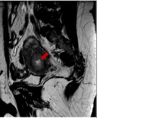
Figure 3 Magnetic Resonance findings
:Sagittal T2-weighted image shows an
heterogeeous thicker endometrium (arrow).:
Area of Interest: Genital / Reproductive
system female;
Imaging Technique: MR;
:Sagittal T2-weighted image shows an
heterogeeous thicker endometrium (arrow).:
Area of Interest: Genital / Reproductive
system female;
Figure 4 Post-treatment magnetic resonance imaging
:Very good response to antituberculosis therapy
instituted. Currently, thin endometrium, without
visualization of the lesion of exuberant
endometritis detected in the previous study.:
Area of Interest: Pelvis;
Imaging Technique: MR;
:Mild downsizing of bilateral tubo-ovarian
abscesses, with some content liquid filling
both tubes.:
Area of Interest: Genital / Reproductive
system female; Pelvis;
MeSH
[C13.371.056.750.249] Endometritis
Inflammation of the ENDOMETRIUM, usually caused by intrauterine infections. Endometritis is the most common cause of postpartum fever.
[C13.371.056.390.890] Salpingitis
Inflammation of the uterine salpinx, the trumpet-shaped FALLOPIAN TUBES, usually caused by ascending infections of organisms from the lower reproductive tract. Salpingitis can lead to tubal scarring, hydrosalpinx, tubal occlusion, INFERTILITY, and ectopic pregnancy (PREGNANCY, ECTOPIC)
[C13.371.803.940] Tuberculosis, Female Genital
Tuberculosis of the genitalia in women. [C13.371.056.630.450] Oophoritis
Inflammation of the OVARY, generally caused by an ascending infection of organisms from the endocervix.
References
[1] Kim SH, Kim SH, Yang DM et al (2004) Unusual causes of tubo-ovarian abscess. CT and MR Radiographics 24:1575-1589
imaging findings
[2] Engin G, Acuna B, Acuna G, Tunaci M (2000) Imaging of extrapulmonary tuberculosis RAdioGraphics 20:471-88
AJR 194:555-61
[4] Chen CJ, Yao WJ, Chou CY, et al (2008) Peritoneal tuberculosis with elevated serum CA125 Ann Nucl Med 22:525-527
mimicking peritoneal carcinomatosis on F-18 FDG-PET/CT