ORIGIN
AL RESEAR
CH
158
Low-intensity laser favors muscle regeneration in a
malnourished and recovered experimental model
Laser de baixa intensidade favorece a regeneração muscular em modelo experimental
desnutrido e recuperado
Láser de baja intensidad favorece la regeneración muscular en modelo experimental
desnutrido y recuperado
Maisa Cardoso da Silva1, Rafael Schimith da Silveira1, Cintia Yuri Matsumura2, Adriana Pertille3
1Student of the Master’s Program in Human Movement Sciences of Universidade Metodista de Piracicaba (Unimep) – Piracicaba (SP), Brazil. 2Assistant professor at the Department of Anatomy of the Bio-Sciences Institute of Universidade Júlio de Mesquita Filho (Unesp) –
Botucatu (SP), Brazil.
3Professor of the Graduate Program in Human Movement Sciences of Universidade Metodista de Piracicaba (Unimep) – Piracicaba (SP), Brazil.
Corresponding address: Adriana Pertille – Universidade Metodista de Piracicaba (Unimep), Programa de Pós-Graduação em Ciências do Movimento Humano – Rodovia do Açúcar, 7000 – Piracicaba (SP), Brasil – CEP 13400-911 – E-mail: [email protected] – Finance source: CNPq 423505/2016-5 – Conflict of interests: Nothing to declare – Presentation: Feb. 5th, 2017 – Accepted for publication: Jan. 12th,2018 – Approved by the Ethics Committee on the Use of Animals of Universidade Metodista de Piracicaba under protocol no. 03/2016.
ABSTRACT | Low-Level Laser Therapy – LLLT is used frequently on muscle lesions, but needs to be investigated in a malnutrition model. The aim of this study was to analyze the effects of LLLT on muscle regeneration of rats subjected to malnutrition and protein recovery. Forty recently weaned Wistar rats were used, divided into control group (C), subjected to a normal-protein diet (14% casein), and the malnourished group (D), subjected to a low-protein diet (6% casein) for 45 days and to a normal-protein diet until the end of the experiment. Subsequently, the right tibialis anterior muscle was subjected to cryogenic cooling and treated with LLLT (830 nm AsGaAl, 30 mW, 20 J/cm²), three times a week, for 7 and 21 days. There was a reduction of the inflammation/regeneration area in the C21 group compared to D21 (p<0.05), which became more evident
with the LLLT (C21L and D21L). The TNF-α contents were
reduced after 21 days of the injury. The connective tissue density area (CTDA) was lower in the C21 and C21L groups compared to the respective malnourished groups (p<0.05). LLLT reduced the CTDA in group D21L in comparison to D21
(p<0.05), but the TGF-β1 contents were not influenced. The
cross-sectional area (CSA) of the muscle fiber increased in the 21-day groups. Higher levels of m-TOR were found in the C21L group when compared to D21L (p<0.05). It was concluded that LLLT favored muscle regeneration in the late stage of the experimental model of postnatal malnutrition and subsequent protein recovery.
Keywords | Malnutrition; Muscles/Injuries; Low-Level Laser Therapy.
RESUMO | A terapia por laser de baixa intensidade (
Low-Level Laser Therapy – LLLT) é utilizada com frequência nas
lesões musculares, mas precisa ser investigada em modelo de desnutrição. O objetivo desse estudo foi analisar os efeitos da LLLT na regeneração muscular de ratos submetidos à desnutrição e recuperação proteica. Foram utilizados 40 ratos
Wistar, recém-desmamados, divididos em grupo controle (C),
que consumiu ração normoproteica (14% caseína), e grupo desnutrido (D), que consumiu ração hipoproteica (6% caseína) por 45 dias e ração normoproteica até o final do experimento. Posteriormente, o músculo tibial anterior direito foi criolesado e tratado com LLLT (AsGaAl 830nm, 30mW, 20J/cm²), três vezes por semana, por 7 e 21 dias. Houve redução da área de inflamação/regeneração no grupo C21 comparado ao D21 (p<0,05), sendo mais evidente com a
LLLT (C21L e D21L). O conteúdo de TNF-α foi reduzido após
21 dias da lesão. A área de densidade de tecido conjuntivo (ADTC) foi menor nos grupos C21 e C21L comparados aos respectivos grupos desnutridos (p<0,05). A LLLT reduziu a ADTC no grupo D21L quando comparado do D21 (p<0,05),
porém o conteúdo de TGF-β1 não foi influenciado. A área
de secção transversa (AST) da fibra muscular aumentou nos grupos 21 dias. A m-TOR apresentou maior conteúdo no grupo C21L quando comparado ao D21L (p<0,05). Concluiu-se que a LLLT favoreceu a regeneração muscular na faConcluiu-se tardia no modelo experimental de desnutrição pós-natal e posterior recuperação proteica.
RESUMEN | La terapia por láser de baja intensidad
(Low-Level Laser Therapy – LLLT) es utilizada con frecuencia en
las lesiones musculares, sin embargo, precisa ser investigada en modelo de desnutrición. El objetivo de ese estudio fue analizar los efectos de la LLLT en la regeneración muscular de ratones sometidos a la desnutrición y a la recuperación proteica. Fueron
utilizados 40 ratones Wistar, recién-destetados, divididos en grupo
control (C), que consumió ración normoproteica (el 14% caseína), y grupo desnutrido (D), que consumió ración hipoproteica (el 6% caseína) por 45 días y ración normoproteica hasta el final del experimento. Posteriormente, el músculo tibial anterior derecho que tuvo criolesión y fue tratado con LLLT (AsGaAl 830nm, 30mW, 20J/cm²), tres veces a la semana, por 7 y 21 días. Hubo reducción del área de inflamación/regeneración en el grupo C21 comparado
al D21 (p<0,05), siendo más evidente con la LLLT (C21L y D21L). El
contenido de TNF-α fue reducido después de 21 días de la lesión.
El área de densidad de tejido conjuntivo (ADTC) fue más pequeña en los grupos C21 y C21L comparados a los respectivos grupos desnutridos (p<0,05). La LLLT redujo la ADTC en el grupo D21L cuando comparado del D21 (p<0,05), sin embargo, el contenido
de TGF-β1 no fue influenciado. El área de sección transversa
(AST) de la fibra muscular incrementó en los grupos 21 días. La m-TOR presentó contenido más grande en el grupo C21L cuando comparado al D21L (p<0,05). Se concluyó que la LLLT favoreció la regeneración muscular en la etapa tardía en el modelo experimental de desnutrición posnatal y posterior recuperación proteica.
Palabras clave | Desnutrición; Músculos/Lesiones; Terapia por Luz de Baja Intensidad.
INTRODUCTION
Muscle injuries are common and the inabilities generated by them are directly related to the intrinsic
properties of muscle recovery1. These involve
the presence of satellite cells that are capable of proliferation and differentiation of new fibers and
formation of scars2, which are composed primarily
of collagen, due to the accumulation of extracellular
matrix3.
Low-Level Laser Therapy is used as treatment option for recovery of muscle injuries, because it modifies
cellular metabolism4 and has a series of advantages
over conventional treatments, such as: decreasing scar formation time and ensuring better healing of the injury in patients with a systemic condition, such as malnutrition, diabetes and hypothyroidism, that impairs
this process5.
Malnutrition is a condition that results from the insufficient intake of nutrients and energy or from the inadequate biological utilization of the food ingested, not being necessarily related to the
individual’s condition of hunger6. In malnutrition,
the tissue repair process is hindered because there are changes in the process of protein synthesis and
breakdown of collagen7.
Positive effects of LLLT on the repair of skin wounds of malnourished animals were observed with different wavelengths and energy densities, however, its effects on muscle regeneration of malnourished animals have been little investigated.
The aim of this study was to analyze the effects of LLLT (830 nm) on muscle regeneration of rats subjected to malnutrition and protein recovery.
METHODOLOGY
Forty young Wistar mice were used, having been kept in the Vivarium of the School of Health Sciences of Faculdade de Ciências da Saúde da Universidade Metodista de Piracicaba (Unimep) at 23 ± 2° C, under a 12-hour light/dark cycle and with food and water
ad libitum. The study was approved by the Ethics
Committee on the Use of Animals of Unimep, under protocol No. 03/2016.
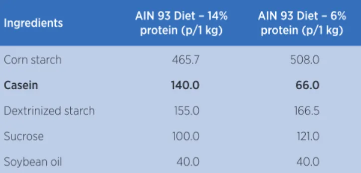
The normal-protein (AIN 93M – 14%) and low-protein (AIN – 6%) diets commercialized by Prag Soluções Serviços e Comércio Ltda. were used, as described in Table 1.
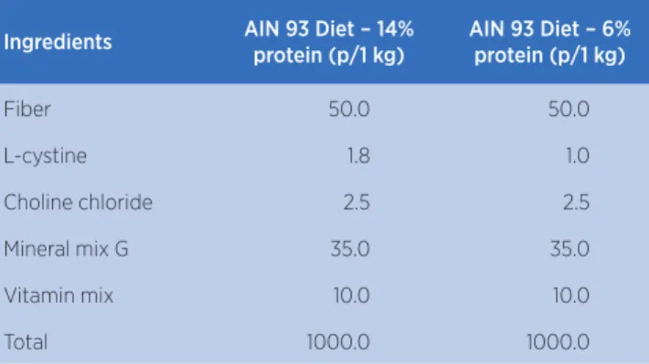
Table 1. Composition of the normal-protein (14%) and low-protein (6%) diets used by the groups
Ingredients AIN 93 Diet – 14% protein (p/1 kg)
AIN 93 Diet – 6% protein (p/1 kg)
Corn starch 465.7 508.0
Casein 140.0 66.0
Dextrinized starch 155.0 166.5
Sucrose 100.0 121.0
Soybean oil 40.0 40.0
Ingredients AIN 93 Diet – 14% protein (p/1 kg)
AIN 93 Diet – 6% protein (p/1 kg)
Fiber 50.0 50.0
L-cystine 1.8 1.0
Choline chloride 2.5 2.5
Mineral mix G 35.0 35.0
Vitamin mix 10.0 10.0
Total 1000.0 1000.0
Source: Reeves, Nielsen e Fahey Jr10
Initially, the animals with 21 days of life were divided randomly into two groups – Control (C, n=20): received the normal-protein diet; and Malnourished/ Recovered (D, n=20): received the low-protein diet for 45 days and later were recovered with the normal-protein diet until the end of the experiment. At the completion of 90 days of diet (111 days of life), all animals suffered a cryogenic lesion and were further divided in 8 groups (n=5): 7-day injury (C7/D7); 7-day injury+LLLT (C7L/D7L); 21-day injury (C21/D21); 21-day injury+LLLT (C21L/D21L), having been sacrificed at the end of the treatment.
For the cryogenic lesion, the animals were anesthetized with intraperitoneal injection (1.16 g/10 ml ketamine hydrochloride and 2 g/100 ml xylazine hydrochloride, 0.09 dose and 0.06 mL/100 g body weight, respectively). The tibialis anterior muscle (TA) was exposed and pressed with a 1 cm × 0.5 cm metal bar cooled in liquid nitrogen for 10 seconds. The procedure was performed twice,
according to the protocol created by Miyabara et al.11
AsGaAl 830 nm low-intensity diode laser was used for the treatment, with 30 mW power and 20 J/cm² energy density, through the trigger point technique over the injured area, the animals having been manually restrained by one researcher while another applied the laser. The treatment began 24 hours after the injury, three times a
week, every two days12. The animals of groups C7L and
D7L received three sessions and the animals of the C21L and D21L groups received nine treatment sessions.
After the experiment’s period, the animals were anesthetized as previously described and sacrificed. The TA muscle was removed, weighed and divided transversely into two equal parts for light microscopy and immunoblotting. The frozen muscles were cut transversely (10μm) using a cryostat (HYRAX C 25 – Zeiss), and the sections were stained with hematoxylin and eosin. The blades
were used to measure the cross-sectional area (CSA), the inflammation/regeneration area (%Infl/Reg) and the connective tissue density area (CTDA), using an optical microscope with a camera attached to it, with 20× objective and connected to a computer with the Image-Pro Plus 6.0 software (Media Cybernetics).
For the CSA of muscle fiber, 200 regenerating fibers were analyzed per animal, characterized by their centralized core. For the measurement of the CTDA, 15 images were evaluated by animal, and a grid containing 88 intersections was superimposed over the images, those which were covering the connective tissue having been counted and, later, the result was transformed into percentage.
The inflammation and regeneration area was characterized as featuring intense inflammatory infiltrate and fibers in initial stage of regeneration. These fibers have small diameter, low quantities of strongly basophilic
cytoplasm and a central core13. For this analysis, the
optical microscope with a camera attached to it was used, its 4× objective having been employed for taking pictures of the cross-section. Later, the images were analyzed in the Image J program, the total area of the muscle and the area with inflammatory infiltrate and fibers in initial process of regeneration having been calculated.
For immunoblotting, another part of the muscles was cut into small pieces and homogenized in a specific buffer, at 4ºC, using Polytron PTA 20S-type homogenizer (Brinkmann Instruments, Westbury, NY, USA) operated at maximum speed for 30 seconds. The extracts were centrifuged at 11,000 rpm at 4ºC for 20 minutes and the supernatant was used for quantitation of the total protein. The samples were treated with Laemmli buffer and heated in dry bath for 5 minutes. Then, 50 g of protein were applied in SDS-polyacrylamide gel at 12% in an electrophoresis equipment from Bio-Rad (mini-Protean, Bio-Rad Laboratories, Richmond, CA, USA). The electrotransfer of the gel to the nitrocellulose membrane was carried out in 90 minutes at 120V. The membranes were washed with a basal solution and incubated with 10 g
of primary antibody (TGF-β1 (transforming growth factor
beta), mouse monoclonal, Sigma-Aldrich, T7039; TNF-α
(tumor necrosis factor alfa), mouse monoclonal, Sigma,
T0157; m-TOR (mammalian target of rapamycin), rabbit
polyclonal, Sigma, T2949; GAPDH (Glyceraldehyde
3-phospate dehydrogenase), mouse monoclonal, Santa
Cruz, SC-59540) diluted in 10 ml of basal solution containing 1% skimmed milk at 4°C during the night. The next day, the membranes were washed with a
basal solution and incubated in 10 ml of basal solution containing 1% skimmed milk and 2.5 g of secondary
antibody (Goat Anti-Rabbit IgG-HRP, Santa
Cruz: sc-2004; Goat Anti-Mouse IgG-HRP, Santa
Cruz: sc-2005) for 2 hours at room temperature. Subsequently, the membranes were washed with basal solution and exposed to the chemiluminescence solution (Pierce) for 5 minutes, and then, the fluorescent signal was captured in the G-Box equipment (GeneSys).
After obtaining the buffers, the membranes were washed with basal solution and incubated with 10 ml
of Stripping Buffer (10mM Tris-HCl 7.5 pH; 0.1M
β-Mercaptoethanol; 8M Urea) for 1 hour at 60°C,
and incubated in 1M Tris-HCl with 7.5 pH for 30 minutes, washed with basal solution and processed as described previously for marking the GAPDH protein, an internal control protein which does not change in quantity under different physiological conditions. The buffers were scanned and quantified through optical densitometry using the Image J program (The National Institute of Health, USA).
The data were analyzed using the Bioestat software version 5.0, and normalcy was assessed through the Shapiro-Wilk test. One Way ANOVA test was used for the analysis of variance, with Tukey’s post-test. P-value<0.05 was considered as significant.
RESULTS
After weaning, all animals had the same body mass (40.2±2.7 g). At the end of the first 45 days of the protocol, group D exhibited statistically significant reduction in body weight when compared to group
C (53±7.2 versus 293.5±18.6 g, p<0.05). After the
nutritional recovery phase (90 days), group D’s body mass increased, however, it did not reach group C’s values (305±20.4 versus 389.7±33 g, p<0.05).
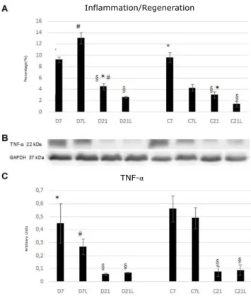
In the histological section, a large inflammatory infiltrate and connective tissue may be noted in the 7-day groups, especially in group D7L, with small presence of muscle fibers in the early stage of regeneration. In the C7L group, reduction of the inflammatory infiltrate and connective tissue may be noted. In the 21-day groups, increase in the CSA of the regenerated fibers (with centralized core) may be noted in all groups, mainly in the C21L group (Figure 1).
In the acute phase of the muscle regeneration process, it was noted that groups C7 and D7 showed the same behavior in relation to the inflammatory process. However, in the chronic phase, there is a larger inflammation area in group D21 compared to C21 (p<0.01, Figure 2 A and C). LLLT favored the reduction of the inflammation/ regeneration area in group C7L compared to C7 (p<0.01), without changing the muscle content of the
TNF-α cytokine. In group D7L there was increase in
the inflammation/regeneration area and decrease in the
TNF-α content compared to D7 (p<0.05; Figure 2A and
C). In the chronic phase, LLLT favored the reduction in the inflammation area, however, the interference of the type of diet in this context was not clear.
Figure 1. Histological cross-sections stained with HE of the tibialis anterior muscle of the groups assessed. Notice the inflammatory infiltrate and increased connective tissue in the 7-day groups and the increased CSA in fibers with centralized core in the 21-day groups
P
ercentage(%)
Arbitr
a
ry
Units
Inflammation/Regeneration
TNF-α
Figure 2. Analysis of inflammation in the groups evaluated
In A: mean and standard deviation of the percentage of the inflammation and regeneration area; In B: TNF-α buffers; In C: Mean and standard deviation of the TNF-α content in arbitrary units. C: control; D: malnourished/recovered; L: group treated with LLLT. § differs from the respective 7-day group; * differs from the respective L group; # differs from the respective C group; p<0.01
In relation to the quantification of the connective tissue, in the early stages of regeneration, LLLT favored the reduction of the CTDA only in group C7L when compared to C7 (p<0.01; Figure 3A). The contents of
the TGF-β1 cytokine were similar in all 7-day groups
(Figure 3C). Throughout the course of the regeneration process there was reduction of the CTDA in the control group (C21 and C21L) and in group D21L when compared to the respective 7-day groups (p<0.01). The positive effect of LLLT was evident in group D21L, with reduction in the CTDA when compared to group D21 (p<0.01; Figure 3A). However, the contents of the
TGF-β1 cytokine showed reduction only in group C21
when compared to C7 (p<0.01; Figure 3C.)
The CSA and the contents of the m-TOR protein were similar among all 7-day groups (Figure 4A and C), however, after 21 days of the injury, only group C21 showed a larger CSA and increase in the m-TOR content when compared to C7 (p<0.01; Figure 4A and C). LLLT favored the increase in the CSA of muscle fiber and in the m-TOR content in group C21L when compared to groups C21 and D21L (p<0.01; Figure 4A and C). In malnourished animals, the contents of the m-TOR protein were similar between the groups.
P
ercentage(%)
Arbitr
a
ry
Units
CTDA
Figure 3. Quantification of the connective tissue in the groups evaluated
In A: mean and standard deviation of the percentage of the area of connective tissue density. In B: TGF-β1 buffers. In C: mean and standard deviation of the TGF-β1 content in arbitrary units. C: control; D: malnourished/recovered; L: group treated with LLLT. § differs from the respective 7-day group; * differs from the respective L group; # differs from the respective C group; p<0.01
Arbitr
a
ry
Units
CSA
Figure 4. Analysis of muscle fiber in the groups evaluated
DISCUSSION
In this study, the gradual response of the control animals’ regeneration process, with reduction of
inflammation, decrease of muscle TNF-α and
of TGF-β1 and increase of m-TOR protein and
muscle fiber CSA were noted, the process having been favored by the irradiation from the LLLT. The results of the malnourished groups suggest that the process of regeneration happens more slowly, with accumulation of connective tissue and deficit in the recovery of the CSA of regenerated muscle fibers, which are also minimized by the irradiation from the LLLT.
The post-weaning malnutrition protocol used in this study was effective, undermining the animals’ development, which was evidenced by the reduction in body mass of the malnourished group. These
results corroborate Escriva et al.14, who claim that the
reduction in the body mass of young rats caused by a low-protein diet is the result of functional changes
of insulin in the tissues. Ihemelandu15 claims that
malnutrition affects the growth and differentiation of cells, and that damages to the muscle system with reduction in proteins are fundamental to decrease body weight in adulthood.
In the process of regeneration of a normally nourished individual’s muscle fiber, after 48 hours of the injury, the necrotic parts of the muscle fibers are removed by macrophages, and, at the same time, the formation of connective tissue by fibroblasts begins. On the third day, the activation of satellite cells occurs, and on the fifth day, the fusion of myoblasts begins, and the connective tissue becomes denser. On the seventh day, the muscle cells regenerated begin to invade the scar’s region and, around the 21st day, the myofibrils merge, with little connective tissue
between them16.
LLLT is used to speed up the process of
regeneration as noted by Renno et al.17, who evaluated
rats submitted to cryolesion and treated with AsGaAl
laser (808 nm, 50 mw, and 10 J/cm2 and 50 J/cm2
energy densities) and found reduction in the areas of cellular infiltrate and injury compared to the control group after 13 days. A similar result was found after 21 days of cryolesion in the control and malnourished groups in this work.
Aimbire et al.18 observed in rats with lung injury that
LLLT (AsGaAl, with 1.0, 2.5 and 5 mW power and
650 nm wavelength) significantly reduced serum levels
of TNF-α in the animals that received the irradiation
when compared to the control group, the effect having been dose-dependent.
In this study we observed no such effect on the
muscle content of TNF-α, probably due to it not
representing the plasma content of the cytokine evaluated in most works.
The delay in the regeneration of malnourished and recovered animals after 21 days, suggested by the larger inflammation area in group D21, confirms what was
observed by Pertille et al.19 14 days after the cryolesion
with the same malnutrition protocol.
Lee et al.20 conducted a histopathological and
morphometric analysis of the soleus muscle of rats submitted to a low-protein diet in the first few days of life, with subsequent recovery. In the normally nourished group, the connective tissue analysis showed a predominance of type I collagen distributed in an organized manner. The malnourished group showed a predominance of type III collagen in a disorganized manner, and there was return of type I collagen in the recovered group, but in a partially organized manner. The type I collagen is responsible for forming parallel
fibers which confer tensile strength and rigidity21, and
laser therapy accelerates the process of tissue repair with increased production and improvement in the
organization of the collagen fibers22.
The positive effect of LLLT was evident in group D21L, with the reduction in the CTDA. However, the
contents of the TGF-β1 cytokine showed reduction
only in the 21-day control group. This cytokine is important in the synthesis and remodeling of the extracellular matrix, which is, therefore, commonly used
to investigate the formation of fibrosis23.
In addition, LLLT favored the increase of CSA in the 21-day groups, mainly in the C21L group, in which there was an increase of the m-TOR protein, a kinase protein found in two multi-protein complexes,
one of them being mTORC124, the central regulator
of cellular growth, for controlling the RNAm translation and, consequently, the synthesis of
proteins25.
The increase of CSA in fibers with LLLT confirms what has been noted in earlier works on the TA muscle,
but with different LLLT parameters26,27.
CONCLUSION
Low-intensity laser therapy with the parameters used favored muscle regeneration in the late phase (21 days) of the experimental model of post-natal protein malnutrition and subsequent nutritional recovery.
REFERENCES
1. Mann CJ, Perdiguero E, Kharraz Y, Aguilar S, Pessina P, Serrano AL et al. Aberrant repair and fibrosis development in skeletal muscle. Skelet Muscle. 2011;1(1):1-21. doi: 10.1186/2044-5040-1-21
2. Tidball JG, Villalta SA. Regulatory interactions between muscle and the immune system during muscle regeneration. Am J Physiol Regul Integr Comp Physiol. 2010;298(5):R1173-87. doi: 10.1152/ajpregu.00735.2009
3. Shin EH, Caterson EJ, Jackson WM, Nesti LJ. Quality of healing: defining, quantifying, and enhancing skeletal muscle healing. Wound Repair Regen. 2014; 22(Supl 1):18-24. doi: 10.1111/wrr.12163
4. Dortbudak O, Haas R, Mallath-Pokorny G. Biostimulation of bone marrow cells with a diode soft laser. Clin Oral Implants Res. 2000;11(6):540-5.
5. Meireles GCS, Silva CA, Marques AMCM, Pinheiro ALB. A efetividade da fototerapia laser no reparo tecidual em portadores de desordem funcional sistêmica. Rev Eletr Fainor. 2014;7(2):71-84.
6. Monteiro CA. A dimensão da pobreza, da desnutrição e da fome no Brasil. Estud Av. 2003;17(48):7-20. doi: 10.1590/S0103-40142003000200002
7. Silveira IS, Raiser AG, Polydoro AS, Santos MN. Efeitos da dieta protéica na cicatrização de fraturas distais de fêmur imobilizadas com pinos intramedulares em cão. Acta Cirurg Bras. 1997;12(3):178-81. doi: 10.1590/S0102-86501997000300008
8. Pinheiro ALB, Meireles GC, Vieira ALB, Almeida D, Carvalho CM, Santos JN. Phototherapy improves healing of cutaneous wounds in nourished and undernourished wistar rats. Braz Dent J. 2004;15(Special issue):SI21-8.
9. Pinheiro ALB, Meireles GC, Carvalho CM, Ramalho LM, Santos JN. Biomodulative effects of visible and IR laser light on the healing of cutaneous wounds of nourished and undernourished Wistar rats. Photomed Laser Surg. 2009;27(6):947-57. doi: 10.1089/pho.2009.2607
10. Reeves PG, Nielsen FH, Fahey Jr GC. AIN-93 purified diets for laboratory rodents: final report of the American Institute of Nutrition ad hoc writing committee on the reformulation of the AIN-76A rodent diet. J Nutr. 1993;123(11):1939-51.
11. Miyabara EH, Martin JL, Griffin TM, Moriscot AS, Mestril R. Overexpression of inducible 70 kDa heat shock protein in mouse attenuates skeletal muscle damage induced by cryolesioning. Am J Physiol Cell Physiol. 2006;290(4):C1128-38. doi: 10.1152/ajpcell.00399.2005
12. Pertille A, Macedo AB, Oliveira CP. Evaluation of muscle regeneration in aged animals after treatment with low-level laser therapy. Rev Bras Fisioter. 2012;16(6):495-501.
13 Marques MJ, Machado RV, Minatel E, Santo Neto H. Disodium cromoglycate protects dystrophin-deficient muscle fibers from leakiness. Muscle Nerve. 2008;37(1):61-7. doi: 10.1002/mus.20892
14. Escriva F, Kergoat M, Bailbe D, Pascual-Leone AM, Portha B. Increased insulin action in the rat after protein malnutrition early in life. Diabetologia. 1991;34(8):559-64.
15. Ihemelandu EC. Fibre number and sizes of mouse soleus muscle in early postnatal protein malnutrition. Acta Anat. 1985;121(2):89-93.
16. Jarvinen TA, Jarvinen TL, Kaariainen M, Kalimo H, Jarvinen M. Muscle injuries: biology and treatment. Am J Sports Med. 2005;33(5):745-64. doi: 10.1177/0363546505274714
17. Renno ACM, Assis L, Peres B, Rodrigues NC, Brunelli RM,
TomaRL, et al. The effects of low level laser therapy on injured
skeletal muscle. Braz Arch Biol Technol. 2014;57(1):48-54. doi: 10.1590/S1516-89132014000100008
18. Aimbire F, Albertini R, Pacheco MT, Castro-Faria-Neto HC, Leonardo PS, Iversen VV, et al. Low-level laser therapy induces dose-dependent reduction of TNFalpha levels in acute inflammation. Photomed Laser Surg. 2006;24(1):33-7. doi: 10.1089/pho.2006.24.33
19. Pertille A, Moura KF, Matsumura CY, Ferretti R, Ramos DM, Petrini AC, et al. Evaluation of skeletal muscle regeneration in experimental model after malnutrition. Braz J Biol. 2017;77(1):83-91. doi: 10.1590/1519-6984.10415
20. Lopes TS, Quintana HT, Bortolin JA, Alves PHM, Matos RSB, Liberti EA, et al. Protein malnutrition pre- and postnatal and nutritional rehabilitation modulates the morphology of muscle fibers in wistar rats. J Diet Suppl. 2017;14(3):278-87. doi: 10.1080/19390211.2016.1212960
21. Lehto M, Duance VC, Restall D. Collagen and fibronectin in a healing skeletal muscle injury. An immunohistological study of the effects of physical activity on the repair of injured gastrocnemius muscle in the rat. J Bone Joint Surg Br. 1985;67B(5):820-8.
22. Reis SRA, Medrado AP, Marchionni AMT, Figueira C, Fracassi LD, Knop LAH. Effect of 670-nm laser therapy and dexamethasone on tissue repair: a histological and ultrastructural study. Photomed Laser Surg. 2008;26:307-13. doi: 10.1089/pho.2007.2151
23. Heldin CH, Miyazono K, Dijke P. TGF-β signaling from cell
24. Laplante M, Sabatini DM. mTOR signaling in growth control and disease. Cell. 2012; 149:274-93. doi: 10.1016/j.cell.2012.03.017
25. Mahoney SJ, Dempsey JM, Blenis J. Cell signaling in protein synthesis ribosome biogenesis and translation initiation and elongation. Prog Mol Biol Transl Sci. 2009;90C:53-107. doi: 10.1016/S1877-1173(09)90002-3
26. Oliveira NM, Parizzotto NA, Salvini TF. GaAs (904-nm) laser radiation does not affect muscle regeneration in mouse skeletal muscle. Lasers Surg Med. 1999; 25(1):13-21.