C
AS
E
S
TUD
Y
Correspondence to: Ana Claudia Mattiello Sverzut − Universidade de São Paulo − Faculdade de Medicina de Ribeirão Preto − Avenida Bandeirantes, 3.900 − Campus Universitário − CEP: 14049-900 − Ribeirão Preto (SP), Brazil − E-mail: [email protected]
Presentation: Oct. 2013 – Accepted for publication: Feb. 2014 – Financing source: none – Conflict of interests: nothing to declare – Approval of the Ethics Committee No. 6017/2013 ABSTRACT | This case study aimed to verify the model
of Rose et al.1 as a feasible to assess energy expenditure
in gait of children with Duchenne muscular dystrophy (DMD). Three DMD patients aged 6, 7 and 8 years old participated of this study. It was obtained weight, height, leg length measurement (LLM), resting and gait heart rate (HR) held on as 55-meter oval circuit performed dur-ing a two-minute test at each speed. Energy expenditure was calculated using the HR. It was performed a descrip-tive analysis (average) and these were compared, indi-vidually, to normative data. The average gait speed of these three patients was similar to the normative data for slow speed and lower considering comfortable and fast speed. The energy expenditure to slow speed of the patients 2 and 3 was similar to the normality, and lowest for patient 1; at comfortable speed, the energy expendi-ture obtained for all patients was similar; at fast speed, the patients 1 and 2 presented similar to normal values, but the patient 3 presented higher energy expenditure. It was concluded that the energy expenditure evaluation using HR was easily executed in the clinical practice and it can help therapeutic choices. For patient 3, an aerobic training could be indicated and for the others, they could keep the routine assessments.
Keywords | Muscular Dystrophy, Duchenne; Energy Metabolism; Gait; Child.
Gait energy expenditure in children with Duchenne
muscular dystrophy: case study
Gasto energético na marcha de crianças com distroia muscular de Duchenne: estudo de caso
El gasto energético durante la marcha de los niños con distroia muscular de Duchenne:
un estudio de caso
Mariana Angélica de Souza¹, Marília Ester Ferreira², Cyntia Rogean de Jesus Alves de Baptista³, Ana Claudia Mattiello Sverzut4
Study conducted at the Structure and Function of Skeletal Muscle – Laboratory of Scientific Research. Department of Biomechanics, Medicine and Rehabilitation of the Locomotor Apparatus and at the Center of Physical Education, Sports and Recreation of the Ribeirao Preto Medicine School of the Universidade de São Paulo (FMRP-USP) − Ribeirão Preto (SP), Brazil.
¹Gradute Program in Rehabilitation and Functional Performance of the FMRP-USP − Ribeirão Preto (SP), Brazil.
²Laboratory of Scientific Research − Structure and Function of Skeletal Muscle of the FMRP-USP − Ribeirão Preto (SP), Brazil. ³Department of Biomechanics, Medicine and Rehabilitation of the Locomotor Apparatus of the FMRP-USP − Ribeirão Preto (SP), Brazil. 4Physical Therapy course of the Department of Biomechanics, Medicine and Rehabilitation of the Locomotor Apparatus of the FMRP-USP − Ribeirão Preto (SP), Brazil.
RESUMO | Este estudo de caso objetivou verificar se o modelo de Rose et al.1 é factível para avaliar o gasto
ener-gético na marcha em crianças com distrofia muscular de Duchenne (DMD). Participaram três crianças com DMD e idades de 6, 7 e 8 anos. Foram avaliados peso, altura, compri-mento dos membros inferiores (CMMII), frequência cardíaca (FC) de repouso e de marcha realizada em circuito oval de 55 m durante um teste de 2 minutos em cada velocidade. O gasto energético foi calculado pela FC. Foi realizada análise descritiva dos dados (média) e estes foram comparados, in-dividualmente, com dados normativos. A velocidade média (Vm) da marcha dos três pacientes foi igual aos dados nor-mativos na etapa velocidade lenta e menor nas etapas de velocidade confortável e rápida. O gasto energético na velo-cidade lenta dos pacientes 2 e 3 foi similar à normalidade, e menor para o paciente 1; na velocidade confortável, o gasto energético de todos os pacientes foi similar; na velocidade rápida, os pacientes 1 e 2 apresentaram valores similares ao normal, porém o paciente 3 teve maior gasto energético. Concluiu-se que a avaliação do gasto energético pela FC foi facilmente executada na clínica, podendo auxiliar na eleição de condutas. Para o paciente 3 poderia ser indicado um trei-namento aeróbio e para os demais manter esse protocolo de avaliação nas visitas subsequentes.
INTRODUCTION
he Duchenne muscular dystrophy (DMD) is a ge-netic myopathy characterized by the progressive loss of muscle strength, from proximal to distal, irst af-fecting the lower limbs and upper limbs later. his progressive muscle weakness cause postural and gait
changes, leading the patient to loss of ambulation2-4.
hus, the rehabilitation of patients with DMD aims to slow the progress of deformities and to provide a better quality of life, especially for the maintenance of
gait for as long as possible4.
herefore, several are the instruments to assess the gait, which aim at understanding the biomechanical
changes and also the energy cost to run it5. Seeking
greater ease of measurement of energy expenditure
dur-ing the gait of children, Rose et al.6 compared the
ener-gy expenditude index (EEI) by oxygen with the rate of energy expenditure by heart rate (HR) and concluded that, although both of them may be used in the evalua-tion of normal children and children with cerebral palsy, it is preferable to use the index obtained by HR, given its ease of data acquisition.
he EEI, whose inal result is expressed as number
of heartbeats per meter walked5, is usually reported in
children with cerebral palsy6-9, however restricted to
works in patients with DMD. In these patients, the resting energy expenditure, measured by calorimetry, is
the target of most studies10-13.
Taktak and Bowker14 compared the energy
expen-diture of gait in patients with DMD, between two types of orthoses. he EE was calculated by the HR
at gait and at rest, and the average velocity (Vave) of
gait in a circuit of 8 m. Other authors15 assessed
en-ergy expenditure in patients with DMD by the values
of HR and Vave obtained in the 6-minute walk test.
However, no study has compared the energy expen-diture during gait in patients with DMD with data
from Rose et al.1.
Assessing the energy expenditure of gait by adopting HR may contribute to the monitoring of the disease’s progression and, thus, favor the election of therapeutic approaches in patients with DMD. hus, this case study aimed to verify whether the model
of Rose et al.1 is feasible to be applied in clinic as an
indirect parameter to indicate the energy expenditure of gait in children with DMD.
METHODOLOGY
Sample
hree patients, seen at the Infant Myopathies’s
Ambulatory, Rehabilitation Center of Clinics Hospital of the Ribeirao Preto Medicine School, University of São
Paulo (CER-HCFMRP-USP), participated in the
study. he inclusion criteria were: conirmed diagno-sis of DMD, to be over six years of age, independent community ambulation, similar functional score (up to two points of diference) in Dimension 1 (D1) of the
Motor Function Measure (MFM)16 and the signing
the Informed Consent Form (ICF) by the guardian.
RESUMEN | Este estudio de caso tuvo como objetivo verificar si el modelo de Rose et al es factible para evaluar el gasto de energía en la marcha de niños con distrofia muscular de Duchenne (DMD). Participaron tres niños con DMD con eda-des de 6, 7 y 8 años. Fueron evaluados peso, altura, longitud de las extremidades inferiores (LEI), frecuencia cardiaca (FC) en reposo y la marcha realizada en el circuito ovalado de 55 m durante un exámen de 2 minutos en cada velocidad. El gasto energético fue calculado por la FC. Se realizó un análisis des-criptivo de los datos (media) y éstos fueron comparados de forma individual con los datos normativos. La velocidad media (VM) de la marcha de los tres pacientes fue igual a los datos normativos en la etapa velocidad lenta y menor en las etapas
de velocidad confortable y rápida. El gasto de energía en la velocidad lenta de los pacientes 2 y 3 fue similar a la norma-lidad, y menor para el paciente 1; en la velocidad confortable, el gasto de energía de todos los pacientes fue similar; en la velocidad rápida, los pacientes 1 y 2 tuvieron valores similares a lo normal, pero el paciente 3 tuvo mayor gasto energético. Se concluyó que la evaluación del gasto energético por la FC se llevó a cabo fácilmente en la clínica, y pudo ayudar en la elección de conductas. Para el paciente 3 podría ser indicado un entrenamiento aeróbico y para los demás mantener ese protocolo de evaluación en las visitas siguientes.
he exclusion criteria were: presence of cardiopulmo-nary diseases or cognitive insuiciency to understand the commands of the physical therapist.
he normative data on energy expenditure in gait by indirect analysis via HR for healthy children, proposed
by Rose et al.1, were used I order to compare the results
obtained with patients with DMD. herefore, these data
were broken down here as normative data of Rose et al.1.
Procedures
he clinical history of patients and the scores of MFM
were obtained in the database of the Infant Myopathies’s
Ambulatory, Rehabilitation of the CER-HCFMRP-USP. he patients selected, according to the inclusion criteria, were invited to participate in the study and the assessments were scheduled for those whose guardians signed Informed Consent Form.
Patients were evaluated at the CEFER of the campus
of Ribeirão Preto, USP. here were collected: weight, height, leg length measurement (LLM) and energy expenditure during gait. his case study has a cross-sectional nature and each test (described below) was performed by the same examiner.
he weight was assessed on a Welmy® W200/5 elec-tronic scale and the height on a stadiometer (Tonelli®). hese measurements were obtained with the barefoot child in a standing position. he LLM was obtained, by a measuring tape, with the child in the supine position, the distance between the anterior superior iliac spine and the medial malleolus. he body mass index (BMI) was calculated by dividing weight by height squared. he body composition was assessed by bioelectrical im-pedance (Biodynamics 450).
For the assessment of the energy expenditure in gait,
a methodology similar to that described by Rose et al.1,
was described, in which children ambulate in an oval circuit of 55 m, in 3 steps, lasting 2 minutes each.
Initially, with the child in a seating position, it was collected the resting heart rate (RHR) through the HR monitor (Polar®). In the irst stage, the child was asked to walk, through the circuit, at a comfortable speed,
with the verbal command “walk normally, as you do every day”. In the second stage, the child was asked to walk slowly: “walk very slowly”. In the third one, they received the command: “walk as fast as you can, but without running”.
he walking heart rate (WHR) was collected in the last 30 seconds of each stage. Between the stages, chil-dren rested for 3 minutes, or until the HR would return to its resting value +/- 5 beats/min.
he Vave of the gait and the EEI rate, at each
stage, were calculated by the formulas Vave = d/t and
EE=(HHR-RHR)/Vave, respectively, since d is the total
distance, in meters, and t is the time duration of each step of the test (2 min).
Statistical analysis
A descriptive analysis (average) of the data was per-formed. hese were compared, individually, to the nor-mative data (average and standard deviation) of Rose
et al. (comprising typical children of 6-8 years of age) in the three stages of the test (slow, comfortable and fast speeds).
RESULTS
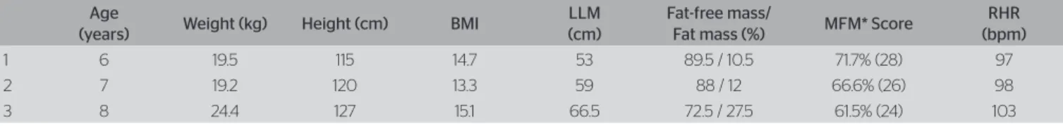
he measures of the anthropometric variables (weight, height, LLM), RHR and the percentage of fat increased with age. here was no diference between the length of the right and left lower limb. he score of MFM de-creased with the advancing age (Table 1).
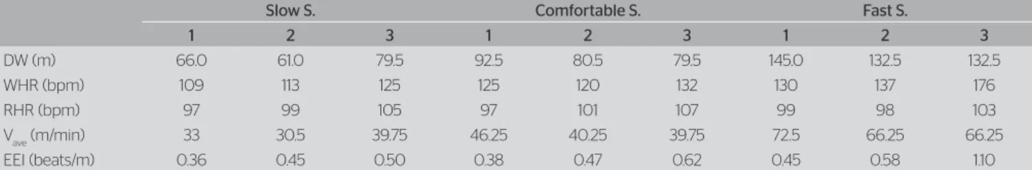
he Table 2 shows that the distance traveled, the
resting and gait HR, the Vave and the energy
expen-diture increased according to the progression of the stage step in the test, for all the patients assessed, except for patient 3, who traveled the same distance
and, thus, kept the Vave during the steps of slow and
comfortable pace.
he older patients had lower scores of MFM and higher energy consumption (Tables 1 and 2).
Table 1. Characteristics of the sample in relation to the anthropometric variables, body composition and score of the measure of motor function
Age
(years) Weight (kg) Height (cm) BMI
LLM (cm)
Fat-free mass/
Fat mass (%) MFM* Score
RHR (bpm)
1 6 19.5 115 14.7 53 89.5 / 10.5 71.7% (28) 97
2 7 19.2 120 13.3 59 88 / 12 66.6% (26) 98
Considering the averages and the standard
devia-tions of healthy children1, the weight and the LLM
of patients 1 and 2 were lower, however, the values of patient 3 were normal. As for height, patient 1 was the only one who got a result lower than the ones of the control group, patients 2 and 3 had similar results
to the healthy group studied by Rose et al.1. he RHR
of the three patients was higher than that obtained
in the healthy group studied by Rose et al.1. he
height, the length of the lower limbs and the RHR increased with age (Table 3).
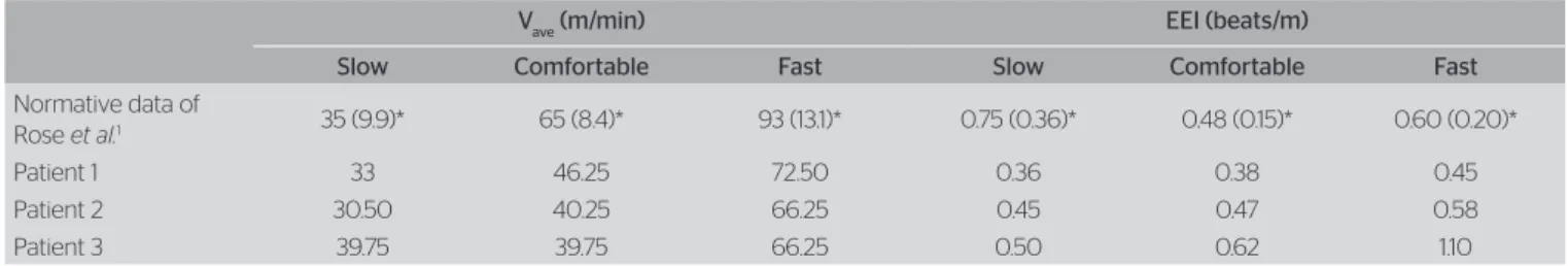
Table 4 shows the comparison between the average speeds and the energy expenditure in patients 1, 2 and 3
and the normative data of Rose et al.1.
All patients had Vave within the interval (average and
standard deviation) of the data presented by Rose et al.1,
in the slow speed stage, and lower Vave in comfortable
and fast speeds.
he energy expenditure of patients, when compared to
normality standards1, behaved diferently according
to the gait speed.
Patient 1 had an energy expenditure within the range (average and standard deviation) of the normative data
of Rose et al.1, at comfortable and fast speeds, and lower
energy expenditure interval (average and standard de-viation) in the control group at slow speed.
Patient 2 had energy expenditure within the interval range (average and standard deviation) of the normative
data of Rose et al.1 at the three speeds (slow,
comfort-able and fast).
Patient 3 had energy expenditure within the range (mean and standard deviation) of the normative data of
Rose et al.1, at slow and comfortable speeds, and higher
energy expenditure in fast speed.
Figure 1 shows the values for the energy expenditure of patients with DMD in three speeds (slow, comfortable and fast). here was an increase in energy expenditure with age progression. It is observed that the energy expenditure of patients 1 and 2 similarly increased according to the speed progression. However, the increased energy expenditure of patient 3 was more pronounced, especially at comfortable to fast speed.
DISCUSSION
Given the scarcity of studies on energy expenditure of gait in patients with DMD, this study aimed to verify
whether the model of Rose et al.1 is feasible in order to
assess the energy expenditure during gait in children with DMD.
As in the study by Rose et al.1, anthropometric data
were obtained, and the values of height and weight of the evaluated patients were similar to the indings
of other authors14,17. Based on the normative data of
Rose et al.1, the weight and length of the lower limbs
of patients 1 and 2 were lower, and of patient 3 were similar. As for the height, only patient 1 had an inferior result, patients 2 and 3 had similar results. As opposed to that, other authors have shown that DMD patients
have lower height and weight than healthy children2.
he progression of DMD leads to a functional de-cline. In this sense, the MFM is the ideal monitoring
tool, and its Portuguese version features high reliability16.
According to the literature18, the score of the MFM
de-creased with age. In this case study, an inde-creased energy expenditure with functional decline of the patients ana-lyzed (scores of patients 1, 2 and 3 difered by 2 points among themselves) may be observed. hese data may suggest an inverse relationship between the score of the
Table 3. Comparison of the anthropometric variables between patients and the normative data of Rose et al.1
Normative data of Rose et al.1
Patients
1 2 3
Age (years) 6–8 6 7 8
Weight (kg) 28 (5.7)* 19.5 19.2 24.4
Height (cm) 127 (9.9)* 115 120 127
LLM (cm) 66 (6.8)* 53 59 66.5
RHR (bpm) 83 (10)* 97 98 103
*Mean (standard deviation); LLM: leg length measure; RHR (bpm): resting heart rate in beats per minute
Table 2. Average velocity and calculation of the energy expenditure in comfortable, slow and fast speeds, for patients 1, 2 and 3
Slow S. Comfortable S. Fast S.
1 2 3 1 2 3 1 2 3
DW (m) 66.0 61.0 79.5 92.5 80.5 79.5 145.0 132.5 132.5
WHR (bpm) 109 113 125 125 120 132 130 137 176
RHR (bpm) 97 99 105 97 101 107 99 98 103
Vave (m/min) 33 30.5 39.75 46.25 40.25 39.75 72.5 66.25 66.25
EEI (beats/m) 0.36 0.45 0.50 0.38 0.47 0.62 0.45 0.58 1.10
MFM and the energy expenditure, i.e., higher scores on MFM, lower energy expenditure during gait.
For the calculation of the energy expenditure, the
heart rates and the gait Vave were considered. In
accor-dance with the literature19, the RHR of the evaluated
patients was higher than in the healthy group studied by
Rose et al.1. his result may be explained by the
autonom-ic nervous system dysfunction, whautonom-ich occurs precocious-ly in patients with DMD, causing decreased parasym-pathetic activity and/or increased symparasym-pathetic activity
with the progression of the disease20. he decrease in the
comfortable Vave with the advancing age and the lowest
speeds (comfortable and fast), when compared to healthy children, may be explained by changes in the gait of pa-tients with DMD with the progression of the disease.
In another study2, there was no diference between the
Vave of gait in patients with DMD and the control group.
As opposed to that, Gaudreault et al.21 showed that the
walking speed of patients was lower than the control
group, and that the usual Vave in patients with DMD does
not difer from the slow speed of the control group. he total energy expenditure encompasses the en-ergy expenditure while at rest and its spend in activity. In DMD patients, the resting energy expenditure is often reported. Some authors report that, in these pa-tients, the resting energy expenditure is lower than in the control group and emphasize the need for longi-tudinal studies, in order to understand these changes
with the progression of the disease10. On the contrary,
to other authors12, the resting energy expenditure is
higher in patients with DMD than in healthy children, and yet, this expenditure is lower in obese rather than in nonobese patients with DMD. In this sense, it is em-phasized that the analysis of energy expenditure should be related to the body composition analysis.
In healthy children, there is a positive correlation be-tween fat and energy expenditure, and this is inversely
proportional to the lean body mass22. In this study, the
increase in energy expenditure with age is consistent with the accumulation of body fat and the loss of lean mass, characteristic aspects of the progression of the
DMD17. Still, only 3 patients showed higher energy
ex-penditure, when compared to the normative data from
Rose et al.1, which seems to be justiied due to a higher
percentage of fat than in other evaluated patients. hree major factors limit the discussion of the results
obtained here1: the lack of studies on energy expenditure
during gait in patients with DMD2, the methodological
variety of studies on resting energy expenditure and the
number of subjects analyzed here3. For this reason,
gener-alizations would be improper, limited, thus, to the propo-sition of interventions only to the participants patients.
From these results, further studies must be conducted in a larger number of patients. An inverse relationship between the score of the MFM and the energy expenditure is hypothesized, assessed by the HR, which will be able to be minimized by the early indication of therapeutic aerobic training.
CONCLUSION
In the three children with DMD who participated in this study, the assessment of energy expenditure of
Table 4. Comparison of the average velocity and the energy expenditure between patients and the normative data of Rose et al.1
Vave (m/min) EEI (beats/m)
Slow Comfortable Fast Slow Comfortable Fast
Normative data of
Rose et al.1 35 (9.9)* 65 (8.4)* 93 (13.1)* 0.75 (0.36)* 0.48 (0.15)* 0.60 (0.20)*
Patient 1 33 46.25 72.50 0.36 0.38 0.45
Patient 2 30.50 40.25 66.25 0.45 0.47 0.58
Patient 3 39.75 39.75 66.25 0.50 0.62 1.10
Vave (m/min): average velocity in meters per minute; EEI (beats/m): rate of energy expenditure in beats per meter; *Mean (standard deviation)
Figure 1. Energy expenditure of patients 1, 2 and 3 in slow, comfortable and fast speeds
Patient 1 Patient 2 Patient 3 1.2
1.0
0.8
0.6
0.4
EEI (be
at
s/
m)
Stages
Slow S. Comfortable S. Fast S.
gait by adopting HR and pairing it with the data from
healthy children1 were easily implemented in the clinic.
he model used by Rose et al.1 seems to indicate the
energy expenditure of gait in children with DMD and proved to be a tool which may assist the therapist in the election of their conduct. For patient 3, the data obtained in this study suggest that the development of the aerobic training may minimize the deleterious re-sponse in the cardiorespiratory system which tracks the progression of the DMD. he remaining patients could be given only routine assessments to measure energy expenditure of walking at diferent speeds. Still, other patients with diferent scores of MFM should be ana-lyzed, following the proposed methodology, in order to increase the conclusions listed here.
ACKNOWLEDGEMENTS
We are grateful to the CEFER of USP (campus of
Ribeirão Preto), to Prof. Dr. Hugo Celso Dutra de Souza and to the laboratory technician Ana Paula Manio.
REFERENCES
1. Rose J, Gamble JG, Lee J, Lee R, Haskell WL. The energy expenditure index: a method to quantitate and compare walking energy expenditure for children and adolescents. J Pediatr Orthop. 1991;11(5):571-8.
2. Doglio L, Pavan E, Pernigotti I, Petralia P, Frigo C, Minetti C. Early signs of gait deviation in Duchenne muscular dystrophy. Eur J Phys Rehabil Med. 2011;47(4):587-94.
3. Humbertclaude V, Hamroun D, Bezzou K, Bérard C, Boespflug-Tanguy O, Bommelaer C, et al. Motor and respiratory heterogeneity in Duchenne patients: Implication for clinical trials. Eur J Paediatr Neurol. 2012;16(2):149-60.
4. Vuillerot C, Girardot F, Payan C, Fermanian J, Iwaz J, De Lattre C, et al. Monitoring changes and predicting loss of ambulation in Duchenne muscular dystrophy with the Motor Function Measure. Dev Med Child Neurol. 2010;52(1):60-5
5. Tanaka MS, Luppi A, Morya E, Fávero FM, Fontes SV, Oliveira ASB. Principais instrumentos para a análise da marcha de pacientes com distrofia muscular de Duchenne. Rev Neurocienc.2007;15(2):153-9.
6. Rose J, Gamble JG, Burgos A, Medeiros J, Haskell WL. Energy expenditure index of walking for normal children and for children with cerebral palsy. Dev Med Child Neurol. 1990;32(4):333-40.
7. Bratteby Tollerz LU, Olsson RM, Forslund AH, Norrlin SE. Reliability of energy cost calculations in children with cerebral palsy, cystic fibrosis and healthy controls. Acta Paediatr. 2011;100(12):1616-20.
8. Konop KA, Strifling KM, Wang M, Cao K, Eastwood D, Jackson S, et al. Upper extremity kinetics and energy expenditure during walker-assisted gait in children with cerebral palsy. Acta Orthop Traumatol Turc, 2009;43(2):156-64. [Turkish].
9. IJzerman MJ, Nene AV. Feasibility of the physiological cost index as an outcome measure for the assessment of energy expenditure during walking. Arch Phys Med Rehabil. 2002;83(12):1777-82.
10. Shimizu-Fujiwara M, Komaki H, Nakagawa E, Mori-Yoshimura M, Oya Y, Fujisaki T, et al. Decreased resting energy expenditure in patients with Duchenne muscular dystrophy. Brain Dev. 2012;34(3):206-12.
11. Elliott SA, Davidson ZE, Davies PS, Truby H. Predicting resting energy expenditure in boys with Duchenne muscular dystrophy. Eur J Paediatr Neurol. 2012;16(6):631-5.
12. Zanardi MC, Tagliabue A, Orcesi S, Berardinelli A, Uggetti C, Pichiecchio A. Body composition and energy expenditure in Duchenne muscular dystrophy. Eur J Clin Nutr. 2003;57(2):273-8.
13. Gonzalez-Bermejo J, Lofaso F, Falaize L, Lejaille M, Raphaël JC, Similowski T, et al. Resting energy expenditure in Duchenne patients using home mechanical ventilation. Eur Respir J. 2005;25(4):682-7.
14. Taktak DM, Bowker P. Lightweight, modular knee-ankle-foot orthosis for duchenne muscular dystrophy: design, development, and evaluation. Arch Phys Med Rehabil. 1995;76(12):1156-62.
15. McDonald CM, Henricson EK, Abresch RT, Florence J, Eagle M, Gappmaier E, et al. The 6-minute walk test and other clinical endpoints in Duchenne mucular Dystrophy: reliability, concurrent validity, and minimal clinically important diferences from a multicenter study. Muscle Nerve. 2013;48(3):357-68.
16. Iwabe C, Miranda-Pfeilsticker BH, Nucci A. Medida da função motora: versão da escala para o português e estudo de confiabilidade. Rev Bras Fisioter. 2008;12(5):417-24.
17. Caromano FA, Tanaka C, João SMA, Kamisaki AP, Yano KC, Ide MR. Correlação da massa e porcentagem de gordura com a idade na Distrofia Muscular de Duchenne. Fisioter Mov. 2010;23(2):221-7.
18. Fischmann A, Hafner P, Gloor M, Schmid M, Klein A, Pohlman U, et al. Quantitative MRI and loss of free ambulation in Duchenne muscular dystrophy. J Neurol. 2013;260(4):969-74.
19. Thomas TO, Morgan TM, Burnette WB, Markham LW. Correlation of heart rate and cardiac dysfunction in Duchenne muscular dystrophy. Pediatr Cardiol. 2012;33(7):1175-9.
20. Yotsukura M, Fujii K, Katayama A, Tomono Y, Ando H, Sakata K,
et al. Nine-year follow-up study of heart rate variability in patients with Duchenne-type progressive muscular dystrophy. Am Heart J. 1998;136(2):289-96.
21. Gaudreault N, Gravel D, Nadeau S, Houde S, Gagnon D. Gait patterns comparison of children with Duchenne muscular dystrophy to those of control subjects considering the efect of gait velocity. Gait Posture. 2010;32(3):342-7.
22. Goran MI, Nagy TR, Gower BA, Mazariegos M, Solomons N, Hood V,