A KINESTHETIC MOTOR IMAGERY STUDY
IN PATIENTS WITH WRITER’S CRAMP
Vitor Tumas
1, Americo C. Sakamoto
1Abstract – The aim was to determine if patients with writer’s cramp (WC) have abnormalities in kinesthetic motor imagery of hand movements. We timed the execution and simulation of a “finger tap task” and a “writing task” in 9 patients with simple WC and 9 matched healthy controls. In the “finger tap task, patients tended to be slower than controls to execute without vision (p=0.190) and to simulate the movements (p=0.094). In the
“writing task”, patients were slower than controls to execute writing with vision (p=0.0001) and without vision of the movements (p=0.0001) and to mentally simulate it (p=0.04). Patients were slower to execute writing than to simulate it (p=0.021) In general, there were not significant correlations between times of execution and simulation of both tasks. In conclusion, patients with WC seem to have slowing in the processes of mental simulation of hand movements that is not specific for writing.
KEY WORDS: writer’s cramp, task-specific dystonia, motor imagery.
Estudo da imagem motora cinestésica em pacientes com cãibra do escrivão
Resumo – O objetivo do estudo foi determinar se pacientes com cãibra do escrivão (CE) teriam anormalidades na imagem motora de movimentos manuais. Foi cronometrado o tempo gasto para a execução e simulação de uma tarefa de “batida dos dedos” e outra de “escrita” em 9 pacientes com CE simples e 9 controles pareados. Na tarefa de “batida dos dedos” os pacientes apresentaram tendência a serem mais lentos que os controles para executá-la com visão dos movimentos (p=0,190) e para simulá-la (p=0,094). Na tarefa de “escrita”, os pacientes foram mais lentos que os controles para executá-la com visão (p=0.0001) e sem visão dos movimentos (p=0,0001) e também para simulá-la (p=0,04). Os pacientes foram mais lentos para escrever que para simular a escrita (p=0,021). Não encontramos correlação entre os tempos de execução e simulação das tarefas. Pacientes com CE apresentam alentecimento no processo de simulação mental de movimentos manuais. PALAVRAS-CHAVE: cãibra do escrivão, distonia tarefa-específica, imagem motora.
1MD, PhD, Professor of the Department of Neurology, Psychiatry and Medical Psychology, Ribeirão Preto School of Medicine, University of São Paulo, Ribeirão Preto SP, Brazil.
Received 7 October 2008, received in inal form 14 January 2009. Accepted 9 April 2009.
Dr. Vitor Tumas – Department of Neurology, Psychiatry and Medical Psychology / Ribeirão Preto School of Medicine - University of São Paulo - Campus Universitário Monte Alegre - 14049-900 Ribeirão Preto SP - Brasil. E-mail: [email protected]
Kinestethic motor imagery (KMI) is the process of men-tal simulation of voluntary movements. There are many experimental indings demonstrating that a motor image is endowed with almost the same properties as those of the corresponding motor execution1. For instance, the timing of mental simulation of movements is described as closely similar2,3 to the real movement times4. More-over, KMI activates cerebral areas involved in the execu-tion of movements and the basal ganglia are one of the main regions activated1,5. Dysfunctions of the basal gan-glia due to neurological diseases like Parkinson’s disease impair KMI2. It could be expected that other pathologi-cal processes affecting this system could also be
associ-ated with abnormalities in KMI. Writer’s cramp (WC) is a common focal task-speciic dystonia with an incomplete-ly understood pathophysiology, but undoubtley linked to dysfunctions of the basal ganglia system6. WC is charac-terized by involuntary muscular contractions of the up-per limb when the patient writes, that result in abnormal posturing, pain and loss of control of the pen7.
oth-er manual tasks seems to be spared. The task-speciicity of the motor disturbance suggests that the abnormalities in motor control may be linked to disruption of a specif-ic motor plan or of a specispecif-ic linkage between the motor program and its effector6.
It was our aim to search for the presence of KMI ab-normalities in paients with simple WC. We hypothesized that if there was a speciic temporal slowing in the low of motor subroutines during motor planning of writing, we should also expect a proportional time delay in the men-tal simulation of the hand movements.
METHOD
Patients and control subject
Consecutive patients with clinical diagnosis of simple WC with slight disability and legible handwriting that were able to perform the motor tasks carried out in our study were includ-ed. Dystonia was scored using the Burke-Fahn-Marsden dysto-nia rating scale12. For comparison, we evaluated healthy control
subjects matched for age, sex and educational level that were tested under the same experimental conditions than the pa-tients. All subjects gave written informed consent to partici-pate in this study that was previously approved by the Local Ethics Committee.
The motor task
In a single experimental session subjects were requested to execute and simulate with the dominant hand two distinct man-ual motor tasks: the “inger tap task” and the “writing task”. At the beginning of the session all subjects were briely instruct-ed and traininstruct-ed to execute both tasks until they could neatly ex-ecute them. Next, the subjects were trained for imagery of the manual tasks avoiding the execution of the movements. They were instructed to simulate the movements exactly as they had executed them. The training time was extended until subjects rated their movement imagination vividly with a score of at least 7 on a 0 to 10 visual analogue scale.
The performance of each subject was measured by the time spent to execute or mentally simulate the tasks (performance time – PT). During the experimental session, one of the authors (VT) ordered the subject to begin his performance at the same time as he activated a hand-held digital chronograph. When the performance was completed by the subject under assessment, he should say: “end”, which was the signal for the examiner to stop the timing. The subjects were continuously monitored dur-ing their performances. Durdur-ing the execution of the tasks the ex-aminer could reject the performance if he detected faulty exe-cution of the task or if the subject took delayed to give the stop order to the examiner.
Each motor task was performed under three distinct con-ditions: (1) motor execution of the task under visual control of the movements (execution with vision); (2) motor execution of the task with eyes closed (execution without vision); (3) mental
simulation of the task with eyes closed (simulation). Each sub-ject performed the task 5 times in each condition. The simula-tion of the tasks was performed with the subject keeping the same posture as used for motor execution.
At the beginning of the experimental session each subject was randomly assigned to irst perform the “inger tap task” or the “writing task”. Then, the subject was supposed to complete the 5 performances for each of the 3 conditions (execution with vision, execution without vision or simulation), randomly con-cluding a total of 15 trials for each task. After that, he repeated the same procedure for the second task.
The “inger tap task”
Subjects were comfortably seated with the right elbow sup-ported on a table, and they were instructed to touch the pad of the thumb with the pad of the second to the ifth ingers succes-sively and repetitively for ive times. The subject was instructed to open his ingers wide before each closure and to perform the closures accurately but as quickly as possible.
The “writing task”
Subjects were comfortably seated with the right forearm supported on a table and holding a standardized pen in their ha-bitual writing position. A blank sheet of paper (letter size) was ixed to the table with scotch tape and positioned according to each subject’s choice. They were instructed to write using cur-sive style a standardized sentence: “Ribeirão Preto, 1 de janeiro de 2007”, as quickly but as legibly and neatly as possible.
During the execution of the motor tasks the non-dominant hand was allowed to rest in the most confortable position for the subject but without touching any upper body part.
Statistical analysis
The PT were calculated as the mean of the set of 5 measures of performance in each condition. We used the non-parametric Friedman test and the Wilcoxon Signed Rank Tests for compar-isons of PT within groups, and the Mann-Whitney test for com-parisons between groups. We also calculated the correlations between the PTs in each group using the Spearman’s correlation coeficient. Statistical level of signiicance was set at p<0.05.
RESULTS
60 years (mean: 35.1 years) and matched for education-al level.
All subjects were able to perform both motor tasks without complaining of signiicant fatigue. They did not complain of any dificulty for imagery of the tasks. Pa-tients and controls did not present involuntary move-ments of their arms or hands during the mental simula-tion of any of either.
In the course of performing the “inger tap task”, some subjects failed to repeat correctly the 5 consecutive se-quences of tapping the thumb against the second through the ifth ingers. All the wrong sequences were excluded from the trial and the performance was immediately re-peated. There were 90 effective and 23 excluded trials. Ten of these trials were excluded from controls. Most ex-cluded trials involved the execution of the task without vision of the movements (60.86%). There were no fail-ures during the execution of the “writing task” or in giv-ing the order to stop the timgiv-ing after inishgiv-ing the execu-tion of the task.
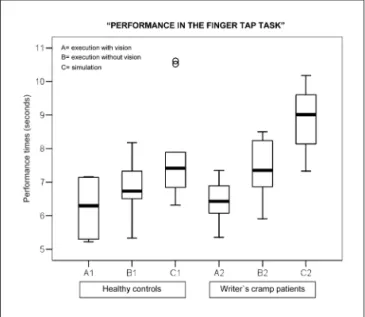
Patients and controls had some similarities in their performances in the “inger tap task” (Fig 1). There were not signiicant differences between them for motor exe-cution of the task with vision of the movements (p=0.666). However, patients with WC had a tendency to be slow-er than controls for the motor execution without vision (p=0.190) and specialy for the mental simulation of the
movements (p=0.094). Patients and controls were signii-cantly slower to mentally simulate (patients p=0.008, con-trols p=0.012) and to execute the “inger tap task” without visual control of the movements (patients p=0.008, con-trols p=0.015), than to execute the task with vision of the movements. Moreover, patients were also slower to men-tally simulate the movements than to execute the task without vision of the movements (p=0.008).
Patients and controls differed significantly in their performances in the “writing task” (Fig 2). Patients with WC were slower than controls to execute writing with vi-sion of the movements (p=0.0001), without vivi-sion of the movements (p=0.0001) and also to mentally simulate writ-ing (p=0.04). The PTs of the control subjects in the “writ-ing task” were not signiicantly different in the three con-ditions of performance (p=0.05) whereas the PTs of the patients with WC patients were longer for motor exe-cution with vision (p=0.021) and without vision (p=0.021) of the movements than for the mental simulation of the “writing task”.
We found strong correlations between the times spent for execution with vision and without vision of the move-mens for the inger tap task and for the writing task for patients and controls (Table). Otherwise, there were not signiicant correlations between the times spent for exe-cution and for simulation of the writing task by patients and controls and for the inger tap task by controls. How-ever, patients with WC had significant correlations for simulation and for execution of the inger tap task. Fig 1. Box plot showing the performances of WC patients and healthy
controls in the “inger tap task”. The performances of patients and controls did not differed signiicantly but patients with WC had a tendency to be slower than controls for the motor execution without vision (p=0.190) and specialy for the mental simulation of the move-ments (p=0.094). Patients and controls were signiicantly slower to mentally simulate and to execute the task without visual control of the movements than to execute the task with vision of the move-ments. Moreover, patients were also slower to mentally simulate the movements than to execute the task without vision of the movements.
DISCUSSION
In this study we found that patients with WC were slower than control subjects to execute and also to simu-late a writing task. Otherwise, they also had a tendency to be slower to simulate a simple repetitive inger tap task, despite their performances in the motor execution of this task were very close to that attained by the healthy con-trol subjects. These indings suggest that in WC there is a signiicant slowing in the mental process enrolled in the KMI, and that motor imagery may unmasks subclinical in-volvement of other apparently unaffected manual tasks. KMI is an introspective kinesthetic feeling of moving the limb as to mentally reproduce its own execution. Its physiological basis shows several parallels with motor ex-ecution1. Functional studies showed that KMI activates al-most the same cerebral areas involved in movement, and some intrinsic physiological features of the movements are also preserved during KMI1,3,13. It is argued that one of these preserved features is the high resemblance between the time spent to execute and to simulate the same move-ment, supporting the view that KMI and motor execution share common neural structures2,3,5,14. It is thought that KMI is encoded in an internal program that relies in the same representations of motor execution and keeps un-touched the temporal relationship between them, so that the time spent to simulate or to execute an action should be similar1,4,15,16. Despite the strong agreement about this topic in the literature, we did not ind signiicant corre-lations between the times spent to execute and to sim-ulate the hand movements in healthy controls. We were not able to explain this unexpected inding. However, our results in healthy subjets were very close to that obtained in previous studies with similar design2.
The main clinical abnormalities in WC are present dur-ing the execution of hand movements, when the
dyston-ic contractions would be produced by an improper func-tioning of a sensorimotor link which could lead to abnor-malities in the control of the movement17. Slowness for writing seems to be mainly related to the clumsiness for the execution of movements due to the abnormal mus-cular contractions18-23. However, during KMI, the motor pathways are only partially activated and probably could not account for the abnormalities that we observed. The fact that the the mental effort to simulate handwriting could not elicit involuntary muscle contractions in our patients, suggest that the mechanisms of muscular activ-ity blockade during simulation of movement was not im-paired in simple WC. So, we would not expect at irst that peripheral inluences could had intervened with the pro-cess of KMI1,3. Then, our indings suggest that part of the motor slowness observed during writing in WC patients may be due to a primary slowing in the motor planning processes.
There are suficient data indicating that patients with WC present many abnormalities preceding the execution of the movements8-11. The task-speciic character of the disease also suggests that the abnormalities may be di-rectly linked to dysfunctions of a speciic motor plan or in the linkage between the motor plan and its proper efec-tor, or both6. The mechanism associated with slowing of kinesthetic imagery in patients with WC is unknown. Func-tional studies suggest that basal ganglia are involved in the process of KMI1,13, and there are reports showing that the pathological involvement of this system may induce abnormalities in KMI. Some studies demonstrated that patients with Parkinson’s disease were slower than nor-mal subjects regarding mental simulation of simple hand movements and rotational tasks2,16. However in WC there are many indings indicating that in addition to the bas-al ganglia there are abnormbas-alities in primary motor
cor-Table. Spearman’s correlation coeficient between the performance times of patients with WC and healthy control subjects of the writing task and the inger tap task.
Patients with WC Control subjects
Execution without vision Simulation Execution without vision Simulation Finger tap task
Execution with vision of movements
0.90 p=0.001*
0.91 p=0.001*
0.88 p=0.002*
0.48 p=0.18 Execution without
vision of movements
1 0.78 p=0.01*
1 0.23 p=0.54 Writing task
Execution with vision of movements
0.91 p=0.001*
–0.11 p=0.76
0.85 p=0.004*
0.25 p=0.51 Execution without
vision of movements
1 0.05 p=0.89
tex, prefrontal motor areas and also in the somatosenso-ry areas24-26, and there appear to be distinct cortico-sub-cortical neural circuits involved in imagined movements including bilateral premotor, prefrontal, supplementary motor and left parietal areas27.
The primary motor cortex is not the main suspected area for KMI slowing since it is not essential for imagery, and we know that damage restricted to the primary mo-tor area does not result in impairment of KMI3,28. Soma-tosensory areas are also dysfunctional in focal hand dy-stonia and we cannot deinitely rule out their participa-tion in KMI deicits, since parietal lesions were found to disturb the chronometry of imagined inger movements despite the fact that parietal activation is often absent during kinesthetic imagery of simple movements3,15. Our preferential hypothesis was that the kinesthetic KMI ab-normalities in WC are due to dysfunctions in the prefron-tal motor areas produced by abnormal striatofronprefron-tal pro-jections since the posterior SMA and the premotor cor-tex seems to be the predominant areas involved in move-ment imagery3.
Previous activation studies in patients with dystonia have failed to show abnormalities during mental simu-lation of movements24. However, recent indings showed that patients with WC were slower than controls in per-forming a visual imagery task involving rotations of the hands but not of the feet29. Visual imagery must be dif-ferentiated from kinesthetic motor imagery since do not activate the motor system as the later3. In addition, func-tional MRI studies have shown abnormalities in cortical activation during imagination of hands movements in sec-ondary dystonia14. These indings are converging to dem-onstrate that patients with WC have a global impairment in KMI that is worst for the task and body area affected by the abnormal movements. The inding that our patients with simple WC also had a tendency for slower simulation of the inger tap task may disclose a subclinical involve-ment of other manual tasks.
In our experiment, the variable evaluated was the time spent to perform and to simulate each motor sequence, what is a very simple way to study a complex cognitive phenomenon. However, we may argue that the expected low sensitivity of this method just strengthened our ind-ings. Our observations in normal subjects were very close to those reported by some previous studies, suggesting that our indings are reliable2. Another critical point is that it is pratically impossible to control for the quality of im-agery. We tried to overcome possible timing errors dur-ing acquisition of KMI by includdur-ing subjects with high ed-ucational level, subjectively checking their sensation of vividness of the simulated movements and closely check the compliance of individuals to promptly advise the ex-act moment when the simulation was ended.
We also need to stand out an alternative hypotheis for our indings that can not be completely ruled out. It is possible that the slowing in the simulation of writing in patients with WC may only represent a proportional re-adaptation of the internal simulation of the movements to the slowing of the execution of the movements. This adaptative response would be due to an internal control system that would mediate a change in the simulation times that would be proportional to the slowing in the execution times16. We can argue against this hypothesis that we disclosed slowing in the simulation of the inger tap task by patients with WC without any accompanying slowing in the execution of the movements. Also, there was not correlation between the times spent to write and to simulate writing by patients, what was predicted by this hypothesis.
In conclusion, patients with WC seem to have slow-ing in the processes of mental simulation of hand move-ments that is not speciic for writing.
REFERENCES
1. Jeannerod M, Decety J. Mental motor imagery: a window into the representational stages of action. Curr Op Neurobiol 1995;5:727-732.
2. Dominey P, Decety J, Broussolle E, Chazot G, Jeannerod M. Motor imagery of a lateralized sequential task is asymmetri-cally slowed in hemi-Parkinson’s patients. Neuropsychologia 1995;33:727-741.
3. Lotze M, Halsband U. Motor imagery. J Physiol (Paris) 2006;99: 386-395.
4. Decety J, Michel F. Comparative analysis of actual and mental movement times in two graphic tasks. Brain Cog 1989;11:87-97. 5. Stephan KM, Fink GR, Passingham RE, et al. Functional anat-omy of the mental representation of upper extremity move-ments in healthy subjects. J Neurophysiol 1995;73:373-386. 6. Hallett M. Pathophysiology of writer’s cramp. Human Mov
Sci 2006;25:454-463.
7. Sheehy MP, Marsden CD. Writers’ cramp-a focal dystonia. Brain 1982;105:461-480.
8. Deuschl G, Toro C, Matsumoto J, Hallett M. Movement-re-lated cortical potentials in writer’s cramp. Ann Neurol 1995; 38:862-868.
9. Van der Kamp W, Rothwell JC, Thompson PD, Day BL, Mars-den CD. The movement-related cortical potential is abnor-mal in patients with idiopathic torsion dystonia. Mov Disord 1995;10(5):630-633.
10. Odergren T, Stone-Elander S, Ingvar M. Cerebral and cerebel-lar activation in correlation to the action-induced dystonia in writer’s cramp. Mov Disord 1998;13:497-508.
11. Hamano T, Kaji R, Katayama M, et al. Abnormal contingent negative variation in writer’s cramp. Clin Neurophysiol 1999; 110:508-515.
Friedman J. Validity and reliability of a rating scale for the pri-mary torsion dystonias. Neurology 1985;35:73-77.
13. Szameitat AJ, Shen S, Sterr A. Motor imagery of complex every-day movements. An fMRI study. NeuroImage 2007;34:702-713. 14. Lehericy S, Gerardin E, Poline JB, et al. Motor execution and
imagination networks in post-stroke dystonia. Neuroreport 2004;15:1887-1890.
15. Sirigu A, Duhamel JR, Cohen L, Pillon B, Dubois B, Agid Y. The mental representation of hand movements after parietal cortex damage. Science (New York) 1996;273:1564-1568. 16. Sabate M, Gonzalez B, Rodriguez M. Adapting movement
planning to motor impairments: the motor-scanning system. Neuropsychologia 2007;45:378-386.
17. Kaji R, Rothwell JC, Katayama M, et al. Tonic vibration relex
and muscle afferent block in writer’s cramp. Ann Neurol 1995; 38:155-162.
18. Von Reis G. Electromyographical studies in writer’s cramp. Acta Med Scand 1954;149:253-260.
19. Hughes M, McLellan DL. Increased co-activation of the upper limb muscles in writer’s cramp. J Neurol Neurosurg Psychiatry 1985;48:782-787.
20. van der Kamp W, Berardelli A, Rothwell JC, Thompson PD, Day BL, Marsden CD. Rapid elbow movements in patients with torsion dystonia. J Neurol Neurosurg Psychiatry 1989; 52:1043-1049.
21. Agostino R, Berardelli A, Formica A, Accornero N, Manfredi M. Sequential arm movements in patients with Parkinson’s
disease, Huntington’s disease and dystonia. Brain 1992;115: 1481-1495.
22. Inzelberg R, Flash T, Schechtman E, Korczyn AD. Kinematic properties of upper limb trajectories in idiopathic torsion dy-stonia. J Neurol Neurosurg Psychiatry 1995;58:312-319. 23. Berardelli A, Hallett M, Rothwell JC, et al. Single-joint rapid
arm movements in normal subjects and in patients with mo-tor disorders. Brain 1996;119:661-674.
24. Ceballos-Baumann AO, Brooks DJ. Activation positron emis-sion tomography scanning in dystonia. Adv Neurol 1998;78: 135-152.
25. Bara-Jimenez W, Catalan MJ, Hallett M, Gerloff C. Abnormal so-matosensory homunculus in dystonia of the hand. Ann Neurol 1998;44:828-831.
26. Tinazzi M, Frasson E, Polo A, et al. Evidence for an abnormal cortical sensory processing in dystonia: selective enhancement of lower limb P37-N50 somatosensory evoked potential. Mov Disord 1999;14:473-480.
27. Gerardin E, Sirigu A, Lehericy S, et al. Partially overlapping neural networks for real and imagined hand movements. Cereb Cortex 2000;10:1093-1104.
28. Sirigu A, Cohen L, Duhamel JR, et al. Congruent unilateral im-pairments for real and imagined hand movements. Neuroreport 1995;6:997-1001.