Arq Neuropsiquiatr 2007;65(1):164-166
1Clínica Neurológica Professor Fernando Pompeu, Rio de Janeiro, Brazil; 2Programa de Epilepsia do Instituto de Neurologia Deolindo Couto e Programa de Pós-Graduação de Psiquiatria e Saúde Mental, Universidade Federal do Rio de Janeiro, Brasil (UFRJ).
Received 21 June 2006, received in final form 9 October 2006. Accepted 17 November 2006.
Dra. Michele Dominici - Avenida Prado Jr 307 / 604 - 22011-040 Rio de Janeiro RJ - Brasil. E-mail: [email protected]
PROBABLE CAUSAL LINK BETWEEN
EPILEPSY AND SLEEP APNEA
Case report
Michele Dominici
1,2, Fernando Pompeu Filho
1, Marleide da Mota Gomes
2ABSTRACT - Patients with epilepsy were reported to have concomitant sleep apnea, but it has been rarely linked to the epilepsy itself. We present a case of a 28-year-old, obese man with secondary medically resist-ant partial complex epilepsy due to a brain trauma, with progressive snoring, and sleep agitation, apneas, and important daytime somnolence. It was noticed in the polysomnographic study that he had several sleep respiratory events, probably due both to the epileptic seizures and the sleep apnea syndrome as a co-morbidity. Apnea and epilepsy will be discussed. A careful video-EEG-polysomnography study is impor-tant in evaluating refractory epileptic patients and/or epileptic patients with snoring.
KEY WORDS: snoring, obstructive sleep apnea, epilepsy.
Provável associação entre epilepsia e apnéia do sono: relato de caso
RESUMO - Pacientes com epilepsia e concomitante apnéia do sono já foram descritos na literatura, mas raramente foram associados à epilepsia como fator causal desta apnéia. Nós apresentamos o caso de um homem com 28 anos, obeso, com epilepsia parcial complexa farmacorresistente secundária a trauma crânio encefálico e roncos progressivos, sono agitado, apnéias, além de importante sonolência diurna. Foram ob-servados, durante estudo polissonográfico, freqüentes eventos respiratórios durante o sono, provavel-mente secundários a crise epilética, além da síndrome de apnéia obstrutiva do sono como uma co-mor-bidade. Um exame cuidadoso de vídeo-EEG-polissonografia do caso é importante na avaliação de pacientes com epilepsia fármaco resistente e/ou epilépticos com roncos.
PALAVRAS-CHAVE: ronco, apnéia obstrutiva do sono, epilepsia.
It is suggested that a third or more of epilepsy patients have obstructive sleep apnea, and it may be even more prevalent in medically resistant epilepsy patients1. Sleep apnea does disrupt sleep and
caus-es exccaus-essive daytime sleepincaus-ess, an inability to con-centrate and headaches. If, however, the person has epilepsy, then the sleep disruption can bring on sei-zures. It has been shown by Devinsky et al.2and
oth-er authors that treating sleep apnea will also help to lessen the frequency of epileptic seizures. Sleep brea-thing disorders fragment sleep causing daytime sleep-iness, migraine, attention deficits and in epileptic patients apnea may facilitate the occurrence of sei-zures. Patients with epilepsy and concomitant obs-tructive sleep apnea have been described in the lit-erature, but epilepsy has rarely been associated “per se” to the sleep breathing apnea. The syndrome of
sudden death of unidentified cause (SUDEP) is an im-portant cause of death among patients with epilep-sy (7-17%), particularly in the group of patients with refractory epilepsy (50%). Studies in this group of patients are growing, but according to Kanner3they
have not entirely addressed the risks of death in the daily clinic patients. Studies trying to correlate SUDEP with the autonomous abnormalities in cardiac and respiratory function observed during seizures were performed4, however the results are not entirely
esta-blished yet. The coexistence of epilepsy and apnea suggests a link between structures of the brain that are responsible for epilepsy and those which control breath like the insula, amygddala, cingulated gyrus, and orbitofrontal cortex5.
intermit-Arq Neuropsiquiatr 2007;65(1) 165
tent apneas were related to both the epilepsy and the obstructive apnea syndrome.
CASE
A 28-year-old obese male was referred to a polysomno-graphic center for his seizures evaluation. The seizures were medically resistant and secondary to a brain trauma that occurred after a vehicle accident. He also had non position-al snoring, arousposition-al, apneas and daytime sleepiness. His cur-rent medications were valproate, carbamazepine, pheno-barbital and clonazepan. The patient complained of pro-gressive daytime somnolence. His Epworth Sleepiness Scale (ESS) was 15 and his weight was 100 kg (BMI=39).
Whole night video-EEG-polysomnography study was performed as part of his epilepsy investigation. It was reg-istered results of a 18 channels EEG, electro-oculogram, electromyogram, electrocardiogram, nasal/oral airflow and
abdominal/thoracic respiratory movements. Sleep stages were visually scored following standard criteria. A magnet-ic resonance image (MRI) demonstrated areas of encephalo-malacia in cortical and sub-cortical frontal lobes. The neu-rologic examination showed only cognitive decline. The video-EEG-polysomnography revealed: inter-ictal multi-focal spikes (frontal left and right independent and mid temporal left and right); ictal EEG seizure pattern alterna-tively beginning in both hemispheres and sometimes spreading to the contra-lateral side (Figs 1, 2, 3). It was reg-istered several auto motor seizures, frequently (58) associ-ated with autonomic changes (central apneas, bradycar-dia) and obstructive sleep apneas and hypopneas (Figs 4, 5, 6). In the whole exam, there were detected 21 obstruc-tive sleep apneas, 29 central apneas and 126 hypopneas with an apnea and hypopnea index (AHI) of 28/h and 226 dessaturations.
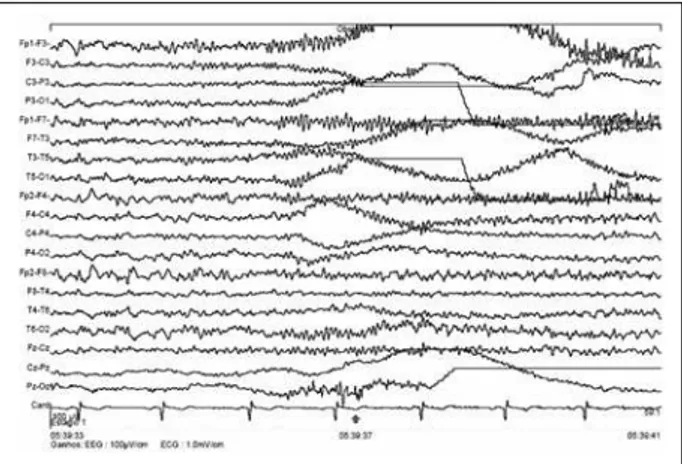
Fig 1. Epoch with 8 s of EEG. The ictal EEG seizure pattern begin in the right hemisphere and spread to the contra-lateral side, with related EEG runs of polyspike in the left anterior areas related to the central sleep apnea (Fig 4).
Fig 3. Epoch with 8 s of EEG. The ictal EEG seizure pattern is dominant in the left hemisphere. Related EEG showing runs of polyspike in the left fronto-polar area compatible with the patient brain injury related to the obstructive sleep apnea (Fig 6).
Fig 4. Epoch with 20 s of standard PSG, showing a central sleep apnea, associated with a seizure rhythm pattern in the EEG ending in a auto-motor seizure recorded by the video-registra-tion. Probable central apnea (polysomnographic view) seizure related.
DISCUSSION
Cases with multiple risk factors such as brain le-sions, exposure to several drugs with respiratory and central nervous system suppressant effects and obe-sity render it difficult to the physician to realize what is cause and effect. Obstructive sleep apnea has been reported in people with epilepsy. Nevertheless, there have been only a limited number of reports of sleep apneas due to epileptic seizures. Thus, we speculate that this condition may be neglected in most instan-ces. Our case presented both types of apnea: central and obstructive, either primary or secondary to sei-zures. Clinicians should investigate apnea in epilep-tic patients when indicated. Polysomnography can help to identify seizure’s related apnea and also per-mits the clinician to provide the best medical care. In our opinion, this study should be performed in all medically resistant epilepsy patient suspected to have apnea or unexplained daytime somnolence. Better understanding the role of these two related events (apnea and epilepsy) in the spectrum of the sudden unidentified death epileptic syndrome (SUDEP), may help to clarify the etiopathogenesis of this syndrome.
In conclusion, we presented a case of a patient with refractory epilepsy, which video-EEG-polysomno-graphy demonstrated a significant amount (33%) of respiratory events associated to seizures. We believe that these apneic related events may help to unrav-el the etiopathogenesis of SUDEP and to better un-derstand the daytime symptoms of epileptic patients. This case shows also the importance of using more frequently a Video- EEG-PSG study in a subset of re-fractory epilepsy patients with or without snoring.
Acknowledgments – We acknowledge the thorough-ly paper English revision made by Ms Livia Sanches Dominici.
REFERENCES
1. Malow BA, Levy K, Maturen K, Bowes R. Obstructive sleep apnea is common in medically refractory epilepsy patients. Neurology 2000;5 5:1002-1007.
2. Devinsky O, Ehrenberg B, Barthlen GM, Abramson HS, Luciano D. Epilepsy and sleep apnea syndrome. Neurology 1994;44:2060-2064. 3. Kanner AM. Unrevealing the secrets of sudden death in epilepsy: is it
possible? Epilepsy Curr 2004;4,225-226.
4. O’Regan ME, Brown JK. Abnormalities in cardiac and respiratory func-tion observed during seizures in childhood. Dev Med Child Neurol 2005;47:4-9.
5. Devinsky O. Effects of seizures on autonomic and cardiovascular func-tion. Epilepsy Curr 2004;4:43-46.
166 Arq Neuropsiquiatr 2007;65(1)
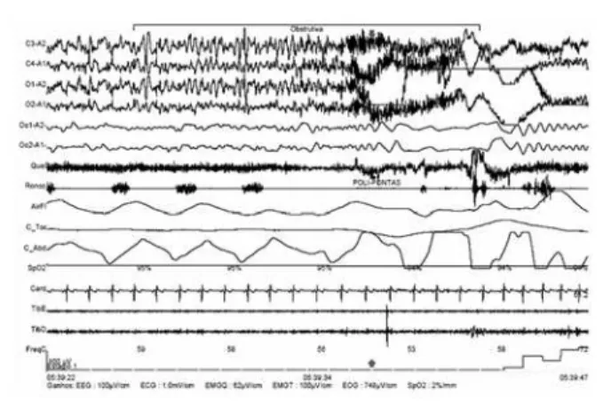
Fig 5. Epoch with 25 s of standard PSG, showing an obstruc-tive sleep apnea associated with an ictal seizures EEG pattern ending with an automotor crises registered in the videomon-itorization. Probable obstructive critical apnea (polysomno-graphic view).