Risk factors for pulmonary hypertension in patients
receiving maintenance peritoneal dialysis
Y. Zeng
1*, D.D. Yang
1*, S. Feng
1, H.Y. Shen
1, Z. Wang
1, S. Jiang
1, Y.B. Shi
1and J.X. Fu
2 1Department of Nephrology, The Second Affiliated Hospital of Soochow University, Suzhou, China 2Department of Hematology, The Second Affiliated Hospital of Soochow University, Suzhou, ChinaAbstract
We investigated the risk factors for pulmonary hypertension (PH) in patients receiving maintenance peritoneal dialysis (MPD). A group of 180 end-stage renal disease patients (124 men and 56 women; mean age: 56.43±8.36) were enrolled in our study, which was conducted between January 2009 and June 2014. All of the patients received MPD treatment in the Dialysis Center of the Second Affiliated Hospital of Soochow University. Clinical data, laboratory indices, and echocardiographic data from these patients were collected, and follow-ups were scheduled bi-monthly. The incidence and relevant risk factors of PH were analyzed. The differences in measurement data were compared byt-test and enumeration data were compared with thew2test. Among the 180 patients receiving
MPD, 60 were diagnosed with PH. The remaining 120 were regarded as the non-PH group. Significant differences were observed in the clinical data, laboratory indices, and echocardiographic data between the PH and non-PH patients (all Po0.05). Furthermore,
hypertensive nephropathy patients on MPD showed a significantly higher incidence of PH compared with non-hypertensive nephropathy patients (Po0.05). Logistic regression analysis showed that the proportion of internal arteriovenousfistula, C-reactive
protein levels, and ejection fraction were the highest risk factors for PH in patients receiving MPD. Our study shows that there is a high incidence of PH in patients receiving MPD and hypertensive nephropathy patients have an increased susceptibility to PH.
Key words: Maintenance peritoneal dialysis; Pulmonary hypertension; End-stage renal disease; Internal arteriovenousfistula; Hypertensive nephropathy; Ejection fraction
Introduction
End-stage renal disease (ESRD) affects 10–16% of
adults worldwide. It is characterized by a significantly
reduced estimated glomerular filtration rate and increased
urinary albumin excretion (1). ESRD is clinically defined as
kidney failure requiring dialysis or transplantation, and is associated with high healthcare costs and mortality. ESRD costs nearly US$23 billion each year in health care costs in the US, and the mortality rates are eight times higher in 20- to 64-year-old ESRD patients treated by dialysis than those individuals of similar age (2). The United States Renal Data System annual data report showed that the mortality of chronic kidney disease (CKD) patients in 2008 was 1.7 times higher than that of non-CKD patients after adjusting for age, gender, race, prior hospitalizations, and comorbidity (3). In-center hemodialysis and home peritoneal dialysis (PD) are the two most common dialysis therapies. Costs associated with PD are almost US$20,000 lower than those of hemo-dialysis, but PD and hemodialysis have similar health outcomes (4). Maintenance peritoneal dialysis (MPD) is a common renal replacement therapy for ESRD patients (3).
However, MPD is complicated by diseases such as pul-monary hypertension (PH).
PH was previously classified into two categories,
primary PH or secondary PH, based on the presence of identified causes or risk factors (5). PH is linked to diverse etiologies, such as left heart failure, chronic hypoxic lung diseases, collagen vascular disease, portal hypertension,
chronic recurrent thromboembolism, human immunodefi
-ciency virus infection, and exposure to drugs and toxins
(6–8). PH is frequently associated with CKD and ESRD,
and is hemodynamically defined as a mean pulmonary
artery pressure (PAP) greater than 25 mmHg. A recent study suggested that left heart dysfunction is the underlying cause of PH in patients with kidney disease (9). The incidence of PH is high in ESRD patients and ranges from 9 to 39% in Stage 5 CKD patients, between 18.8 and 68.8% in hemodialysis patients, and between 0 and 42% in patients receiving PD treatment (10). Several mechanisms have been proposed for the incidence of PH in kidney diseases, such as left ventricular disorders, arteriovenous
Correspondence: H.Y. Shen:<[email protected]>| J.X. Fu:<[email protected]>
*These authors contributed equally to this study.
fistula, volume overload, sleep disorder, dialysis membrane
exposure, endothelial dysfunction, and vascular calcification
(11). Genctoy et al. found that older age, lower ejection fraction, and secondary hyperparathyroidism may contribute to PH in CKD with proteinuria (12). Alhamad et al. studied the clinical characteristics of PH patients receiving hemo-dialysis or peritoneal hemo-dialysis versus patients receiving renal transplantation (13). However, few studies have investi-gated the incidence of PH in patients receiving MPD and the risk factors promoting PH in MPD patients.
In this study, we investigated the incidence of PH in patients receiving MPD. We also examined potential risk factors through a detailed analysis of clinical data, laboratory indices, and echocardiographic data that were collected during bi-monthly follow-up visits.
Material and Methods
Patients
Between January 2009 and June 2014, 180 ESRD patients receiving MPD treatment at the Dialysis Center
of the Second Affiliated Hospital of Soochow University
were selected for this study, which was approved by the
Hospital’s Ethical Committee. The patient group consisted
of 124 men and 56 women (mean age: 56.43±8.36; mean
dialysis time: 36.35±3.38 months). Inclusion criteria for
patients in this study were as follows: i) patients were
treated by continuous ambulatory peritoneal dialysis and their daily cumulative dialysate dose was 6–8 L;ii) patients received renal replacement therapy for longer than 6 months
and were in a stable condition;iii) patients were compliant
and willing to be followed up; andiv) patients were older
than 18 years of age. All of the patients received pulmonary perfusion imaging and showed even perfusion without perfusion defects. Written informed consent was obtained from all patients.
The exclusion criteria of patients were as follows:
i) patients with chronic lung disease that could greatly
affect PAP (14) (e.g., dyspnea and a state of air intake with
arterial blood gas analysis showing a pHo7.35, chronic
thromboembolic disease, which can also greatly affect PAP (15), obvious arteriovenous ischemia, or
thromboem-bolism diagnosed by imaging methodology); ii) patients
having received renal replacement therapy within the past 6 months or who were hospitalized during renal
replace-ment therapy within the past 3 months; iii) patients with
severe left heart disease, such as New York Heart Association class III/IV heart failure and a left ventricular
ejection fraction o50%; andiv) patients having received
prior hemodialysis. Additionally, we excluded patients with the following diseases that cause pulmonary hyperten-sion: congenital heart disease, acute coronary syndrome, postinfarction syndrome, valvular heart disease (valvular regurgitation or grade II or above valvular stenosis), pericardial disease, autoimmune disease, chest wall or lung parenchymal disease, and pulmonary embolism.
Collection of clinical data, laboratory indices, and echocardiographic data
Clinical data including age, gender, smoking (ex-smokers or current (ex-smokers), height, weight, interdialytic
weight gain, total dialysate, average ultrafiltration volume,
liquid removal volume, pulmonary function tests (forced vital capacity and forced expiratory volume in 1 second), dialysis time, systolic pressure, diastolic pressure, mean arterial pressure, and body mass index were recorded. Chronic renal failure and its complications were also recorded. Laboratory indices were recorded, including the serum levels of albumin, total bilirubin, alanine transaminase, aspartate aminotransferase, hemoglobin, brain natriuretic peptide (BNP), parathyroid hormone, calcium, phosphorus, creati-nine, urea nitrogen, and C-reactive protein (CRP). Ultrasonic cardiographic data were recorded, including the right ventricular diameter, right ventricular outflow tract diameter, main pulmonary artery diameter, left atrial diameter, left ventricular diastolic diameter, left ventricular systolic dia-meter, left ventricular outflow tract diameter, interventricular septal thickness, aortic root dimension, ejection fraction, mitral regurgitation, pericardial effusion, and left ventricular mass index.
Follow-up and diagnostic criteria of PH
After collecting clinical data, laboratory indices and data from ultrasonic cardiograms were collected. Patients were followed up bi-monthly. The termination of follow-up included patients with complicated PH or a follow-up time of longer than 36 months. During follow-up, PAP was evaluated by echocardiography. Systolic tricuspid regurgitant jet velocity (V) was measured directly, and PAP was calculated according
to Bernoulli’s equation: PAP = 4 V2+10 mmHg (16,17).
PH was confirmed by a systolic PAPX35 mmHg according
to echocardiographic assessment of the right heart guidelines published by the American Society of Echocardiography in 2010 (18). The date and time of diagnosis of PH were recorded, and patients were then categorized into either the PH group or the non-PH group. The clinical differences between PH and non-PH groups were compared to identify risk factors for PH in patients receiving MPD.
Statistical analysis
Data were analyzed by SPSS 20.0 (IBM Corp., USA).
Measurement data are reported as means±SD and the
differences were measured by the t-test. Enumeration data
are reported as percentage or rate, and the differences were
compared with the w2 test. Factors related to PH were
analyzed by logistic regression analysis. A P value o0.05
was considered to be significant.
Results
Patients receiving MPD diagnosed with PH
group, with PAP ranging from 35.0 to 62.2 mmHg (mean
PAP: 53.6±9.6 mmHg). Therefore, the incidence of PH in
this cohort was 33.3%. In the PH group, 40 patients were
men, 20 were women, and the mean age was 59.65±11.24
years. The remaining 120 patients were categorized as non-PH and included 84 men and 36 women, with a mean age of
61.15±10.61 years. PAP of the non-PH patients ranged from
12.5 to 34.8 mmHg (mean PAP: 21.3±6.4 mmHg).
Clinical data
Smoking, systolic pressure, diastolic pressure, mean arterial pressure, and the proportion of internal arteriovenous
fistula were significantly higher in the PH group than in the
non-PH group (all Po0.05). However, there were no
significant differences in age, gender, body mass index,
interdialytic weight gain, dialysis time, total dialysate, average
ultrafiltration volume, liquid removal volume, forced vital
capacity, and forced expiratory volume in 1 second between
the PH and the non-PH groups (all P40.05; Table 1).
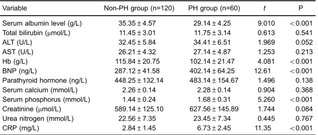
Laboratory indices
Serum BNP, phosphorus, and CRP levels were
significantly higher in the PH group than in the non-PH
group (all Po0.05). However, serum albumin levels and
hemoglobin were significantly lower in the PH group than
in the non-PH group (Po0.05). There were no significant
differences in serum total bilirubin, aspartate aminotrans-ferase, alanine transaminase, parathyroid hormone, cal-cium, creatinine, and urea nitrogen levels between the two
groups (P40.05; Table 2).
Primary disease
With regard to primary diseases in the 180 patients receiving MPD, 35% had chronic glomerulonephritis, 33.3% had hypertensive nephropathy, 18.9% had diabetic nephro-pathy, and 12.8% had other diseases, such as aristolochic acid nephropathy or polycystic kidney disease The incidence
of PH was significantly higher in hypertensive nephropathy
patients than in any of the non-hypertensive nephropathy patients (Po0.05). There was no significant difference in the incidence of PH between chronic glomerulonephritis patients, diabetic nephropathy patients, or other primary
disease patients (P40.05; Table 3), indicating that these
diseases had a minimal influence on PH in MBD patients.
Echocardiography
The PH group showed a significantly higher right
ventricular diameter, right ventricular outflow tract diameter, main pulmonary artery diameter, left atrial diameter, and interventricular septum thickness compared with the non-PH group (all Po0.001). The incidence of mitral regurgita-tion and pericardial effusion was also significantly higher in the PH group compared with the non-PH group (both
Po0.05). However, the ejection fraction was significantly
lower in the PH group compared with the non-PH group (Po0.01). No significant changes in left ventricular diastolic diameter, left ventricular outflow tract, and aortic root dimen-sion were observed between the PH and non-PH groups
(all P40.05). The left ventricular mass index was also not
significantly different between the two groups (both P40.05; Table 4).
Table 1.Clinical data in the pulmonary hypertension (PH) and the non-PH groups.
Variable Non-PH group (n=120) PH group (n=60) t orw2 P
Age (years) 61.15±10.61 59.65±11.24 0.860 0.392
Gender (male/female) 84/36 40/20 0.207 0.649
Smoking 18 (15.0%) 21 (35.0%) 9.427 0.002
BMI (kg/m2) 23.85±4.23 24.75±4.85 0.397 0.692
IDWG 2.93±1.02 2.64±0.98 1.822 0.070
Dialysis time (months) 35.86±2.95 36.12±3.01 0.550 0.583
Total dialysate (L) 8.53±0.65 8.64±0.59 1.103 0.272
Average ultrafiltration volume (mL) 1435.23±378.52 1498.29±355.64 1.075 0.284 Liquid removal volume (mL) 1698.26±425.65 1678.36±465.37 0.287 0.775
FVC (V/mL) 3702±850 3955±812 1.910 0.058
FEV1 (V/mL) 2637±560 2786±694 1.551 0.123
Systolic pressures (mmHg) 140.12±18.54 151.24±20.15 3.583 o0.001 Diastolic pressures (mmHg) 70.41±11.87 80.35±11.01 5.561 o0.001 Mean arterial pressure (mmHg) 106.21±16.6 116.21±17.6 3.662 o0.001 Internal arteriovenousfistula (n) 70 (58.3%) 45 (75%) 4.816 0.028 Non-internal arteriovenousfistula (n) 50 (41.7%) 15 (25%)
Analysis of risk factors for patients receiving MPD complicated by PH
We next examined the risk factors for development of PH using logistic regression analysis of systolic blood pressure, diastolic blood pressure, mean arterial pressure, the proportion
of arteriovenousfistula, serum BNP levels, phosphorus levels,
CRP levels, albumin levels, hemoglobin levels, hypertensive nephropathy, right ventricular diameter, right ventricular outflow tract diameter, main pulmonary artery, left atrial diameter, interventricular septum thickness, ejection fraction, mitral regurgitation, and pericardial effusion. These factors were found to be significantly different in single factor analysis when comparing the PH group with the non-PH group. A forward conditional logistic regression method was used and showed
that the proportion of arteriovenous fistula, CRP levels, and
ejection fraction were the highest risk factors for PH in patients receiving MPD (Table 5).
Discussion
This is thefirst prospective study to analyze patients
receiving MPD for complications leading to PH. We found a high occurrence of PH (33.3%) in this cohort of ESRD patients receiving MPD, which is consistent with previous
studies reporting similar high incidence rates. One previous
study observed a prevalence of PH as high as 30–60% in
cohorts and correlated this high incidence with increased mortality and poor renal transplantation outcome (19). Another study showed that uremic patients receiving chronic dialysis treatment had a high prevalence of PH. This effect was mainly due to elevated pulmonary blood
flow and increased pulmonary vascular resistance, which
was worsened by fluid overload in hemodialysis patients
(20). PH in CKD patients may be induced by left ventricular disorders and some of the typical risk factors of CKD (21). Leung et al. also demonstrated that variation in PH is largely explained by the degree of pulmonary venous hypertension (22). The pathogenesis of PH in ESRD patients might also be due to alterations in endothelial function, increased cardiac output, and myocardial dysfunction, leading to an elevated left heartfilling pressure (9).
To examine the underlying cause of PH in patients receiving MPD, we analyzed clinical data, laboratory indices, and echocardiographic data. We also used echocardiogra-phy in the PH and non-PH groups during follow-up visits. We found that smoking, systolic pressure, diastolic pressure, mean arterial pressure and the proportion of internal
arteriovenous fistula were significantly higher in the PH
Table 2.Laboratory indices in the pulmonary hypertension (PH) and the non-PH groups.
Variable Non-PH group (n=120) PH group (n=60) t P
Serum albumin level (g/L) 35.35±4.57 29.14±4.25 9.010 o0.001 Total bilirubin (mmol/L) 11.45±3.01 11.75±3.14 0.613 0.541
ALT (U/L) 32.45±5.84 34.41±6.51 1.969 0.052
AST (U/L) 26.21±4.32 27.14±4.87 1.253 0.213
Hb (g/L) 115.84±20.75 102.14±21.47 4.081 o0.001
BNP (ng/L) 287.12±41.58 402.14±64.25 12.61 o0.001
Parathyroid hormone (ng/L) 448.25±132.14 483.14±154.67 1.496 0.138
Serum calcium (mmol/L) 2.26±0.14 2.28±0.14 0.904 0.368
Serum phosphorus (mmol/L) 1.44±0.24 1.68±0.31 5.260 o0.001 Creatinine (mmol/L) 589.14±125.10 627.56±145.89 1.744 0.084 Urea nitrogen (mmol/L) 22.56±7.35 23.45±7.34 0.445 0.767
CRP (mg/L) 2.84±1.45 6.73±2.45 11.35 o0.001
Data are reported as means±SD. ALT: alanine transaminase; AST: aspartate aminotransferase; Hb: hemoglobin; BNP: brain natriuretic peptide; CRP: C-reactive protein. Thet-test was used for statistical analysis.
Table 3.Primary diseases in the pulmonary hypertension (PH) and the non-PH groups.
Chronic glomerulonephritis
Diabetic nephropathy
Hypertensive nephropathy
Other disease
Total cases (n=180) 63 (35.0) 34 (18.9) 60 (33.3) 23 (12.8) non-PH group (n=120) 45 (37.5) 27 (22.5) 30 (25.0) 18 (15.0)
PH group (n=60) 18 (30.0) 7 (11.7) 30 (50.0) 5 (8.3)
w2 0.578 2.160 5.333 1.259
P 0.447 0.141 0.021 0.261
group than in the non-PH group. Smoking remains a significant risk factor for lung disease and is also related to PH (23,24). Estimated systolic pressure and mean pulmon-ary arterial pressure are the two main echocardiographic
variables used to evaluate PH patients (25). PH is defined
as a mean PAP of at least 25 mmHg at rest (19). Internal
arteriovenous fistula remains the best choice for vascular
access in hemodialysis patients (26). After successfully
creating an arteriovenous fistula, arterial blood flow
in-creases 10- to 20-fold, which is associated with arterial and venous dilation, possibly leading to PH (27).
In the current study, laboratory indices showed that BNP,
serum phosphorus, and CRP levels were significantly higher
in the PH group than in the non-PH group. This finding
suggests that these factors might be involved in the development of PH in patients receiving MPD. BNP released from ventricular myocytes is a powerful predictor in cardio-vascular disease patients with atrialfibrillation, correlating with higher post-stroke mortality and cardio-embolic stroke (28). Additionally, elevated serum BNP levels are also reported in acute pulmonary embolism patients (29). Plasma BNP has also been reported by many studies as a prognostic indicator in patients with primary PH due to some degree of right ventricular dysfunction (30,31). Serum phosphorus levels play an important role in energy production, membrane transport,
and signal transduction (32). However, a report showed no
significant differences in serum phosphorus between
hemo-dialysis and PH (33). CRP has pro-inflammatory and
anti-inflammatory actions, and its pro-inflammatory effects include
induction of inflammatory cytokines and tissue factor. The
level of CRP correlates with PH and is an independent predictor of PH (34), as observed in this study.
In the current study, the incidence of PH in
hyperten-sive nephropathy patients was significantly higher than
that in non-hypertensive nephropathy patients, indicating that hypertensive nephropathy could lead to the develop-ment of PH. Hypertensive nephropathy associated with a
progressive decline in glomerular filtration rate, despite
reduced blood pressure (35), is the second leading cause of complete kidney failure. This condition is associated
with significant morbidity and mortality (36).
In our study, based on echocardiography results, the right ventricular diameter, right ventricular outflow tract diameter, main pulmonary artery diameter, left atrial diameter, and
interventricular septum thickness were significantly higher
in the PH group compared with the non-PH group, along
with a significantly higher incidence of mitral regurgitation
and pericardial effusion in the PH group. However, the
ejection fraction was significantly lower in the PH group
than in the non-PH group. PH is closely linked with right Table 4.Echocardiography in the pulmonary hypertension (PH) and the non-PH groups.
Variable non-PH group
(n=120)
PH group (n=60)
t P
Right ventricular diameter (mm) 20.54±2.21 24.87±2.08 12.890 o0.001 Right ventricular outflow tract diameter (mm) 27.54±4.02 32.45±4.54 7.100 o0.001 Main pulmonary artery diameter (mm) 23.64±3.10 25.47±2.84 3.951 o0.001 Left atrial diameter (mm) 41.86±6.06 46.82±5.57 5.467 o0.001 Left ventricular diastolic diameter (mm) 46.41±5.94 48.76±9.94 1.687 0.096 Left ventricular systolic diameter (mm) 34.54±5.12 35.35±6.10 0.885 0.379 Left ventricular outflow tract diameter (mm) 26.35±3.14 27.14±3.31 1.535 0.128 Interventricular septal thickness (mm) 11.01±2.00 12.20±2.01 3.751 o0.001 Aortic root dimension (mm) 30.58±3.12 31.02±3.15 0.886 0.377
Ejection fraction (%) 60.4±8.5 56.4±7.5 3.224 0.002
Mitral regurgitation (%) 44.2±4.6 76.4±7.5 30.510 o0.001
Pericardial effusion (%) 7.2±2.1 8.0±1.9 2.570 0.011
Left ventricular mass index 117.14±51.02 141.74±56.40 1.303 0.197
Data are reported as means±SD. Thet-test was used for statistical analysis.
Table 5. Forward conditional logistic regression analysis for risk factors of patients receiving maintenance peritoneal dialysis complicated by pulmonary hypertension.
Variable Regression coefficient Standard error Wald P EXP (B) 95% CI
Arteriovenousfistula proportion 0.762 0.351 4.716 0.030 2.143 1.077–4.263
C-reactive protein 1.619 0.273 35.101 0.000 5.049 2.955–8.628
Ejection fraction 0.434 0.104 17.512 0.000 1.543 1.259–1.890
ventricular dysfunction and left ventricular systolic dys-function, as well as left-sided valvular disease with chronic elevation in left ventricularfilling pressure (37). Ventricular hypertrophy, such as increased ventricular diameter, occurs in response to pressure overload in PH (38). Our
results of logistic regression analysis further confirmed
that the proportion of arteriovenous fistula, CRP levels,
and ejection fraction are the highest risk factors for PH in patients receiving MPD.
There are some limitations in the present study. First, we used an echocardiogram for the diagnosis of PH, which is not as accurate as right cardiac catheterization (39). PAP is conventionally evaluated by a right cardiac catheterization procedure. However, in our study, PAP was evaluated by echocardiogram because of the need to perform repeated PAP evaluations over an extended period of time. Addition-ally, the right cardiac catheterization procedure is traumatic
and might affect the patient’s compliance with examinations.
Second, we used laboratory indices, such as BNP and serum phosphorus, to analyze the potential mechanism of PH in patients receiving MPD. BNP is mainly used as a prognostic indicator for PH patients. The relationship between serum phosphorus and the development of PH is controversial.
Taken together, our results provide an important clinical reference for the risk of PH in patients receiving MPD. Further investigation is likely to reveal early diagnostic and treatment methods in patients receiving MPD, which are urgently needed, as well as the precise mechanisms of development of PH in patients receiving MPD.
Acknowledgments
We would like to thank the director of the Dialysis
Center of the Second Affiliated Hospital of Soochow
University for his support of our study.
References
1. Fox CS, Matsushita K, Woodward M, Bilo HJ, Chalmers J, Heerspink HJ, et al. Associations of kidney disease measures with mortality and end-stage renal disease in individuals with and without diabetes: a meta-analysis.
Lancet 2012; 380: 1662–1673, doi: 10.1016/S0140-6736 (12)61350-6.
2. Villar E, McDonald SP, Couchoud C. Incidence of treatment for end-stage renal disease among individuals with diabetes in the U.S. continues to decline: response to Burrows, Li, and Geiss. Diabetes Care 2010; 33: e69, doi: 10.2337/ dc10-0074.
3. An X, Mao HP, Wei X, Chen JH, Yang X, Li ZB, et al. Elevated neutrophil to lymphocyte ratio predicts overall and cardiovascular mortality in maintenance peritoneal dialysis patients. Int Urol Nephrol 2012; 44: 1521–1528, doi: 10.1007/s11255-012-0130-3.
4. Mehrotra R, Chiu YW, Kalantar-Zadeh K, Bargman J, Vonesh E. Similar outcomes with hemodialysis and peritoneal dialy-sis in patients with end-stage renal disease. Arch Intern Med2011; 171: 110–118, doi: 10.1001/archinternmed.2010. 352.
5. Simonneau G, Gatzoulis MA, Adatia I, Celermajer D, Denton C, Ghofrani A, et al. Updated clinical classification of pulmonary hypertension. J Am Coll Cardiol 2013; 62: D34–D41, doi: 10.1016/j.jacc.2013.10.029.
6. Lundgren J, Radegran G. Pathophysiology and potential treatments of pulmonary hypertension due to systolic left heart failure.Acta Physiol2014; 211: 314–333, doi: 10.1111/ apha.12295.
7. Seeger W, Adir Y, Barbera JA, Champion H, Coghlan JG, Cottin V, et al. Pulmonary hypertension in chronic lung diseases.J Am Coll Cardiol 2013; 62: D109–D116, doi: 10.1016/j.jacc.2013. 10.036.
8. Dai HL, Zhang M, Xiao ZC, Guang XF, Yin XL. Pulmonary arterial hypertension in HIV infection: a concise review.
Heart Lung Circ2014; 23: 299–302, doi: 10.1016/j.hlc.2013. 10.088.
9. Sise ME, Courtwright AM, Channick RN. Pulmonary hyperten-sion in patients with chronic and end-stage kidney disease.
Kidney Int2013; 84: 682–692, doi: 10.1038/ki.2013.186. 10. Nasri H. Unexplained pulmonary hypertension in peritoneal
dialysis and hemodialysis patients.Rev Port Pneumol2013; 19: 238–239, doi: 10.1016/j.rppneu.2013.05.004.
11. Li Z, Liang X, Liu S, Ye Z, Chen Y, Wang W, et al. Pulmonary hypertension: epidemiology in different CKD stages and its association with cardiovascular morbidity.PLoS One2014; 9: e114392, doi: 10.1371/journal.pone.0114392.
12. Genctoy G, Arikan S, Gedik O. Secondary hyperparathyroid-ism is associated with pulmonary hypertension in older patients with chronic kidney disease and proteinuria.Int Urol Nephrol2015; 47: 353–358, doi: 10.1007/s11255-014-0889-5. 13. Alhamad EH, Al-Ghonaim M, Alfaleh HF, Cal JP, Said N. Pulmonary hypertension in end-stage renal disease and post renal transplantation patients.J Thorac Dis2014; 6: 606–616, doi: 10.3978/j.issn.2072-1439.2014.04.29. 14. Zhang R, Tan X, He Q, Chen Q, Gai J, Wei J, et al.
Comparison of symptom and risk assessment methods among patients with chronic obstructive pulmonary disease.
Chin Med J2014; 127: 2594–2598.
15. Taboada D, Pepke-Zaba J, Jenkins DP, Berman M, Treacy CM, Cannon JE, et al. Outcome of pulmonary endarterectomy in symptomatic chronic thromboembolic disease.Eur Respir J
2014; 44: 1635–1645, doi: 10.1183/09031936.00050114. 16. Tokushima T, Utsunomiya T, Yoshida K, Ogawa T, Kido K,
Ohtsubo Y, et al. Estimation of the systolic pulmonary arterial pressure using contrast-enhanced continuous-wave Dop-pler in patients with trivial tricuspid regurgitation.Jpn Heart J
1999; 40: 311–320, doi: 10.1536/jhj.40.311.
18. Mansour IN, Lang RM, Aburuwaida WM, Bhave NM, Ward RP. Evaluation of the clinical application of the ACCF/ASE appropriateness criteria for stress echocardiography.J Am Soc Echocardiogr 2010; 23: 1199–1204, doi: 10.1016/ j.echo.2010.07.008.
19. Kawar B, Ellam T, Jackson C, Kiely DG. Pulmonary hypertension in renal disease: epidemiology, potential mechanisms and implications. Am J Nephrol 2013; 37: 281–290, doi: 10.1159/000348804.
20. Zhao LJ, Huang SM, Liang T, Tang H. Pulmonary hyperten-sion and right ventricular dysfunction in hemodialysis patients.Eur Rev Med Pharmacol Sci2014; 18: 3267–3273. 21. Bolignano D, Rastelli S, Agarwal R, Fliser D, Massy Z, Ortiz A, et al. Pulmonary hypertension in CKD.Am J Kidney Dis
2013; 61: 612–622, doi: 10.1053/j.ajkd.2012.07.029. 22. Leung CC, Moondra V, Catherwood E, Andrus BW.
Prevalence and risk factors of pulmonary hypertension in patients with elevated pulmonary venous pressure and preserved ejection fraction.Am J Cardiol2010; 106: 284– 286, doi: 10.1016/j.amjcard.2010.02.039.
23. El-Kersh K, Perez RL, Smith JS, Fraig M. Smoking-related interstitialfibrosis (SRIF) and pulmonary hypertension.BMJ Case Rep2013; 2013: doi: 10.1136/bcr-2013-008970. 24. Seimetz M, Parajuli N, Pichl A, Veit F, Kwapiszewska G,
Weisel FC, et al. Inducible NOS inhibition reverses tobacco-smoke-induced emphysema and pulmonary hypertension in mice.Cell2011; 147: 293–305, doi: 10.1016/j.cell.2011.08.035. 25. Pyxaras SA, Pinamonti B, Barbati G, Santangelo S, Valentincic M, Cettolo F, et al. Echocardiographic evaluation of systolic and mean pulmonary artery pressure in the follow-up of patients with pulmonary hypertension. Eur J Echocardiogr
2011; 12: 696–701, doi: 10.1093/ejechocard/jer127.
26. Asano M, Thumma J, Oguchi K, Pisoni RL, Akizawa T, Akiba T, et al. Vascular access care and treatment practices associated with outcomes of arteriovenousfistula: interna-tional comparisons from the Dialysis Outcomes and Practice Patterns Study.Nephron Clin Pract2013; 124: 23–30, doi: 10.1159/000353733.
27. Allon M, Litovsky S, Young CJ, Deierhoi MH, Goodman J, Hanaway M, et al. Medialfibrosis, vascular calcification, intimal hyperplasia, and arteriovenousfistula maturation.Am J Kidney Dis2011; 58: 437–443, doi: 10.1053/j.ajkd.2011.04.018. 28. Rost NS, BiffiA, Cloonan L, Chorba J, Kelly P, Greer D, et al.
Brain natriuretic peptide predicts functional outcome in ischemic stroke.Stroke 2012; 43: 441–445, doi: 10.1161/ STROKEAHA.111.629212.
29. Winkler BE, Schuetz W, Froeba G, Muth CM. N-terminal prohormone of brain natriuretic peptide: a useful tool for the detection of acute pulmonary artery embolism in post-surgical patients.Br J Anaesth2012; 109: 907–910, doi: 10.1093/bja/ aes315.
30. Harbaum L, Hennigs JK, Baumann HJ, Luneburg N, Griesch E, Bokemeyer C, et al. N-terminal pro-brain natriuretic peptide is a useful prognostic marker in patients with pre-capillary pulmonary hypertension and renal insufficiency.PLoS One
2014; 9: e94263, doi: 10.1371/journal.pone.0094263. 31. Surie S, Reesink HJ, van der Plas MN, Hardziyenka M,
Kloek JJ, Zwinderman AH, et al. Plasma brain natriuretic peptide as a biomarker for haemodynamic outcome and mortality following pulmonary endarterectomy for chronic thromboembolic pulmonary hypertension. Interact Cardio-vasc Thorac Surg2012; 15: 973–978, doi: 10.1093/icvts/ ivs415.
32. Dhingra R, Sullivan LM, Fox CS, Wang TJ, D’Agostino RB Sr, Gaziano JM, et al. Relations of serum phosphorus and calcium levels to the incidence of cardiovascular disease in the community.Arch Intern Med2007; 167: 879–885, doi: 10.1001/archinte.167.9.879.
33. Tarrass F, Benjelloun M, Medkouri G, Hachim K, Benghanem MG, Ramdani B. Doppler echocardiograph evaluation of pulmonary hypertension in patients undergoing hemodialysis.
Hemodial Int 2006; 10: 356–359, doi: 10.1111/j.1542-4758. 2006.00129.x.
34. Kwon YS, Chi SY, Shin HJ, Kim EY, Yoon BK, Ban HJ, et al. Plasma C-reactive protein and endothelin-1 level in patients with chronic obstructive pulmonary disease and pulmonary hypertension.J Korean Med Sci2010; 25: 1487–1491, doi: 10.3346/jkms.2010.25.10.1487.
35. Mahajan A, Simoni J, Sheather SJ, Broglio KR, Rajab MH, Wesson DE. Daily oral sodium bicarbonate preserves glomerular filtration rate by slowing its decline in early hypertensive nephropathy.Kidney Int 2010; 78: 303–309, doi: 10.1038/ki.2010.129.
36. Hart PD, Bakris GL. Hypertensive nephropathy: prevention and treatment recommendations. Expert Opin Pharmac-other2010; 11: 2675–2686, doi: 10.1517/14656566.2010. 485612.
37. Agarwal R, Shah SJ, Foreman AJ, Glassner C, Bartolome SD, Safdar Z, et al. Risk assessment in pulmonary hypertension associated with heart failure and preserved ejection fraction.J Heart Lung Transplant2012; 31: 467– 477, doi: 10.1016/j.healun.2011.11.017.
38. Piao L, Fang YH, Parikh KS, Ryan JJ, D’Souza KM, Theccanat T, et al. GRK2-mediated inhibition of adrenergic and dopami-nergic signaling in right ventricular hypertrophy: therapeutic implications in pulmonary hypertension.Circulation2012; 126: 2859–2869, doi: 10.1161/CIRCULATIONAHA.112.109868. 39. Gavilanes F, Alves JL Jr, Fernandes C, Prada LF, Jardim
CV, Morinaga LT, et al. Left ventricular dysfunction in patients with suspected pulmonary arterial hypertension.