The Endoplasmic Reticulum Stress Response
in Neuroprogressive Diseases: Emerging Pathophysiological Role
and Translational Implications
Gerwyn Morris
1,2&Basant K. Puri
3&Ken Walder
4&Michael Berk
2,5,6,7,8&Brendon Stubbs
9,10,11&Michael Maes
2,12&André F. Carvalho
13,14Received: 29 January 2018 / Accepted: 20 March 2018 #The Author(s) 2018
Abstract
The endoplasmic reticulum (ER) is the main cellular organelle involved in protein synthesis, assembly and secretion.
Accumulating evidence shows that across several neurodegenerative and neuroprogressive diseases, ER stress ensues,
which is accompanied by over-activation of the unfolded protein response (UPR). Although the UPR could initially
serve adaptive purposes in conditions associated with higher cellular demands and after exposure to a range of
pathophysiological insults, over time the UPR may become detrimental, thus contributing to neuroprogression.
Herein, we propose that immune-inflammatory, neuro-oxidative, neuro-nitrosative, as well as mitochondrial pathways
may reciprocally interact with aberrations in UPR pathways. Furthermore, ER stress may contribute to a deregulation
in calcium homoeostasis. The common denominator of these pathways is a decrease in neuronal resilience, synaptic
dysfunction and even cell death. This review also discusses how mechanisms related to ER stress could be explored
as a source for novel therapeutic targets for neurodegenerative and neuroprogressive diseases. The design of
randomised controlled trials testing compounds that target aberrant UPR-related pathways within the emerging
framework of precision psychiatry is warranted.
Keywords
Neurodegeneration . Neuroprogression . Unfolded protein response . Mood disorders . Endoplasmic reticulum stress .
Molecular neurobiology
* Basant K. Puri
1
Tir Na Nog, Bryn Road seaside 87, Llanelli, Wales SA15 2LW, UK
2
IMPACT Strategic Research Centre, School of Medicine, Deakin University, Geelong, Australia
3 Department of Medicine, Imperial College London, Hammersmith
Hospital, London, England W12 0HS, UK
4
The Centre for Molecular and Medical Research, School of Medicine, Deakin University, P.O. Box 291, Geelong 3220, Australia
5
Department of Psychiatry, University of Melbourne, Melbourne, Australia
6 Orygen, the National Centre of Excellence in Youth Mental Health,
Parkville, Australia
7
Centre for Youth Mental Health, University of Melbourne, Melbourne, Australia
8
Florey Institute for Neuroscience and Mental Health, Melbourne, Australia
9
Physiotherapy Department, South London and Maudsley NHS Foundation Trust, London, UK
10
Health Service and Population Research Department, Institute of Psychiatry, Psychology and Neuroscience, King’s College London, London, UK
11
Faculty of Health, Social Care and Education, Anglia Ruskin University, Chelmsford, UK
12
Department of Psychiatry, Chulalongkorn University, Bangkok, Thailand
13
Department of Psychiatry, Faculty of Medicine, University of Toronto, Toronto, ON, Canada
14
Centre for Addiction & Mental Health (CAMH), Toronto, ON, Canada
Introduction
The endoplasmic reticulum (ER) is a cell organelle that plays
an indispensable role in protein synthesis, folding and sorting,
as well as the delivery of proteins to their ultimate cellular
destination. This role is facilitated by the presence of a
multi-tude of chaperone proteins capable of binding to hydrophobic
areas of newly synthesised, but as yet unfolded, proteins to
facilitate optimal protein folding and prevent protein–protein
aggregation. Under physiological conditions, protein folding
and function are also facilitated by
N
-linked glycosylation and
the formation of disulphide bonds by reaction mechanisms
favoured by the highly oxidative environment of the ER [
1
,
2
].
However, in pathophysiological circumstances, the
accu-mulation of misfolded or unfolded proteins may ensue [
2
,
3
].
Several mechanisms may contribute to the accumulation of
unfolded proteins, including an excessive biosynthesis of
re-active oxygen species (ROS), a lowered efficiency of cellular
anti-oxidant defences [
2
,
4
], as well as disturbances in calcium
homoeostasis [
2
,
3
]. In addition, in diseases like amyloidosis
and Huntington’s disease the accumulation of misfolded
pro-teins appears to be a pivotal pathophysiological event. In such
circumstances, the ER initially elicits an adaptive or protective
response described as the unfolded protein response (UPR)
aimed at restoring homoeostasis within the organelle and the
cell through the re-establishment of protein homeostasis
[
5
–
7
]. Nevertheless, in some pathophysiological situations,
the homeostatic capacity of the ER and the UPR may not meet
cellular demands and may even become detrimental (vide
in-fra), a condition referred to as ER stress. While severe and
prolonged ER stress may trigger apoptotic cell death [
8
,
9
],
there is an accumulating body of evidence supporting the
proposition that sub-lethal ER stress and the consequent
chronic upregulation of the UPR are involved in the
patho-genesis and pathophysiology of several diseases [
10
–
12
].
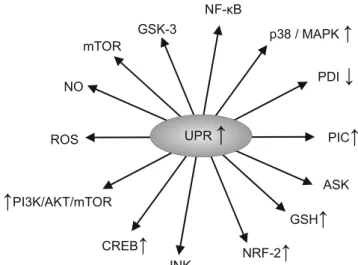
Figure
1
summarises the effects of upregulation of the UPR.
Exemplars of such illnesses include Alzheimer’s disease
[
13
,
14
], Parkinson’s disease [
15
,
16
], multiple sclerosis [
17
,
18
] and amyotrophic lateral sclerosis [
19
,
20
]. More recently,
a putative role of ER stress for psychiatric disorders in which
neuroprogression may occur, including bipolar disorder [
12
,
21
,
22
], major depressive disorder [
23
,
24
] and schizophrenia,
[
25
] has been disputed. It is noteworthy that the chronic
up-regulation of the UPR may lead to the development of chronic
inflammation [
26
,
27
], oxidative stress [
11
,
28
,
29
] and
mul-tiple dimensions of mitochondrial dysfunction [
30
–
33
] and
that these elements appear to be shared factors involved in
the pathogenesis and pathophysiology of neurodegenerative
and neuroprogressive disease, although disease-specific
ele-ments also seem to be involved [
34
–
39
]. There is also some
evidence to suggest that the detrimental effects of ER stress
and chronic UPR upregulation could be
B
druggable
^
and
hence inhibition of pathways involved in the UPR may confer
neuroprotection. For example, there are reports demonstrating
that inhibition of ER stress pathways could protect against
neuronal injury [
40
–
42
].
Thus, this review has two overarching aims: first, to detail
putative pathways whereby activation of the UPR may
insti-gate or exacerbate chronic inflammation, oxidative/nitrosative
stress and multiple dimensions of mitochondrial dysfunction
that are observed across neuroprogressive illnesses and,
sec-ond, to examine therapeutic options targeting ER stress and
t h e U P R a s n o v e l n e u r o t h e r a p e u t i c t a r g e t s f o r
neuroprogressive diseases. Initially, processes stemming from
ER stress and UPR activation which may lead to the initiation
or exacerbation of chronic neuroinflammation will be
critical-ly examined before moving on to a consideration of putative
pathways leading to the initiation or exacerbation of oxidative
and nitrosative stress, and multiple dimensions of
mitochon-drial dysfunction.
ER Stress, Activation of the UPR
and the Development of Chronic
Inflammation
Processes Involved in the Activation of the UPR
conformational changes, thus inducing highly specific
down-stream signalling cascades [
44
,
45
].
Activation of PERK and the Development of Chronic
Inflammation
PERK phosphorylates eukaryotic translation initiation
factor-2α
(eIF2α) leading to an inhibition of general protein
transla-tion and promotransla-tion of the preferential translatransla-tion of
transcrip-tion factor ATF4 [
7
,
46
]. ATF4 in turn translocates to the
nucleus whereupon it induces the transcription of additional
UPR target genes and, in an environment of extreme ER
stress, ATF4 targets the promoter of the gene that encodes
the transcription factor CHOP, which plays a major role in
the instigation of apoptotic cell death [
47
] (see [
48
] for a
review).
Activation of PERK leads to upregulation of the
JAK1/STAT3 signalling axis and subsequent increments in
the transcription and translation of IL-6 and oncostatin, thus
forming a feed-forward loop driving escalating levels of
in-flammation [
49
]. It is noteworthy that activation of PERK in
astrocytes, and subsequent paracrine activation of microglia,
is now recognised as a relevant mechanism in the initiation
and perpetuation of neuroinflammation [
49
]. PERK activation
leads to phosphorylation of eIF2α, which also suppresses the
translation of IκB, resulting in translocation of the cytosolic
transcription factor NF-κB to the nucleus, whereupon it may
induce the expression of genes involved in instigating and
regulating inflammatory pathways [
50
]. Furthermore, PERK
may also regulate cellular redox homoeostasis via the
activa-tion of nuclear factor erythroid 2-related factor 2 (Nrf2) and
the subsequent upregulation of reduced glutathione [
51
–
53
]. It
is also noteworthy that PERK-activated ATF4 also regulates
the cellular redox state and may also act independently of
PERK to induce the production of pro-inflammatory
cyto-kines [
50
].
Activation of ATF6 and the Development of Chronic
Inflammation
Upregulation of monomeric ATF6 also exerts a range of
com-plex, broadly pro-inflammatory effects via the upregulation of
NF-κB via mechanisms involving activation of the CREB and
PI3K/Akt/mTOR signalling pathways [
50
,
54
]. The
upregula-tion of this UPR pathway also exerts direct effects on
inflam-mation via the upregulation of toll-like receptor activity on
macrophages [
55
].
Activation of IRE1
α
and the Development of Chronic
Inflammation
IRE1α
functions both as a kinase and as an endonuclease,
which is activated via a process of oligomerisation in the
absence of GRP78 inhibition. Evidence suggests that this
en-zyme could play a major role in regulating the splicing of
several mRNAs and its activity is an indispensable player in
the translation and activation of transcription factor X-box
binding protein-1 (XBP-1) [
56
,
57
]. XBP-1, in turn, increases
the transcription of several UPR target genes including the one
encoding GRP78 [
58
,
59
]. The activated IRE1α
can also form
a multiprotein complex with apoptosis signal-regulating
ki-nase 1 (ASK1), resulting in the upregulation of various
intra-cellular signalling systems such as c-Jun N-terminal kinase
(JNK) [
60
], p38/MAPK [
61
,
62
], NF-κB [
63
,
64
], glycogen
synthase kinase 3 (GSK-3) [
65
,
66
], mammalian target of
rapamycin (mTOR) [
67
,
68
] and the phosphatidylinositol
3-kinase/protein kinase B/mTOR (PI3K/AKT/mTOR) pathway
[
69
–
71
]. These pathways also play a major role in determining
the balance between cell survival and cell death, generally
promoting cell survival in an environment of chronic
oxida-tive stress. Yet it is important to note that their effects on cell
survival are pleiotropic, and activation of these pathways may
also drive cellular death in other circumstances, particularly
when ER stress is severe [
72
,
73
]. The net effect of these
signalling systems is somewhat unpredictable as they engage
in a complex pattern of mutual cross-talk with the UPR and
each other, and their relative activities appear to influence the
balance between cell proliferation and cell death [
74
–
76
].
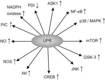
Figure
2
illustrates the actions of the UPR.
For example, the UPR activates GSK-3, possibly via a
route involving increased autophagy of the inactive kinase
phosphorylated at serine 219 [
65
]. This kinase in turn also
appears to play a role in the regulation of the UPR by
influenc-ing the phosphorylation status of CHOP and caspase-3 [
66
,
77
]. There is also evidence of a bidirectional feedback
be-tween UPR activity and levels of mTOR signalling [
78
–
80
].
Similarly, the activation of NF-κB by the UPR also acts to
reduce ER stress by accelerating the clearance of misfolded
proteins via the modulation of autophagic activity [
72
].
Readers interested in a detailed consideration of the
mecha-nisms enabling and regulating such
B
cross-talk
^
, and how
such communication leads to variations in biochemical and
immunological profiles over time, are referred to previous
scholarly reviews [
81
,
82
]. Importantly, from the perspective
of the aims of this paper, changes in the activity of p38/
MAPK, JNK, NF-κB, mTOR, GSK-3 and PI3K/AKT have
pivotal roles in instigating and/or modulating inflammatory
and immune pathways and the activity of peripheral
mononu-clear blood cells such as macrophages [
83
–
87
].
result of upregulated p38/MAPK activity induces
transcrip-tional activation of tumour necrosis factor-α
(TNF-α), IL-6
and other pro-inflammatory cytokines [
83
,
89
]. Similarly,
there is copious evidence that activation of JNK signalling
plays a major role in cytokine production and the subsequent
development of inflammation [
84
,
90
].
The NF-κB pathway also regulates the production of
pro-inflammatory cytokines and several other processes
driving the inflammatory response, such as leukocyte
re-cruitment and the survival of peripheral mononuclear
blood cells, which are important contributors to the
in-flammatory response [
91
,
92
]. Furthermore, the complex
bidirectional signalling between pro-inflammatory
cyto-kines (notably TNF-
α
) allows for the development of a
self-amplifying inflammatory response [
93
,
94
]. However,
it should be noted that the anti-apoptotic activity of
NF-κB may protect cells against the ravages of
inflamma-tion and in certain circumstances the pro-apoptotic
prop-erties of this signalling system can also contribute to the
resolution of inflammation by contributing to the
immu-nologically silent destruction of infiltrating leucocytes and
macrophages [
91
].
The activity of GSK-3 influences the balance between the
production of pro- and anti-inflammatory cytokines, T cell
differentiation, toll-like receptor responses and the
prolifera-tion and activity of transcripprolifera-tion factors which are known to
play a regulatory role in the duration and magnitude of the
immune response, such as signal transducer and activator of
transcription (STAT), nuclear factor of activated T cells
(NFAT), T-box transcription factor (Tbet) and NF-κB
[
95
–
97
]. A recent review further illustrates the
immunoregu-latory role of GSK [
98
]. Much of this regulatory activity
oc-curs in concert with mTOR and PI3K/AKT pathways [
99
].
These interactions are complex but are essentially connected
with the role of mTOR as a metabolic sensor and its capacity
to integrate metabolic and immune processes, thereby
regulating the activation and proliferation of T cells, B cells
and antigen presenting cells. Readers interested in a detailed
consideration of the processes involved are invited to consult
the work of Powell et al. [
100
] and Weichhart et al. [
85
]. It
should also be noted that the PI3K/AKT/mTOR signalling
axis has a broadly restraining effect on the development of
chronic inflammation by limiting the production of type 1
interferons, while increasing the production of IL-10, and
hence, its downregulation during chronic UPR activation
may contribute to the development and perpetuation of an
inflammatory state [
86
].
Given the above data, accumulating evidence supporting
an association between the chronic upregulation of the UPR
and the development of chronic inflammation is perhaps
unsurprising [
26
,
65
,
70
,
101
]. It is also noteworthy that
the chronic upregulation of pro-inflammatory cytokines in
tandem with upregulation of NF-κB and p38/MAPK may
enhance the biosynthesis of ROS and nitric oxide (NO), and
thus may promote or otherwise aggravate oxidative and
nitrosative stress [
87
,
102
–
104
], and hence provides a
mechanism for the development of chronic oxidative stress
accompanying acute or chronic upregulation of the UPR
[
11
,
28
,
105
]. Moreover, the complex interplay of NF-κB,
p38/MAPK and ROS may lead to a self-amplifying pattern
of redox dyshomoeostasis [
106
–
108
]. However, there are a
number of other mechanisms which may also contribute to
the development of oxidative and nitrosative stress
follow-ing ER stress and over-activation of the UPR which seem
underdiscussed, and we will now turn to a consideration of
these factors.
UPR Activation and the Development
of Oxidative and Nitrosative Stress
Mechanisms Involved in the Development of Chronic
Oxidative and Nitrosative Stress
ER stress and the subsequent activation of the UPR may
lead to an increased production of ROS and subsequently
to oxidative stress via a number of mechanisms other than
the upregulation of MAPK and NF-κB [
105
]. Such
mech-anisms involve an upregulation of protein disulphide
isomerase (PDI) resulting in the activation of NADPH
oxidase isomers, notably NOX-2 and NOX-4 [
109
], and
the upregulation of oxidative protein folding in the ER,
which rivals mitochondrial respiration as a source of
cel-lular ROS [
110
,
111
]. Other factors involved in the
devel-opment of oxidative stress in such circumstances include
the oxidation of reduced glutathione (GSH), an increased
S
-nitrosylation of proteins and an increase in Ca
2+efflux
from the ER into the mitochondria [
28
,
112
].
Upregulation of PDI and Activation of NADPH Oxidase
Isoforms
The development of ER stress and activation of the UPR may
lead to upregulation of PDI [
113
–
116
]. This is an important
event in the context of the development of oxidative stress, as
PDI is associated with NOX isoforms and acts as a
redox-sensitive protein which regulates their activation [
10
,
109
,
117
]. The change in cellular redox status
B
sensed
^
by PDI
thus may activate NOX-2 and NOX-4 [
10
,
118
,
119
], leading
to the production of superoxide ions [
120
,
121
].
The effects of PDI in activating NOX enzymes appear to be
of pathophysiological relevance since these enzymes are a
major source of ROS in several cell types [
122
,
123
], and
ROS production by NOX isoforms may even exceed
mito-chondria as the prime source of ROS in some cell types
[
124
]. However, while ROS production within mitochondria
stems from the integral architecture and membrane
organisa-tion, NOX signalling is dependent on multiple protein
inter-actions and post-translational modifications leading to the
as-sembly of a functional NOX complex and the subsequent
trafficking to specific subcellular locations [
122
,
123
]. The
assembly of subunits and the translocation NOX enzymes to
sites of activity appears to be met by the chaperone rather than
the isomerase activity of PDI via hydrophobic rather than
electrostatic or covalent associations [
109
,
125
].
Nevertheless, both the chaperone and isomerase activities of
PDI are required to fulfil its role in the oxidative folding of
proteins within the ER [
126
].
Upregulation of PDI and Increased Rate of ROS
and RNS Production from Oxidative Protein Folding
The ER contains numerous molecules whose task is to ensure
that proteins secreted from the organelle have acquired the
prerequisite post-translational modifications and the correct
conformation [
127
]. One important process involved in
ensur-ing optimal protein foldensur-ing is the acquisition of disulphide
bonds. The interaction between PDI and oxidoreductin-1α
(Ero1α) is probably the most important vehicle for oxidative
protein folding in the ER [
128
,
129
]. Hence PDI has the
ca-pacity to supply, isomerise or, in some circumstances, reduce
disulphide bonds in target proteins [
130
], while its activity is
dependent upon the existence of two distinct remote active
sites which are directly or indirectly oxidised by Ero1α
to
form disulphide bridges [
130
–
132
]. Such oxidation provokes
a conformational change allowing for the entry of unfolded
protein substrates in the reduced state [
133
,
134
]. Once in situ,
key thiol groups on these proteins are oxidised to form
di-sulphide bridges, resulting in the reduction of PDI; these target
proteins then
B
receive
^
disulphide bonds from PDI; Ero1α
then re-oxidises the reduced PDI and transfers electrons from
the reduced PDI to molecular oxygen, which is subsequently
reduced to hydrogen peroxide (H
2O
2) [
135
,
136
], thus
resulting in the re-oxidation of the oxidoreductase [
128
,
137
]. The capacity of Ero1α
to reduce molecular oxygen is
dependent on the existence of a helical structure containing
flavin adenine dinucleotide (FAD) sealed by a disulphide
bridge between Cys(208)-Cys(241). This
B
seal
^
may be
disrupted via the formation of a mixed disulphide bridge
be-tween PDI and one of these cysteines, which underpins the
capacity of this chaperone to regulate the activity of its
co-oxidoreductase [
135
,
136
]. In addition, Ero1α
activity in the
ER is upregulated by the UPR and hence H
2O
2levels may
increase as a result of ER stress [
135
,
136
]. Initially, such
upregulation may have an adaptive purpose as the glutathione
peroxidase isoform GPx7 may utilise H
2O
2to accelerate the
oxidative folding of substrates in vivo [
65
]. Briefly, evidence
suggests that H
2O
2oxidises the Cys57 residue of GPx7 to
produce sulfonic acid, which in turn may react with its
Cys86 to form a disulphide bond. Both the disulphide and
the sulfonic acid forms of GPx7 may oxidise PDI to catalyse
oxidative folding [
138
]. However, the accumulation of ROS
and reactive nitrogen species (RNS) following activation of
the ER [
139
,
140
] leads to
S
-nitrosylation and the subsequent
inactivation of PDI, thus leading to a loss of its chaperone and
isomerase activities [
140
–
142
]. This loss of activity may have
meaningful pathophysiological consequences; the
accumula-tion of misfolded proteins within the ER may further enhance
the UPR, leading to self-amplifying increases in inflammation
as well as oxidative and nitrosative stress [
143
,
144
].
Importantly, such increases in ROS and RNS may also
pro-mote ER stress, which leads to an increase in Ca
2+efflux from
the ER into mitochondria [
28
,
112
] which is enabled by
tubu-lar channels tethering the organelles described as
mitochon-drial associated molecular membranes (MAMs) [
5
,
145
]; an
increase in Ca
2+within the mitochondria may ultimately lead
to the development of multiple dimensions of mitochondrial
dysfunction as discussed below.
ER Stress, UPR Activation and Mitochondrial
Dysfunction
Initial Increase in Mitochondrial Respiration
anion channels (VDACs) are among the most important
mol-ecules for enabling and regulating ER–mitochondria Ca
2+transfer, and are located in the ER and mitochondrial sides
of MAMs, respectively, and may complex with the chaperone
GRP75, thus forming a channel connecting the two organelles
and enabling mutual exchange between membrane and
lumi-nal components [
149
,
150
]. Mitofusin 2 (Mfn2) is another
important protein present on the ER and mitochondrial
sur-faces, which plays an indispensable role in ER–mitochondria
tethering as well as in the modulation of inter-mitochondrial
contacts [
5
,
151
,
152
]. The composition of MAMs adapts in
response to multiple internal and external stimuli [
153
,
154
],
while the formation or dissolution of contact areas between
mitochondria and the ER is further regulated by other aspects
of organelle dynamics [
5
,
148
,
154
]. Importantly, in the
adap-tive phase of ER stress, there is an increased number of
phys-ical contacts between the ER and mitochondrial networks at
the perinuclear regions enabling increased transfer of Ca
2+from the ER into the mitochondria [
5
,
43
,
146
,
155
].
An increase in Ca
2+uptake by the mitochondria may
in-crease transmembrane potential and ATP production aimed at
promoting cellular survival as part of an adaptive response to
ER stress [
156
]. Such an increase in energy production is
accompanied by increases in the production of mitochondrial
proteases such as LON, which are induced by the activation of
the PERK pathway, which in turn regulates the structural
in-tegrity and assembly of cytochrome
c
oxidase (COX) [
157
,
158
]. In this scenario, elevated expression of LON protease
may increase mitochondrial performance by stimulating the
assembly and increasing stabilisation of COX II [
157
,
158
].
However, elevated calcium levels may also increase the
pro-duction of ATP and ROS [
159
–
161
], leading to the activation
of mitochondrial nitric oxide synthase (mtNOS) [
162
,
163
],
and the production of NO, leading to the inhibition of
mito-chondrial function via a number of direct and indirect
mech-anisms including the reversible
S
-nitrosylation of key
struc-tural and functional mitochondrial proteins and enzymes
[
163
–
165
].
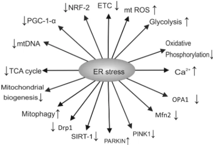
Figure
3
summarises the effects of ER stress.
Elevated Levels of NO and Mitochondrial Function
The nitrosylation of mitochondrial structural proteins and
en-zymes may play a major role in the redox-based regulation of
mitochondrial respiration [
166
,
167
]. While nitrosylation in
response to modest increases in NO levels may initially act
as a defence mechanism aimed at maintaining protein
struc-ture and function [
168
–
170
], further increases in this RNS
may lead to the inhibitory nitrosylation of crucial functional
enzymes such as complex I of the electron transport chain
[
165
,
171
]. Furthermore, the inhibition of complex I by
S
-nitrosylation is another initially cytoprotective response,
which also leads to decreased ATP production and defects in
energy homoeostasis over time [
169
,
170
]. Persistently
elevat-ed cellular concentrations of NO may also lead to the
inhibi-tory nitrosylation of crucial functional cysteine thiols of COX
and complex II of the electron transport chain, thus leading to
chronically suppressed activity of the former and transiently
reduced activity of the latter [
172
,
173
]. Such inhibition may
ultimately impair oxidative phosphorylation and hence
de-crease ATP production and GSH levels within the organelle
[
173
–
175
]. Furthermore, the prolonged inhibition of COX
activity also provokes an increase in ATP production via
gly-colysis in a wide range of cell types as a defensive response
aimed at preventing apoptosis or necrosis [
176
–
178
].
Importantly, the inhibition of complex III and complex IV
by
S
-nitrosylation may further increase the production of
ROS [
179
,
180
], which combined with reduced ATP
genera-tion may contribute to the release Ca
2+from the ER
[
181
–
183
], which may further decrease the biosynthesis of
ATP and also increase the generation of ROS in a positive
feedback loop [
110
,
184
]. This process is of relevance, as an
increased production of ROS may increase the misfolding of
mitochondrial proteins, which coupled with impaired
oxida-tive phosphorylation and ATP production may trigger another
response aimed at restoring mitochondrial homoeostasis,
namely the mitochondrial unfolded protein response
(mtUPR) [
185
–
188
]. Thus, in the section below, we also
dis-cuss the putative pathophysiological relevance of the mtUPR.
Impaired Mitochondrial Performance Following
Activation of the mtUPR
The mtUPR is a multidimensional transcriptional response
initiated and maintained by retrograde
mitochondrial-to-nuclear signalling following increases in protein misfolding
in the mitochondrial matrix and inner membrane space and/
or decreased efficiency of protein importation into
mitochon-dria aimed at restoring mitochonmitochon-drial function and preventing
organellar death [
189
–
192
].
The initiation of the mtUPR is mediated by sensory quality
control proteases with LON or ClpCP being the prime
activa-tors in the matrix [
191
] and the mitochondrial serine protease
HTRA2 playing the same role at the inner membrane space
[
193
–
195
]. Interestingly, the initial upregulation of HTRA2 is
provoked by an overproduction of ROS and the subsequent
phosphorylation of Akt, which in turn activates the oestrogen
receptor in the outer mitochondrial membrane leading to
up-regulation of the transcription factor nuclear respiratory factor
1 (NRF-1) and ultimately leading to increased mitochondrial
production of HTRA-2 [
193
]. This is an illustrative example
of retrograde mitochondrion to nucleus signalling and is
sim-ilar in principle to the retrograde ER to nucleus signalling that
facilitates the UPR response (reviewed in [
196
]). Another
ex-ample involves the upregulation of CHOP, which is an
indis-pensable player in the regulation of mitochondrial proteases
and chaperones, in an attempt to restore intra-mitochondrial
protein folding homoeostasis [
197
,
198
]. However, despite
recent evidence suggesting that signals of protein unfolding
within mitochondria are transduced to the nucleus via
activa-tion of c-Jun, JNK and the activator protein 1 (AP-1) [
64
,
191
], thus sharing some characteristics with the ER UPR,
the precise details underpinning this mechanism remain to
be elucidated [
190
]. It should also be noted that while there
is some evidence that the factors involved in initiating and
regulating the mtUPR are similar in principle to those that
regulate the ER UPR, there is another regulatory mechanism
governing the mtUPR, namely decreased mitochondrial
im-port efficiency, which is unique to the mtUPR, and some
background information is required to understand its genesis
and implications.
The vast majority of mitochondrial proteins originate from
nuclear DNA and hence must be recruited to the mitochondria
and thereafter imported. In most circumstances, this
recruit-ment is initially achieved via the mitochondrial targeting
se-quence (MTS) [
199
]. Once in situ at the outer mitochondrial
membrane (OMM), the protein is directed via a myriad of
regulatory processes to either the OMM, the intermembrane
space, the inner mitochondrial membrane (IMM) or the
ma-trix. Importantly, in order to enter the matrix, the protein must
cross the IMM via the translocase of inner membrane complex
(TIM), which requires the optimal activity of chaperones
lo-cated at the matrix as well as physiological tricarboxylic acid
(TCA) cycle and oxidative phosphorylation activities [
199
,
200
]. Hence, mitochondrial protein import efficiency may
provide a proxy measure of diverse aspects of mitochondrial
performance [
191
,
201
,
202
]. Importantly, a lowered import of
proteins into the mitochondria leads to the accumulation in the
cytoplasm of proteins normally destined for the organelle [
64
,
203
]. Most such proteins are detected and targeted for
proteasomal degradation [
204
,
205
]. However, in lower
ani-mals, at least one mitochondrial protein, the transcription
fac-tor ATFS-1, which regulates the mtUPR in the worm
Caenorhabditis elegans
, has both a MTS, which enables its
mitochondrial import in normal physiological conditions, and
a nuclear localisation sequence (NLS), which enables its
trans-location to the nucleus in conditions of mitochondrial stress
whereupon it activates the mtUPR [
187
]. There are excellent
reviews detailing this process [
190
,
203
]. Notwithstanding
that evidence of such a transcription factor in mammals is
lacking, Fiorese et al. [
206
] have recently reported the
exis-tence of ATF5 in mammalian cells which is regulated
similar-ly to ATFS-1 and may induce a similar transcriptional
response.
The mtUPR is activated by a range of stressors other than
the presence of unfolded proteins, which may lead to a
de-crease in mitochondrial protein import efficiency. In addition
to the presence of heavy metals or other substances acting as
DNA adducts, contaminants in sulphide bonds, or otherwise,
such stressors include a depletion of mtDNA [
186
,
207
], high
levels of ROS [
185
,
186
], mitochondrial ribosome impairment
[
208
,
209
], inhibition of mitochondrial proteases and
chaper-ones [
186
], impaired oxidative phosphorylation and ATP
pro-duction [
187
,
188
] and abnormally high glucose consumption
indicating a switch to the glycolytic pathway as a source of
energy generation [
210
].
It should be stressed that while one facet of the mtUPR
involves the upregulation of genes aimed at increasing
mito-chondrial proteases as well as chaperones, thereby promoting
protein homoeostasis within the mitochondrial protein folding
environment [
186
,
211
], another facet includes changes in the
transcription patterns of genes governing cellular metabolism
[
190
]. In particular, the mtUPR may increase the expression of
genes governing the rate of glycolysis and the catabolism of
amino acids with a concomitant suppression of genes enabling
the optimal performance of the TCA cycle and oxidative
phosphorylation [
212
,
213
].
Furthermore, the progressive decline in mitochondrial ATP
production and mitochondrial membrane potential in such
cir-cumstances coupled with an increase in aerobic glycolysis can
activate another very specific mitochondrial quality control
mechanism involving retrograde mitochondrion to nucleus
signalling referred to as mitophagy [
217
,
218
]. Such a
physi-ologically abnormal elevation in the rate of mitophagy has
adverse bioenergetic consequences as this process appears to
play a relevant role in the regulation of energy homoeostasis
and mitochondrial dynamics [
219
,
220
]. It is also noteworthy
that ER stress and the UPR accompanied by increased levels
of Ca
2+and ROS can also exert detrimental effects on multiple
regulatory processes governing mitochondrial dynamics
di-rectly [
221
–
223
].
Key reactions and pathways associated with the mtUPR are
summarised in Fig.
4
.
UPR Activation and Impaired Mitochondrial
Dynamics
Background
Accumulating evidence indicates that the balance of activity
between pathways regulating mitophagy and those regulating
mitochondrial dynamics (mitochondrial biogenesis, fusion,
fission and motility) may influence mitochondrial mass,
mor-phology and function and thus the cellular capacity to generate
energy and to adjust energy production in the face of changing
metabolic demands [
219
,
224
,
225
]. In particular, changes in
mitochondrial dynamics enable these organelles to maintain a
balance between energy production and changes in energy
demand by generating highly fused networks of mitochondria
or otherwise favouring the formation of more discrete and
isolated organelles [
226
–
228
]. In addition, pathways and
pro-teins governing mitochondrial dynamics may regulate energy
supply and distribution at both the whole organism and
cellu-lar levels [
229
]. Therefore, the targeted manipulation of these
processes may open a relevant therapeutic perspective for
neuroprogressive disorders. Therefore, facets of
mitochondri-al dynamics as well as the pathophysiologicmitochondri-al influence of the
chronic upregulation of the UPR on these processes will now
be discussed as the final mechanistic section of this paper.
ER Stress and UPR Activation as a Source of Impaired
Mitochondrial Mitophagy
Mitophagy is mediated by the cooperative action of the two
proteins parkin and PINK. There are excellent reviews
detail-ing the processes involved in the delivery and regulation of
mitophagy [
203
,
219
,
225
]. Therefore, we provide a brief
description of this process in order to explain the putative
adverse effects of the UPR upon mitophagy.
between these processes is an essential element in regulating
cellular energy homoeostasis [
244
,
245
]. Importantly, changes
in the rate of mitophagy may deregulate mitochondrial
bio-genesis [
219
,
246
], thus compromising cellular energy
homoeostasis.
UPR Activation and Impaired Mitochondrial
Biogenesis
Under physiological conditions, mitochondrial biogenesis is
regulated by a sophisticated interplay between the coactivator
peroxisome proliferator-activated receptor gamma
coactivator-1 alpha (PGC-1α) and the transcription factors
NRF-2 and SIRT-1, which enable coupling between changes
in cellular metabolism to changes in mitochondrial mass and
number [
34
,
214
,
247
]. However, in an environment of ER
stress and UPR activation, elevated levels of NF-κB, MAPK
and PKB/Akt as well as higher NO signalling provoke an
increase in PGC-1α, NRF-1, NRF-2 and SIRT-1, which in
turn induce an increase in mitochondrial biogenesis as a
puta-tive adapputa-tive (i.e. pro-survival) response [
248
,
249
]. However,
with increasing levels of inflammation, and increased levels of
oxidative and nitrosative stress, the upregulation of PGC-1α
is
inhibited by TNF-α
[
104
] and the activity of NRF-2, SIRT-1
and NF-κB may be inhibited by
S
-nitrosylation or
over-oxidation of cysteine residues which normally enable their
function [
171
,
214
], ultimately leading to a chronic state of
decreased mitochondrial biogenesis. The processes governing
mitochondrial biogenesis and those governing mitochondrial
fusion and fission also engage in a complex bidirectional
cross-talk which also plays a role in regulating cellular energy
homoeostasis [
250
], and hence, impaired mitochondrial
bio-genesis can provoke adverse changes in processes governing
mitochondrial fusion and fission which also have the effect of
dysregulating cellular energy generation. In addition, the
mo-lecular players generating inflammation and oxidative stress
also lead to compromised activity of proteins and pathways
regulating fusion and fission, which may lead to decreased
energy production at cellular and whole organism levels.
UPR Activation and Impaired Activity
of Proteins and Processes Governing Fusion
and Fission
Background
Mitochondrial fusion and fission processes are regulated and
enabled by dynamin family GTPases [
251
]. Readers
interest-ed in a detailinterest-ed explanation of the mechanisms underpinning
the actions of these molecular motors are referred to the work
of Ferguson and De Camilli [
252
]. In mammals, the fusion of
OMMs is mediated by Mfn1 and Mfn2, whereas the fusion of
inner membranes is mediated via the protein optic atrophy 1
(OPA1) [
151
,
253
–
256
]. We will focus on their role in
mito-chondrial respiration and how their activities may be
compro-mised in an environment of ER stress and chronic activation
of the UPR.
Role of Mitofusins in Energy Production
and Consequences of UPR Upregulation
Mfn2, and to a lesser extent Mfn1, plays pivotal roles in the
regulation of mitochondrial respiration and energy
homoeostasis [
228
,
257
,
258
]. This role is perhaps
unsurpris-ing given that Mfn2 is an indispensable player in tetherunsurpris-ing
mitochondria and ER stress and enabling high fidelity and
rapid calcium signalling cross-talk between the two organelles
in environments of stress and changing metabolic demands for
energy [
259
,
260
]. While modulation of calcium signalling
appears to be one element underpinning the capacity of
Mfn2 to regulate mitochondrial respiration energy and
adap-tation to increased cellular demands for energy, other
mecha-nisms are clearly involved. Such mechamecha-nisms involve the
in-hibition of ROS production and the regulation of glucose
homoeostasis via mechanisms which are not related to effects
on calcium signalling, although the precise details of such
mechanisms remain to be fully delineated [
257
,
258
].
Crucially, the capacity of this enzyme to adapt the production
of ATP by hypothalamic neurones is a major factor in
regu-lating whole body metabolism and whole body energy
homoeostasis [
257
,
258
]. In this context, it is of paramount
importance that the activity of this enzyme may be inhibited in
an environment of chronic inflammation and nitrosative
stress. For example, MAPK upregulation suppresses Mfn2
activity [
261
] and there is some evidence that this enzyme is
inhibited in an environment in which the production of
pro-inflammatory mediators is elevated [
257
]. It is also of interest
that the capacity of Mfn2 to stimulate mitochondrial function
is dependent on the activation of the PI3K/Akt pathway.
Role of OPA1 in Energy Generation and Consequences
of UPR Upregulation
in vitro data supplied by Kao and fellow workers who reported
that inactivation of OPA1 results in the fragmentation of
established mitochondrial networks as well as a reduction in
oxygen consumption, uncoupling of oxidative
phosphoryla-tion to ATP producphosphoryla-tion and a shift to aerobic glycolysis as the
main mode of energy generation [
265
].
OPA1 has several other regulatory roles in mitochondrial
function such as maintaining the integrity of the quaternary
structures of electron transport chain enzymes and preventing
depolarisation of the IMM. In addition, OPA1-dependent
stabilisation and remodelling of mitochondrial cristae
in-creases the efficiency of energy production by the electron
transport chain, while also reducing the production of ROS
[
266
]. OPA1-driven cristae remodelling is another essential
factor enabling cells to meet energy production in the context
of enhanced energy demands [
267
]. The importance of OPA1
in this domain is further emphasised by the existence of data
demonstrating that its targeted inactivation leads to
detrimen-tal changes in crista morphology and reduces the stability and
performance of the electron transport chain, thereby
compromising oxidative phosphorylation and ATP production
[
268
,
269
].
Other roles include stabilising the association between
cardiolipin and COX, thereby acting as an anti-apoptotic
pro-tein [
266
,
268
–
270
]. Perhaps predictably, there is
experimen-tal evidence that
OPA1
transcription and translation are
upreg-ulated in an environment of chronic nitro-oxidative stress
[
271
], which is also supported by data demonstrating that
mitochondrial dynamics in general, and OPA1 levels in
par-ticular, appear to be under the control of the non-canonical
NF-κB pathway [
272
]. In addition, evidence indicates that
levels of this enzyme are elevated following activation of the
Akt/mTOR pathway [
273
] and more indirectly by ROS and
Ca
2+and by upregulation of PGC-1α
[
274
]. The involvement
of AKT and NF-κB in the regulation of OPA1 activity is
particularly germane as both molecules are inactivated by
S
-nitrosylation in an environment of nitro-oxidative stress
[
275
–
278
], and such inactivation may compromise the ability
of mitochondria to cope with elevated cellular requirements
for energy.
The Role of Drp1 in Energy Generation
and the Consequences of UPR Upregulation
The activity of the mitochondrial fission protein Drp1 is
reg-ulated by a plethora of factors such as Ca
2+concentrations,
ROS levels and a range of post-translational modifications
[
279
–
281
] (see [
282
] for a review). Chronically elevated
levels of ROS and RNS lead to changes in Drp1 activity
and/or rates of mitochondrial fission via a number of routes.
One such route involves the inactivation of Drp1 by AMPK,
whose activity is upregulated in an environment of chronically
elevated ROS generation [
214
,
283
,
284
]. This is of
importance as there is evidence that inhibition of this
GTPase may disrupt mitochondrial networks, thus leading to
adverse changes in organelle morphology accompanied by a
reduction in Mfn1 and Mfn2 as well as a compromised
pro-teolytic processing of OPA1 isoforms [
285
] and hence
pre-sents yet another route by which the chronic upregulation of
the UPR could compromise energy generation. It is also
note-worthy that
S
-nitrosylation of Drp1 in an environment of
chronically upregulated nitro-oxidative stress could increase
the rate of mitochondrial fission [
286
], thereby creating an
imbalance between fusion and fission which may lead to
det-rimental net alterations in mitochondrial morphology and
en-ergy production [
287
–
289
]. The precise mechanisms
under-pinning such an increase in fission rates is still a matter of
ongoing debate. However, it may not be a direct consequence
of nitrosylation-induced increases in the enzymatic activity of
Drp1 [
290
]. Lastly, mitophagy relies on a synergistic interplay
between parkin and the dynamin family kinase Drp1, with the
fission activity of the latter required to generate small
mito-chondria, thereby enabling efficient engulfment by
autophagosomes [
239
,
291
]. Hence, inhibition of this enzyme
may also lead to disrupted mitophagy, which in turn has the
capacity to dysregulate mitochondrial dynamics, and
ultimate-ly ATP production, further compromising energy generation.
Having reviewed the multiple mechanisms involved in
driving the advent or exacerbation of chronic inflammation,
oxidative stress and mitochondrial dysfunction, we will now
consider possible therapeutic targets for the management of
neuroprogressive and neurodegenerative diseases. Based on
the mechanisms highlighted, it seems reasonable to suggest
that molecules with the capacity to target the mechanisms
driving ER stress and the UPR and to ameliorate the adverse
downstream events following the activation of these
path-ways, would be desirable, and this consideration forms the
basis of the approaches suggested below.
Possible Neurotherapeutic Targets
Melatonin
Moreover, melatonin may shift the pattern of mitochondrial
dynamics towards a decrease in fission and an increase in
fusion [
296
,
298
,
301
]. This activity has been demonstrated
in a wide range of cell types [
297
,
301
]. From a mechanistic
perspective, the weight of evidence suggests that melatonin
may attenuate the translocation of Fis1, Drp1 and Bax from
the cytosol to mitochondria and may also upregulate
mito-chondrial fusion proteins Mfn1, Mfn2 and OPA1 [
295
,
300
,
302
,
303
].
Melatonin supplementation also exerts multiple protective
effects on mitochondria via a number of different mechanisms
which may mitigate against the development of maladaptive
processes within these organelles which initially stem from
the upregulation of the UPR (i.e. ER stress). Such mechanisms
include a reduction of mitochondrial oxidative stress [
304
,
305
]; an increased efficiency of ATP production [
306
,
307
];
a reduction in mitochondrial NOS expression [
308
,
309
]; an
amelioration of calcium dyshomoeostasis [
310
,
311
]; the
pres-ervation of mitochondrial membrane potential [
307
,
312
]; and
a reduced release of cytochrome
c
into the cytosol
accompa-nied by the inhibition of caspase-3 activity [
313
]. Several
authors have demonstrated protective effects of melatonin
supplementation against damage to mitochondria caused by
a myriad of different insults including, but not limited to,
sepsis [
314
,
315
], ischaemia/reperfusion [
316
,
317
] and
chal-lenge with neurotoxic compounds such as
1-methyl-4-phenylpyridinium ion (MPP
+) [
302
],
β-amyloid peptide (Aβ
25
–
35) [
318
,
319
], 4-hydroxynonenal [
320
] and
lipopolysac-charide [
309
] .
Melatonin therapy also inhibits the DNA binding activity
and activation of NF-κB, with concomitant reductions in
NLRP3 activity and the synthesis of pro-inflammatory
cyto-kines [
321
–
323
]. These anti-inflammatory effects are
consid-ered to underpin the promising results obtained from studies
investigating the use of melatonin in animal models of
neuro-degenerative diseases and are the motivation for an increased
focus on the use of the molecule as a therapeutic agent
targeting the pathogenesis and pathophysiology of diseases
such as Alzheimer’s disease and Parkinson’
s disease at doses
ranging from 50 to 100 mg daily [
305
,
324
]. In addition, it has
been proposed that melatonin treatment could be useful for
cognitive dysfunction associated with mood disorders [
325
].
However, evidence remains inconclusive [
326
].
CoQ
10Converging preclinical and clinical evidence suggest that
co-enzyme Q
10(CoQ
10) supplementation may offer therapeutic
benefits in a range of neurodegenerative and neuroprogressive
disorders, at least partly owing to its effects on ER stress and
adverse downstream effects. For example, Yubero-Serrano
et al. [
327
] reported that supplementation of CoQ
10in tandem
with a Mediterranean diet effectively suppressed the
expression of genes encoding proteins involved in the UPR.
Furthermore, CoQ
10also appeared to exert a positive effect on
mitochondrial dynamics by exerting a direct effect on ATP
production [
328
], while this therapeutic target may also
pre-serve the structure of mitochondrial cristae, with an
accompa-nying increase in mitochondrial biogenesis [
329
]. In addition,
CoQ
10may also restore endogenous anti-oxidants, such as
vitamin E [
330
], and is also an essential player in enabling
the optimal performance of the electron transport chain and
stabilisation of the mitochondrial permeability transition pore
[
331
] (reviewed by [
332
]).
NAC
Animal and clinical studies have reported beneficial effects of
N
-acetylcysteine (NAC) supplementation on levels of ER
stress [
344
–
347
]. For example, rats supplemented for
2 months with drinking water containing 600 mg NAC per
litre displayed reduced levels of PDI and GRP78 compared
with rats which did not receive NAC [
345
]. Similar findings
indicate that NAC supplementation at 100 or 300 mg/kg for
20 weeks promote significant reductions in ROS levels [
346
,
347
]. Moreover, NAC-related benefits upon ER stress appear
to be dose-dependent, with 20 mmol/L of NAC being more
effective than 10 mmol/L in reducing levels of GRP78 and
ROS [
348
]. It should be noted, however, that NAC is a
pleio-tropic agent and several mechanisms other than direct effects
on the UPR may also contribute to its therapeutic effects
across neurodegenerative and neuroprogressive diseases
[
349
,
350
], while evidence suggests that adjunctive NAC
treatment may mitigate cognitive dysfunction in a range of
such disorders [
351
].
Conclusions and Future Directions
This review indicates that pathways related to the UPR may
reciprocally interact with immune-inflammatory,
neuro-oxi-dative, neuro-nitrosative, as well as mitochondrial
mecha-nisms, which are thought to play a major shared
pathophysi-ological role across several neuroprogressive and
neurodegen-erative diseases. Therefore, the chronic upregulation of the
UPR may interact with a range of cell death mechanisms
underpinning neurodegeneration and neuroprogression [
352
]
and hence represents a novel neurotherapeutic target.
Moreover, this review also opens relevant directions for
fur-ther research. First, the involvement of mechanistic pathways
related to the UPR in separate disorders deserves further
inves-tigation. Second, the extent to which effects upon the UPR
could contribute to therapeutic benefits of novel therapeutic
targets (for example, melatonin, CoQ
10and NAC) is a matter
of ongoing research efforts. Lastly, the identification of patients
who could benefit from therapies targeting ER stress pathways,
taking in account the emerging framework of precision
psychi-atry [
353
], could represent a relevant road of research.
Acknowledgements We should like to thank Myrela O. Machado, MD, PhD, for her kind assistance with the figures.
Authorships All authors contributed to the writing up of the paper.
Compliance with Ethical Standards
Conflict of Interest The authors declare that they have no conflicts of interest.
Open Access This article is distributed under the terms of the Creative C o m m o n s A t t r i b u t i o n 4 . 0 I n t e r n a t i o n a l L i c e n s e ( h t t p : / / creativecommons.org/licenses/by/4.0/), which permits unrestricted use, distribution, and reproduction in any medium, provided you give appro-priate credit to the original author(s) and the source, provide a link to the Creative Commons license, and indicate if changes were made.
References
1. Schroder M (2008) Endoplasmic reticulum stress responses. Cell Mol Life Sci 65(6):862–894. https://doi.org/10.1007/s00018-007-7383-5
2. Lindholm D, Wootz H, Korhonen L (2006) ER stress and neuro-degenerative diseases. Cell Death Differ 13(3):385–392.https:// doi.org/10.1038/sj.cdd.4401778
3. Görlach A, Klappa P, Kietzmann DT (2006) The endoplasmic reticulum: folding, calcium homeostasis, signaling, and redox control. Antioxid Redox Signal 8(9–10):1391–1418.https://doi. org/10.1089/ars.2006.8.1391
4. Fedoroff N (2006) Redox regulatory mechanisms in cellular stress responses. Ann Bot 98(2):289–300.https://doi.org/10.1093/aob/ mcl128
5. Marchi S, Patergnani S, Pinton P (2014) The endoplasmic reticu-lum–mitochondria connection: one touch, multiple functions. Biochim Biophys Acta 1837(4):461–469.https://doi.org/10. 1016/j.bbabio.2013.10.015
6. Torres-Peraza JF, Engel T, Martin-Ibanez R, Sanz-Rodriguez A, Fernandez-Fernandez MR, Esgleas M, Canals JM, Henshall DC et al (2013) Protective neuronal induction of ATF5 in endoplasmic reticulum stress induced by status epilepticus. Brain 136(Pt 4): 1161–1176.https://doi.org/10.1093/brain/awt044
7. Hetz C (2012) The unfolded protein response: controlling cell fate decisions under ER stress and beyond. Nat Rev Mol Cell Biol 13(2):89–102
8. Cheng Y, Yang JM (2011) Survival and death of endoplasmic-reticulum-stressed cells: role of autophagy. World J Biol Chem 2(10):226–231.https://doi.org/10.4331/wjbc.v2.i10.226 9. Benbrook DM, Long A (2012) Integration of autophagy,
proteasomal degradation, unfolded protein response and apopto-sis. Exp Oncol 34(3):286–297
10. Laurindo FR, Fernandes DC, Amanso AM, Lopes LR, Santos CX (2008) Novel role of protein disulfide isomerase in the regulation of NADPH oxidase activity: pathophysiological implications in vascular diseases. Antioxid Redox Signal 10(6):1101–1113. https://doi.org/10.1089/ars.2007.2011
11. Santos CX, Tanaka LY, Wosniak J, Laurindo FR (2009) Mechanisms and implications of reactive oxygen species genera-tion during the unfolded protein response: roles of endoplasmic reticulum oxidoreductases, mitochondrial electron transport, and NADPH oxidase. Antioxid Redox Signal 11(10):2409–2427. https://doi.org/10.1089/ars.2009.2625
12. Pfaffenseller B, Wollenhaupt-Aguiar B, Fries GR, Colpo GD, Burque RK, Bristot G, Ferrari P, Ceresér KMM et al (2014) Impaired endoplasmic reticulum stress response in bipolar disor-d e r : c e l l u l a r e v i disor-d e n c e o f i l l n e s s p r o g r e s s i o n . I n t J Neuropsychopharmacol 17(09):1453–1463.https://doi.org/10. 1017/s1461145714000443
14. Viana RJ, Nunes AF, Rodrigues CM (2012) Endoplasmic reticu-lum enrollment in Alzheimer’s disease. Mol Neurobiol 46(2):522– 534.https://doi.org/10.1007/s12035-012-8301-x
15. Wang HQ, Takahashi R (2007) Expanding insights on the involve-ment of endoplasmic reticulum stress in Parkinson’s disease. Antioxid Redox Signal 9(5):553–561.https://doi.org/10.1089/ ars.2006.1524
16. Cali T, Ottolini D, Brini M (2011) Mitochondria, calcium, and endoplasmic reticulum stress in Parkinson’s disease. Biofactors 37(3):228–240.https://doi.org/10.1002/biof.159
17. Stone S, Lin W (2015) The unfolded protein response in multiple sclerosis. Front Neurosci 9:264.https://doi.org/10.3389/fnins. 2015.00264
18. Getts MT, Getts DR, Kohm AP, Miller SD (2008) Endoplasmic reticulum stress response as a potential therapeutic target in mul-tiple sclerosis. Therapy 5(5):631–640.https://doi.org/10.2217/ 14750708.5.5.631
19. Lautenschlaeger J, Prell T, Grosskreutz J (2012) Endoplasmic re-ticulum stress and the ER mitochondrial calcium cycle in amyo-trophic lateral sclerosis. Amyotroph Lateral Scler 13(2):166–177. https://doi.org/10.3109/17482968.2011.641569
20. Tadic V, Prell T, Lautenschlaeger J, Grosskreutz J (2014) The ER mitochondria calcium cycle and ER stress response as therapeutic targets in amyotrophic lateral sclerosis. Front Cell Neurosci 8:147. https://doi.org/10.3389/fncel.2014.00147
21. Hayashi A, Kasahara T, Kametani M, Toyota T, Yoshikawa T, Kato T (2008) Aberrant endoplasmic reticulum stress response in lymphoblastoid cells from patients with bipolar disorder. Int J Neuropsychopharmacol 12(01):33.https://doi.org/10.1017/ s1461145708009358
22. Bengesser SA, Fuchs R, Lackner N, Birner A, Reininghaus B, Meier-Allard N, Stracke A, Kapfhammer HP et al (2016) Endoplasmic reticulum stress and bipolar disorder—almost for-gotten therapeutic drug targets in the unfolded protein response pathway revisited. CNS Neurol Disord Drug Targets 15(4):403– 413
23. Gold PW, Licinio J, Pavlatou MG (2013) Pathological parainflammation and endoplasmic reticulum stress in depres-sion: potential translational targets through the CNS insulin, klotho and PPAR-[gamma] systems. Mol Psychiatry 18(2):154– 165
24. Timberlake MA, Dwivedi Y (2015) Altered expression of endo-plasmic reticulum stress associated genes in hippocampus of learned helpless rats: relevance to depression pathophysiology. Front Pharmacol 6:319.https://doi.org/10.3389/fphar.2015.00319 25. Rubio MD, Wood K, Haroutunian V, Meador-Woodruff JH (2013) Dysfunction of the ubiquitin proteasome and ubiquitin-like sys-tems in schizophrenia. Neuropsychopharmacology 38(10):1910– 1920.https://doi.org/10.1038/npp.2013.84
26. Grootjans J, Kaser A, Kaufman RJ, Blumberg RS (2016) The unfolded protein response in immunity and inflammation. Nat Rev Immunol 16(8):469–484.https://doi.org/10.1038/nri.2016.62 27. Garg AD, Kaczmarek A, Krysko O, Vandenabeele P, Krysko DV, Agostinis P (2012) ER stress-induced inflammation: does it aid or impede disease progression? Trends Mol Med 18(10):589–598. https://doi.org/10.1016/j.molmed.2012.06.010
28. Eletto D, Chevet E, Argon Y, Appenzeller-Herzog C (2014) Redox controls UPR to control redox. J Cell Sci 127(Pt 17): 3649–3658.https://doi.org/10.1242/jcs.153643
29. Cao SS, Kaufman RJ (2013) Targeting endoplasmic reticulum stress in metabolic disease. Expert Opin Ther Targets 17(4):437– 448.https://doi.org/10.1517/14728222.2013.756471
30. Rocha M, Diaz-Morales N, Rovira-Llopis S, Escribano-Lopez I, Banuls C, Hernandez-Mijares A, Diamanti-Kandarakis E, Victor VM (2016) Mitochondrial dysfunction and endoplasmic reticulum stress in diabetes. Curr Pharm Des 22(18):2640–2649
31. Morais KL, Pacheco MT, Berra CM, Bosch RV, Sciani JM, Chammas R, de Freitas SR, Iqbal A et al (2016) Amblyomin-X induces ER stress, mitochondrial dysfunction, and caspase activa-tion in human melanoma and pancreatic tumor cell. Mol Cell Biochem 415(1–2):119–131. https://doi.org/10.1007/s11010-016-2683-4
32. Grimm S (2012) The ER–mitochondria interface: the social net-work of cell death. Biochim Biophys Acta 1823(2):327–334. https://doi.org/10.1016/j.bbamcr.2011.11.018
33. Scaini G, Rezin GT, Carvalho AF, Streck EL, Berk M, Quevedo J (2016) Mitochondrial dysfunction in bipolar disorder: evidence, pathophysiology and translational implications. Neurosci Biobehav Rev 68:694–713.https://doi.org/10.1016/j.neubiorev. 2016.06.040
34. Morris G, Berk M (2015) The many roads to mitochondrial dys-function in neuroimmune and neuropsychiatric disorders. BMC Med 13(1):68
35. Moylan S, Maes M, Wray NR, Berk M (2013) The neuroprogressive nature of major depressive disorder: pathways to disease evolution and resistance, and therapeutic implications. Mol Psychiatry 18(5):595–606.https://doi.org/10.1038/mp.2012. 33
36. Bhat AH, Dar KB, Anees S, Zargar MA, Masood A, Sofi MA, Ganie SA (2015) Oxidative stress, mitochondrial dysfunction and neurodegenerative diseases; a mechanistic insight. Biomed Pharmacother 74:101–110.https://doi.org/10.1016/j.biopha. 2015.07.025
37. Kohler CA, Freitas TH, Maes M, de Andrade NQ, Liu CS, Fernandes BS, Stubbs B, Solmi M et al (2017) Peripheral cytokine and chemokine alterations in depression: a meta-analysis of 82 studies. Acta Psychiatr Scand 135(5):373–387.https://doi.org/ 10.1111/acps.12698
38. Lai KSP, Liu CS, Rau A, Lanctot KL, Kohler CA, Pakosh M, Carvalho AF, Herrmann N (2017) Peripheral inflammatory markers in Alzheimer’s disease: a systematic review and meta-analysis of 175 studies. J Neurol Neurosurg Psychiatry 88(10): 876–882.https://doi.org/10.1136/jnnp-2017-316201
39. Kohler CA, Freitas TH, Stubbs B, Maes M, Solmi M, Veronese N, de Andrade NQ, Morris G et al (2017) Peripheral alterations in cytokine and chemokine levels after antidepressant drug treatment for major depressive disorder: systematic review and meta-analy-sis. Mol Neurobiol.https://doi.org/10.1007/s12035-017-0632-1 40. Qi X, Hosoi T, Okuma Y, Kaneko M, Nomura Y (2004) Sodium
4-phenylbutyrate protects against cerebral ischemic injury. Mol Pharmacol 66(4):899–908. https://doi.org/10.1124/mol.104. 001339
41. Sokka AL, Putkonen N, Mudo G, Pryazhnikov E, Reijonen S, Khiroug L, Belluardo N, Lindholm D et al (2007) Endoplasmic reticulum stress inhibition protects against excitotoxic neuronal injury in the rat brain. J Neurosci 27(4):901–908.https://doi.org/ 10.1523/jneurosci.4289-06.2007
42. Maly DJ, Papa FR (2014) Druggable sensors of the unfolded protein response. Nat Chem Biol 10(11):892–901.https://doi. org/10.1038/nchembio.1664
43. Bravo R, Gutierrez T, Paredes F, Gatica D, Rodriguez AE, Pedrozo Z, Chiong M, Parra V et al (2012) Endoplasmic reticu-lum: ER stress regulates mitochondrial bioenergetics. Int J Biochem Cell Biol 44(1):16–20.https://doi.org/10.1016/j.biocel. 2011.10.012
44. Wang M, Wey S, Zhang Y, Ye R, Lee AS (2009) Role of the unfolded protein response regulator GRP78/BiP in development, cancer, and neurological disorders. Antioxid Redox Signal 11(9): 2307–2316.https://doi.org/10.1089/ars.2009.2485