CLASS II AND CLASS III DENTOFACIAL DEFORMITIES
Postura de cabeça nas deformidades dentofaciais Classe II e Classe III
Marcela Ralin de Carvalho Deda(1), Francisco Veríssimo de Mello-Filho(2),Samuel Porfírio Xavier(3), Luciana Vitaliano Voi Trawitzki(4)
(1) Physiotherapist; PhD student in Medical Sciences by
Facul-dade de Medicina de Ribeirão Preto da UniversiFacul-dade de São Paulo, FMRP/USP, Ribeirão Preto, São Paulo, Brazil; Master Degree in Medical Sciences by Faculdade de Medi-cina de Ribeirão Preto da Universidade de São Paulo.
(2) Physician; Professor of Faculdade de Medicina de Ribeirão
Preto da Universidade de São Paulo, FMRP/USP, Ribeirão Preto, São Paulo, Brazil; Livre-docente by Faculdade de Medicina de Ribeirão Preto da Universidade de São Paulo.
(3) Dentist; Professor of Faculdade de Odontologia de Ribeirão
Preto da Universidade de São Paulo, FORP/USP, Ribeirão Preto, São Paulo, Brazil; PhD in Maxillo-Facial Surgery and Traumatology by Universidade Estadual Paulista Júlio de Mesquita Filho, UNESP.
(4) Speech Language Pathologist; Professor, Faculdade de
Medicina de Ribeirão Preto da Universidade de São Paulo, FMRP/USP, Ribeirão Preto, São Paulo, Brazil; PhD in Bios-cience applied to Medical Clinic by Faculdade de Medicina de Ribeirão Preto da Universidade de São Paulo.
Conlict of interest: non-existent
According to Grade et al.2, the
temporoman-dibular joint (TMJ) represents the connection of the mandible to the base of the skull, which in turn presents muscle and ligament connections with the cervical region. Together, they form a functional system denoted cranio-cervico-mandibular. Studies concerning this intimate connection have been designed in order to conirm that changes of posture of the head and of other body parts may lead to functional alteration of the masticatory system and vice versa3-8. It has been observed that the molar
relation seems to play an important role in this connection and that certain malocclusion problems may be related to changes in head posture more than others9.
Several authors have studied the presence of changes in head posture in patients with malocclu-sion. Some of them have stated that patients with class II and class III malocclusion present changes in head posture on the sagittal plane1, 10-13, as well as
on the frontal and transverse plane13-15.
Although there is a consensus about the connec-tion existing between the stomatognathic and
ABSTRACT
Purpose: this study investigates whether there is a difference in head posture between groups with different dentofacial deformities (class II and class III) and a group with no deformity. Method: 25 volunteers aged from 16 to 40 year old took part in the study. Ten patients had a diagnosis of class II dentofacial deformity, 15 had a diagnosis of class III skeletal deformity, and 15 healthyvolunteers matched for sex and age to the group with deformity were used as a control group. Head posture was irst checked, followed by evaluation through postural photography (photogrammetry). Results: there was no signiicant difference (p>0.05) between groups regarding postural evaluation by photogrammetry. However, postural evaluation using clinical inspection, revealed anterior head posture among subjects with class II dentofacial deformity compared to subjects with class III deformity (p = 0.001) and to control group (p = 0.001). The percentage of class II dentofacial deformity subjects with neutral head posture was also lower compared to class III dentofacial deformity (p = 0.008) and to control group (p = 0.001). Conclusion: subjects with class II dentofacial deformity may show anteriorization of the head. There is no inluence of the deformity on the increase or reduction of the head-neck angle when analyzed by photogrammetry.
KEYWORDS: Malocclusion; Posture; Head
INTRODUCTION
cervical systems, there is extensive discussion about the type of head posture alteration present in individuals with dentofacial deformity. On this basis, the objective of the present study was to determine whether there is a difference between patients with different dentofacial deformities (class II and class III patterns) regarding head posture observed on the sagittal plane and individuals without deformity, using clinical inspection and photogrammetry.
METHOD
This was a prospective observational study of the case-control type carried out on three groups of subjects. The irst group had class II dentofa-cial deformity characterized by mandibular retrog-nathism and/or excess maxillary growth (GD-II) and the second had class III dentofacial deformity
characterized by mandibular prognathism and/or maxillary deiciency, with the mandible more ante-riorized in relation to the maxilla (GD-III). A third group with no dentofacial deformity was used as control (GC).
GC volunteers were students and employees of various Units of the University of São Paulo, Ribeirão Preto Campus, while GD-II and GD-III volunteers were patients seen at the Outpatient Clinic of Craniomaxillofacial Surgery, Integrated Center for the Study of Facial Deformities (CIEDEF) of the University Hospital, Faculty of Medicine of Ribeirão Preto, University of São Paulo (HCFMRP-USP), selected during the period from September 2007 to October 2009.
Data regarding the number of subjects, gender, weight (kg), height, body mass index (BMI, kg/m2)
and age (years) of GC, GD-II and GD-III subjects are listed in Table 1.
Groups n M Gender F Weight Height BMI Range Age Mean
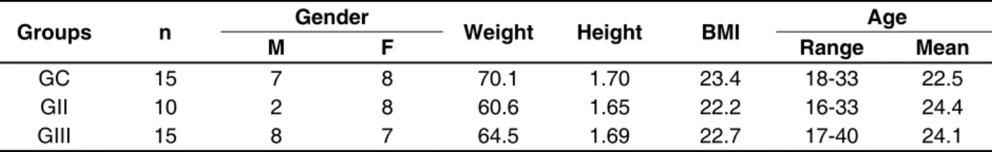
GC 15 7 8 70.1 1.70 23.4 18-33 22.5 GII 10 2 8 60.6 1.65 22.2 16-33 24.4 GIII 15 8 7 64.5 1.69 22.7 17-40 24.1 Table 1 – Group characterization regarding number of subjects, gender, age, weight (kg), height (m) and BMI (kg/m2)
CG: control group; GII: group with class II deformity; GIII: group with class III deformity; n: number of subjects; M: male; F: female; BMI: body mass index.
The groups with dentofacial deformity (GD) consisted of individuals of both genders with maxil-lomandibular skeletal alterations on the sagittal plane classiied as class II or class III regardless of occlusal and skeletal alterations in the horizontal and/or vertical direction (except for marked anterior open bite), wearing ixed upper and lower orthodontic braces, all of them scheduled for surgery for the correction of this deformity. The age range of these groups was 16 to 40 years.
GC consisted of individuals of both genders of similar age as GD subjects, with complete perma-nent dentition (except for the third molars), with no occlusal alterations in either the anteroposterior or transvere and vertical directions; with Angle’s class I molar relationship (occlusal of the mesiovestibular cusp of the irst upper molar in the mesial vestibular sulcus of the irst lower molar).
Subjects with central or peripheral neurological disorders, with traumas and/or tumors in the head and neck regions, or with some genetic syndrome
were excluded from the study. Also excluded were subjects wearing full or partial dentures or with the absence of more than one tooth on the same side of the dental arch, regardless of the interdental space.
The exclusion criteria for GC were: use of orth-odontic braces, including containing braces, signs or symptoms of temporomandibular disorders, or clinical evidence of alterations in face morphology. There were no limitations regarding the race or social level of the participants.
A room in the surgical clinic of the Dental School of Ribeirão Preto, University of São Paulo (FORP-USP) was used for the procedures of the study. The room had natural as well as artiicial lighting and was reserved in order to provide privacy to the subject to be evaluated.
The participants were submitted to two types of postural evaluation.
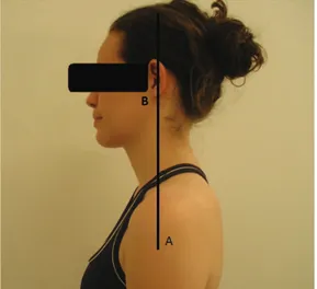
on his lower limbs, looking ahead and with his arms along the body. In a lateral view (sagittal plane), the examiner observed if there was anterioriza-tion or posteriorizaanterioriza-tion of the head in relaanterioriza-tion to the shoulders. To this end, an imaginary vertical line was traced from the center of the shoulder joint to the earlobe. When this imaginary line passed the earlobe posteriorly, head anteriorization was consid-ered to be present and when it passed the earlobe anteriorly, head posteriorization was considered to be present16 (Figure 1).
standardized, but the head of the volunteer was always placed in the center of the frame and perpen-dicular to the facial proile in order to avoid distor-tions. To prevent the participants from remaining in some intermediate oblique position, they were instructed to place their feet immediately behind the tape.
In the present study, three points were estab-lished for the analysis of head posture using the head/neck angle: chin, sternal manubrium and external acoustic meatus11. For better visibility of
the sternal manubrium an adhesive Pimaco label 13 mm in diameter was attached to each partici-pant. All photographic recordings and the marking of the adhesive label were performed by the senior researcher. The greater the head-neck angle, the greater the anteriorization of the head.
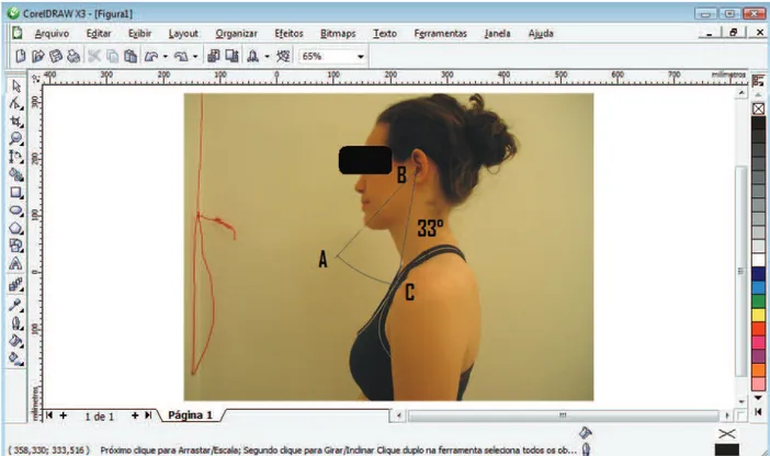
The digital photographs were analyzed with a Pentium Dual-Core/ Windows Vista computer containing the CorelDraw X3 software in order to quantitate the head-neck angle (Figure 2). The soft-ware permits the digital tracing of lines that deter-mine angular values in degrees.
To guarantee reliable measurements, in addi-tion to the senior researcher, two physiotherapists were selected and previously trained for analysis of the angles, for a total of three examiners. The raters were unaware of the group to which a subject belonged. Each rater made three consecutive measurements of each subject at different times in relation to the other raters.
The values were tabulated and intra- and inter-examiner reliability regarding the head-and neck angle in photogrammetry was analyzed. Analysis of the degree of intra- and inter-examiner agreement was performed by calculating the intraclass coefi-cient (ICC) with a 95% conidence interval (CI).
The Fisher exact test was used to determine possible statistically signiicant differences in the evaluation of head posture (during clinical inspec-tion) between GD-II and GD-III, GC and GD-II, and GC and GD-III. The nonparametric Mann-Whitney test was used to determine differences between groups regarding photogrammetry (head-neck angle).
The study was approved by the Research Ethics Committee of HCFMRP-USP (protocol n° 2513/2007) and all subjects gave written informed consent to participate.
All statistical tests were performed using the SPSS (Statistical Package for Social Sciences) soft-ware, version 17.0 for Windows Vista, with the level of signiicance set at p ≤ 0.05.
Figure 1 – (a) center of the shoulder joint; (b) earlobe. The volunteer shows a slightly anteriorized position of the head observed by postural inspection of the head on the sagittal plane
Head posture was then evaluated using postural photography (photogrammetry) with an Olympus digital camera with a resolution of 3.2 megapixels, positioned parallel to the loor on a leveled Weifeng WT 3770 tripod. The digital images obtained were stored on CDs for later analysis.
The volunteers were photographed with both feet lat on the loor,on the sagittal plane and in left proile. They were instructed to keep their habitual posture and to look in the horizontal direction (towards the mirror), without occlusal contact of the teeth (maintaining the functional space free) and with their arms along the body.
RESULTS
Analysis of intra- and inter-examiner reliability regarding photogrammetry for the head-neck angle revealed an excellent level of inter-examiner
reliability with an ICC of 0.976 and a CI of 0.959 to 0.986. Table 2 provides a schematic presentation of the degree of intra-examiner agreement according to the ICC, with an excellent level of reliability being observed.
Figure 2 – Head-neck angle using the CorelDraw X3 software: (a) chin; (b) external acoustic meatus; (c) manubrium
Examiners Mean (°) ICC CI (95%)
1 34.1 0.988 0.980 - 0.993
2 33.9 0.976 0.960 - 0.987
3 34.0 0.959 0.933 - 0.977
Table 2 – Intra-examiner reliability for the determination of the head-neck angle
ICC: intraclass correlation coeficient; CI: conidence interval.
Regarding the inspection of head posture on the sagittal plane, comparison of GD-II and GD-III revealed that GD-II presented a higher percentage (p = 0.001) of patients with head anteriorization than GD-III (100% > 33,3%). GD-III also presented a higher percentage (p = 0.008) of patients with a neutral head posture than GD-II (53.3% > 0%). No signiicant difference was detected in head posteri-orization (p = 0.50).
Comparison of GC and GD-II revealed a greater percentage of patients with head anteriorization (p =
0.001) in GD-II (100% > 26.7%). GC also presented a greater percentage (p = 0.001) of individuals with a neutral head posture than GD-II (73,3% > 0%). No signiicant difference was observed regarding head posteriorization (p = 1.00).
Postural evaluation based on the head-neck angle determined by photogrammetry showed no difference between GC and GD-II (p = 0.49), GC
and GD-III (p = 0.20) or GD-II and GD-III (p= 0.06). These results are presented in Table 4.
Table 3 – Head posture analyzed on the sagittal, frontal and transverse planes of CG, GD-II and GD-III
CG: control group; GII: group with class II deformity; GIII: group with class III deformity; SD: standard deviation; n: number of subjects; * p≤0.05/ Fisher exact test
Head posture GC (n=15) GD-II (n=10) GD-III (n=15)
Anteriorized 4 (26.7%) 4 (26.7%) 4 (26.7%) Neutral position 10 (100%)* 10 (100%)* 10 (100%)*
Posteriorized 5 (33.3%) 5 (33.3%) 5 (33.3%)
GC (n=15) GD-II (n=10) GD-III (n=15)
Mean (± SD) 34.3 (4.3) 36.20 (4.2) 32.3 (5.0) Median (range) 35.0(27.0 to 42.7) 36.4(27.7 to 41.7) 33.3(23.7 to 40.0) Table 4 – Comparison of the head-neck angle between groups
CG: control group; GII: group with class II deformity; GIII: group with class III deformity; SD: standard deviation; n: number of subjects; p > 0.05/ Mann Whitney test.
DISCUSSION
Evaluation of head alignment on the sagittal plane by postural inspection revealed anterior head posture in GD-II patients compared to GD-III and GC. This head anteriorization has been justiied by some authors10. 17, 18.
According to Urbanowicz18, there is an intimate
relationship between head anteriorization and the change in mandibular rest, since occipital extension over the atlas occurs and, according to the theory of Makofsky17, when there is occipital extension
over the atlas, the maxilla accompanies this sliding and the mandible positions itself behind the maxilla. However, it is dificult to establish a causality relation-ship between head posture and mandibular posture since head posture may change in the presence of poor mandibular posture2. According to
Biasotto-Gonzalez10, anterior posture of the head occurs as a
way to compensate for mandibular retrusion, i.e., as a way to compensate for poor mandibular posture.
The results of the present study agree with most of the studies which detected anterior head posture in class II individuals1, 10-12 and normal head posture
in class I individuals1, 11, 12.
However, the present study disagrees with some authors who state that class III patients tend to have head posteriorization1, 10. 12, and with Rosa et
al.13 who, even though they observed head
ante-riorization in class II individuals, noted that there was a higher percentage of subjects with head
anteriorization in the class III group than in the class II group.
As was the case for the present investigation, other studies that assessed head posture by photo-grammetry also obtained good intra- and inter-examiner levels of reliability11, 19-21.
When GC was compared to GD-II and GD-III, no differences were observed between them regarding the head-neck angle determined by photogram-metry. Hower, in the groups with deformity, class II individuals tended to show anteriorization of the head. The results obtained by photographic evalu-ation were quite close to those obtained by postural inspection, which revealed anteriorization of the head in the same individuals despite different points of analysis.
Many ways of quantifying head posture by photo-grammetry exist in the literature19-22. However, the
only study examining photogrammetry in individuals with malocclusion was that by Gadotti et al.11 who
observed anteriorization of the head in class II indi-viduals compared to class I indiindi-viduals.
CONCLUSION
Clinical evaluation of head posture revealed a predominance of anteriorized head posture in class II individuals compared to class III individuals. Regarding the difference between individuals with and without dentofacial deformity, a difference was
observed only between class II individuals and controls since a predominance of head anterioriza-tion was observed in class II individuals.
Regarding photogrammetry, there was no differ-ence between the groups with and without dentofa-cial deformity.
REFERENCES
1. D’Attilio M, Caputi S, Epifania E, Festa F, Tecco, S. Evaluation of cervical posture of children in skeletal class I, II, and III. Cranio. 2005; 23(3): 218-28. 2. Grade R, Caramês J, Pragosa A, Carvalhão J, Sousa S. Postura e disfunção temporomandibular: controvérsias atuais. Rev Port Estomatol Cir Maxilofac. 2008; 49(2): 111-7.
3. Lippold C, Danesh G, Hoppe G, Drerup B, Hackenberg L. Sagittal spinal posture in relation to craniofacial morphology. Angle Orthod. 2006; 76(4): 625-31.
4. Lippold C, Danesh G, Hoppe G, Drerup B, Hackenberg L. Trunk inclination, pelvic tilt and pelvic rotation in relation to the craniofacial morphology in adults. Angle Orthod. 2007; 77(1): 29-35.
5. Savjani D, Wertheim D, Edler R. Change in cranio-cervical angulation following orthognathic surgery. Eur J Orthod. 2005; 27(3): 268-73.
6. Pachì F, Turlà R, Checchi A P. Head posture and lower arch dental crowding. Angle Orthod. 2009; 79(5): 873-9.
7. Tardieu C, Dumitrescu M, Giraudeau A, Blanc Jl, Cheynet F, Borel L. Dental occlusion and postural control in adults. Neurosci Lett. 2009; 450 (2): 221-4. 8. Bergamini M, Pierleoni F, Gizdulich A, Bergamini C. Dental occlusion and body posture: a surface EMG study. Cranio. 2008; 26(1): 25-32.
9. Alkoide EA, Alnamankani E. The association between posture of the head and malocclusion in Saudi subjects. Cranio. 2007; 25(2): 98-105.
10. Biasotto-Gonzalez DAB. Abordagem interdisciplinar das disfunções temporomandibulares. São Paulo: Manole; 2005.
11. Gadotti IC, Berzin F, Biasotto-Gonzalez. Preliminary rapport on head posture and muscle activity in subjects with class I and II. J Oral Rehabil. 2005; 32(11): 794-9.
12. Martesmeier I, Diedrich P. Which correlations between cervical posture and malocclusions? Fortschr Kieferorthop. 1992; 52: 26-32.
RESUMO
Objetivo: este estudoinvestiga se existe diferença entre grupos com diferentes deformidades dento-faciais (padrão classe II e classe III) e o grupo sem a deformidade em relação à postura de cabeça.
Método: participaram deste estudo, voluntariamente, 25 pacientes (entre 16 e 40 anos). Dez pacien-tes com diagnóstico de deformidade dentofacial classe II e 15 pacienpacien-tes com o diagnóstico de classe III esquelética e 15 voluntários sadios, com equivalência em sexo e idade ao grupo de deformidade, formando o grupo controle. Primeiramente foi realizada a inspeção da postura de cabeça. Logo em seguida foi realizada a avaliação postural de cabeça por meio da fotograia postural (fotogrametria).
Resultados: não houve diferença signiicante (p>0,05) entre os grupos em relação à avaliação postu-ral utilizando-se a fotogrametria. Já em relação à avaliação postupostu-ral pela inspeção clínica, observou-se uma postura anterior de cabeça nos indivíduos com a deformidade dentofacial padrão clasobservou-se II, comparados ao padrão classe III (p = 0,001) e ao grupo controle (p = 0,001). Foi visto também que o grupo deformidade classe II apresentou um percentual inferior de indivíduos com posição neutra de cabeça comparado ao grupo deformidade classe III (p = 0,008) e ao grupo controle (p = 0,001).
Conclusão: indivíduos com deformidade dentofacial classe II podem apresentar uma anteriorização de cabeça. Não há inluência da deformidade no aumento ou na redução do ângulo cabeça-pescoço, analisado por meio da fotogrametria.
13. Rosa LP, Moraes LC, Moraes MEL, Filho EM, Castilho JCM. Avaliação da postura corporal associada às maloclusões de classe II e classe III. Rev Odonto Ciênc. 2008; 23(1): 20-5.
14. Michelotti A, Manzo P, Farella M, Martina R. Occlusion and posture: is there evidence of correlation? Minerva Stomatol. 1999; 48(11): 525-34.
15. Tay, DKL. Phisiogonomy in the classiication of individuals with a lateral preference in mastication. J Orofac Pain. 1994; 8(1): 61-72.
16. Kendall FP, McCreary EK, Provance PG. Músculos: provas e funções. 4 ed. São Paulo: Manole; 1995.
17. Makofsky H. The effect of head posture on muscle contact position: the sliding cranium theory. Cranio. 1989; 7(4): 286-96.
18. Urbanowicz M. Alteration of vertical dimension and its effect on head and neck posture. Cranio. 1991; 9(2): 174-9.
19. Iunes DH, Castro FA, Salgado HS, Moura IC, Oliveira AS, Bevilaqua-Grossi D. Coniabilidade intra e interexaminadores e repetibilidade da avaliação postural pela fotogrametria. Rev Bras Fisioter. 2005; 9(3): 327-34.
20. Iunes DH, Bevilaqua-Grossi D, Oliveira AS, Castro FA, Salgado HS. Análise comparativa entre avaliação postural visual e por fotogrametria computadorizada. Rev Bras Fisioter. 2009; 13(4): 308-15.
21. Santos MM, Silva MPC, Sanada LS, Alves CRJ. Análise postural fotogramétrica de crianças saudáveis de 7 a 10 anos: coniabilidade interexaminadores. Rev Bras Fisioter. 2009; 13(4): 350-5.
22. Kussuki MOM, João SMA, Cunha ACP. Caracterização postural da coluna de crianças obesas de 7 a 10 anos. Fisioter Mov. 2007; 20(1): 77-84.
RECEIVED IN: 11/21/2010 ACCEPTED IN: 03/20/2011 Mailing Address:
Marcela Ralin de Carvalho Deda Rua Arnaldo Victaliano,700, apt. 611 Jardim Palma Travassos
Ribeirão Preto – SP CEP: 14091-220