Canine angulation in Class I and Class III
individuals: A comparative analysis with a new
method using digital images*
Objectives: This study aimed to determine the mesiodistal angulation of canine crowns
in individuals with Class III malocclusion in comparison with Class I individuals.
Meth-ods: Measurements were taken from digital photographs of plaster models and imported
into an imaging program (Image Tool). These procedures were repeated to assess random method error (Dahlberg’s formula), and analyze reproducibility by intraclass correla-tion. The sample consisted of 57 patients with complete permanent dentition, untreated orthodontically and divided into two groups according to their malocclusion: Group I consisted of 33 patients with Class I malocclusion, 16 males and 17 females, mean age 27 years; Group II comprised 24 patients with Class III malocclusion, 20 males and 4 females,
mean age 22 years. Results: Random error for canine angulation ranged from 1.54 to 1.96
degrees. Statistical analysis showed that the method presented an excellent reproduc-ibility (p<0.01). Results for canine crown angulation showed no statistically significant difference between maxillary canines in the Class I and Class III groups, although canine angulation exhibited, on average, 2 degrees greater angulation in Class III individuals. Mandibular canines, however, displayed a statistically significant difference on both sides between Class I and Class III groups (p = 0.0009 and p = 0.0074). Compared with Class I patients, angulation in Class III patients was lower in mandibular canines and tended to follow the natural course of dentoalveolar compensation, routinely described in the lit-erature. Conclusion: The results suggest that dental compensation often found in literature involving the incisors region, also affects canine angulation, especially in the lower arch. Abstract
Keywords: Mesiodistal angulation. Canine. Class III malocclusion. Class I malocclusion.
** Specialist in Orthodontics, Brazilian Association of Dentistry, Pará State.
*** Assistant Professor, Department of Orthodontics, School of Dentistry, Federal University of Pará. Coordinator, Specialization Program in Orthodontics, Brazilian Association of Dentistry, Pará State. PhD student, Department of Orthodontics, Rio de Janeiro State University (UERJ).
Lucyana Ramos Azevedo**, Tatiane Barbosa Torres**, David Normando***
IntROduCtIOn
Inclination and angulation have been the subject of orthodontic studies since the days
when Angle4 systemized orthodontic treatment
by developing the edgewise appliance, where inclinations and angulations are controlled through bends in the archwires, which are in-serted in bracket slots.
Some time ago, orthodontists realized the ad-vantages of bracket angulation,10 but no consen-sus has been reached concerning the appropriate amount of angulation for each tooth. Thus, the possibility arose of designing individual brackets for each type of tooth, employing archwires with no bends, or manufacturing brackets tailored for each individual patient.
A key step in this direction was the study on “The Six Keys to Normal Occlusion,” describ-ing six common characteristics of 120 models of optimal natural occlusion, which should be the goals of orthodontic treatment.2 In this study, the second key concerns tooth crown angulation. By analyzing the angle formed by the intersection of the buccal axis of the clinical crown with a line running perpendicular to the occlusal plane and passing through the center of the clinical crown, it was found that clinical crowns are usually an-gulated mesially at varying degrees, depending on the group of teeth being examined. In this study, dental crown angulation was determined by measuring the angle formed between clini-cal crown and occlusal plane. Models were cut beforehand in the center of the clinical crowns with the aid of a plastic protractor. A recent study examined 61 study models with normal, natural occlusion in Brazilians,12 and showed that most individuals exhibited only one to three occlusion keys. The most frequently observed characteris-tics were curve of Spee (100%), tight proximal contacts (42.6%) and proper dental crown in-clinations (34.4%). Mesial angulation of dental crowns was found in 27.9% of the sample.
The Straight-Wire technique makes use of
brackets preadjusted or tailored for each indi-vidual tooth, allowing each tooth to be ideally positioned until treatment completion. Since its inception, the original proposal2 provided, in ad-dition to the use of standard brackets in many patients, for the use of different prescriptions to suit the different types of malocclusion, treat-ments and the desired or possible positioning of the teeth after treatment. In other words, the tailoring of a customized orthodontic appliance according to the features of each malocclusion. The concept of normality and the potential of orthodontics have been redefined since the 1970s, when these precepts were formulated.
Originally, compensations3 were related
to inclinations (torque) on incisor brackets to compensate for the skeletal discrepancies that had not been addressed in their entirety dur-ing orthodontic treatment. In the case of Class III malocclusion, a buccal torque was applied to maxillary incisors and a lingual torque on man-dibular incisors. Changes induced in the arches derive from dental compensation in cases of skeletal malocclusion, as reflected in the buc-colingual tipping of the teeth in the opposite direction of the skeletal error. Thus, many cases of mild skeletal Class III malocclusion, that do not require surgical treatment, could be solved simply by performing dental compensation at the end of treatment. Achieving such outcome would require case customization since each patient has unique skeletal and dental charac-teristics.5 Thus, manipulating canine angulation can play an important part in compensating for orthodontic skeletal error.
at compensating for Class III malocclusions.5 The purpose of these changes was to increase or maintain the perimeter of the upper arch and reduce or maintain the perimeter of the lower arch, thereby encouraging the creation of an an-terior positive overjet, introducing greater com-pensation and increasing the potential for mal-occlusion correction, despite the skeletal error.
Despite growing interest in modifying tooth angulation and inclination described by the study on the six keys to normal occlusion2, few stud-ies have examined the reliability of the measure-ments when employing a particular method. Al-though several methods have been described for measuring tooth inclination (torque),2,6,9,13,14 few investigations have evaluated the error inherent in the method used to analyze tooth angulation.14
A recent study6 described a new method to measure tooth angulation and torque using volumetric computed tomography (VCT). To this end, tomographic slices were made of the anterior teeth of two individuals with facial patterns II and III, respectively. After evalua-tion, it was concluded that computed tomogra-phy (CT) can be a useful means for evaluating tooth torque and angulation, greatly contribut-ing to research involvcontribut-ing tooth positioncontribut-ing as well as orthodontic treatment customization since it enables professionals to check tooth po-sitioning on an individual basis. Furthermore, it is a distortion-free test. However, these tooth angulation measurements on models and CTs should be made with caution because these are relatively new methods that still require further studies to prove their efficacy and, particularly, reliability. The risk radiation and high cost of CT scans should also be emphasized.
A device was recently introduced, which was specifically designed to measure the angulation and inclination of dental crowns.14 Plaster mod-els were attached to a table and the long axis on the crown of each tooth was determined. Once each model had been correctly positioned and
attached, the table was rotated in L shape until the long axis of each tooth crown coincided with a marking made centrally in a magnifying glass, which was fixed to the table. The number of gear teeth, rotated from its zero point (previously de-fined during device calibration), corresponded to the value of each angle, as it was measured. Reproducibility was confirmed by analysis of systematic error using Student’s t-test. The ran-dom error observed in tooth angulation mea-surements ranged between 0.30 and 1.33. With the advent of this new device it became possible to establish mean angulation and inclination val-ues for dental crowns of Brazilian patients with normal occlusion. The results revealed a mean angulation of 7.13° for maxillary canines and 2.43° for mandibular canines.
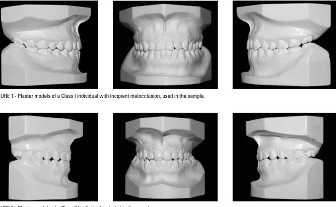
FIGURE 1 - Plaster models of a Class I individual with incipient malocclusion, used in the sample.
FIGURE 2 - Plaster models of a Class III individual included in the sample.
MAtERIAL And MEtHOdS
The sample used in this study was selected from private orthodontic practices and consisted of 57 patients in the stage of permanent dentition.
With the purpose of conducting a compara-tive analysis of permanent canine angulations among Class I and Class III individuals, the sample was divided into two groups. The first group was comprised of 33 Class I patients with incipient orthodontic problems, i.e., cases where orthodontic treatment would be lim-ited to minor movements (closure of diaste-ma, mild crowding, posterior molar crossbite, among others), without previous orthodontic treatment (Fig 1). The second group consisted of 24 individuals with Class III malocclusion (Fig 2). Patients with tooth loss, agenesis, bi-maxillary protrusion, syndromes and moderate or severe crowding were excluded from the sample because these factors might affect ca-nine angulation.
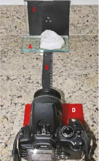
Canine angulations were obtained from stan-dardized digital photographs of each quadrant of the initial plaster models of the sample pa-tients, taken with a digital camera (Canon Rebel 6.0 megapixels, Tokyo, Japan) with a 18-55 mm lens. (Fig 3). These models were placed on a glass plate (A), at a distance of 20 cm from the camera (B). At the bottom of each model a black device was placed with a marking in the center, used as reference to centralize the canines (C). The camera lens was laid on a wax plate in order to optimize lens direction (D).
C
A
B
D
FIGURE 3 - Method used for standardizing photographic snapshots of the plaster models: A= 10 mm glass plate, B= 20 cm millimeter ruler, C= black plastic plate with mark indicating the center of the object (back sleeve of a compact disc/CD), D= wax plate.
FIGURE 4 - Photograph of the study model exported to the imaging pro-gram used to obtain the canine angle measurements.
central incisors to the mesiobuccal cusp of the first permanent molar. Subsequently, Image Tool was used to trace the long axis on the clinical crown of the canine, and from the intersection of these two lines (occlusal plane and long axis) the angulation value for the clinical crown on the plaster model was obtained (Fig 4).
To analyze the method error, the initial plas-ter model quadrants of all patients were photo-graphed again 30 days later and all the steps pre-viously described were repeated to obtain new canine angulation measurements.
The random error was calculated according to Dahlberg’s formula (S²=∑d²/2n) and an analysis of the reproducibility of the measurements was performed using the intraclass correlation test, both with a confidence level of 95%. One outlier with a value far below the other measurements taken for tooth 43, in the Class III group, was excluded from the evaluation.
Means, standard deviations, mean differenc-es, analysis of the normal distribution and inde-pendent t-test were used to detect differences between canine angulations in the Class I and Class III groups.
RESuLtS
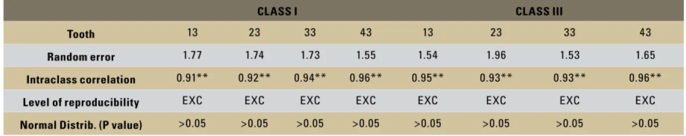
At first, normal distribution was observed for canine angulations in both groups (p> 0.05) (Table 1). Random error difference ranged from 1.54 to 1.96 between measurements (Table 1). Regarding the reproducibility analysis (intraclass correlation), statistical analysis revealed excel-lent method reproducibility
Canine angulations in both groups were ana-lyzed by comparing the measurements of each canine in the Class I groups with its analogue in the Class III group.
Mean angulations of left maxillary canines in the Class I group (x=8.13°) were not statisti-cally different either (p=0.26), when compared with the means for the same teeth in the Class III group (x=10.1°) (Table 2).
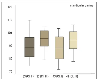
Furthermore, mean angulations of right man-dibular canines in the Class I group (x=3.78°) were statistically different (p=0.007) when compared with the means for the same teeth in the Class III group (x=-1.67°) (Table 2). Mean angulations of left mandibular canines in the Class I group (x=3.27°) were also statistically different (p=0.0009) when compared with the means for the same teeth in the Class III group (x=-2.78°) (Table 2).
In summary, the clinical crowns of maxillary canines were similarly turned mesially in both groups, although slightly more pronounced in Class III individuals. Moreover, mandibular ca-nines in the Class I group had their clinical crowns turned mesially, while their analogues in the Class
III group were either upright or had their clinical crowns turned distally (Figs 5 and 6).
dISCuSSIOn
The primary aim of this study was to exam-ine whether there were differences in permanent canine angulations among individuals present-ing with Class I and Class III malocclusions us-ing a simplified method that made use of photos scanned from plaster models and exported to an image manipulation program for simple angle reading (Image Tool).
There have been few studies on the degree of reliability of measurements taken from models, perhaps because this was originally considered a direct method. However the modifications used in this study showed that the method used to measure canine crown angulations, as well as be-ing very simple to use, is remarkably reproducible, displaying a random error of less than 2º (Table 1).
CLASS I CLASS III
Tooth 13 23 33 43 13 23 33 43
Random error 1.77 1.74 1.73 1.55 1.54 1.96 1.53 1.65
Intraclass correlation 0.91** 0.92** 0.94** 0.96** 0.95** 0.93** 0.93** 0.96**
Level of reproducibility EXC EXC EXC EXC EXC EXC EXC EXC
Normal Distrib. (P value) >0.05 >0.05 >0.05 >0.05 >0.05 >0.05 >0.05 >0.05 TABLE 1 - Random error (Dahlberg’s formula), method reproducibility (intraclass correlation) and normal distribution analysis of values obtained for canine angulations in Class I and Class III groups.
TABLE 2 - Angulation means (angle complement), standard deviations (SD), mean differences and p value (independent t-test) in groups I and Class III. ** p<0.01; EXC= Excellent reproducibility.
ns= non-significant; ** p<0.01.
CLASS I CLASS III CLASS I X CLASS III
Tooth Mean SD Mean SD Diff. between means p-value
13 82.08 (7.92°) 5.81 80.03 (9.97°) 6.61 2.04 0.22(ns)
23 81.87 (8.13°) 6.10 79.90 (10.1°) 6.89 1.97 0.26 (ns)
33 86.73 (3.27°) 6.99 92.78 (-2.78°) 5.48 -6.04 0.0009**
100 120
90 110
80 100
70
90
60
80
70
13 (CI. I) 13 (CI. III) 23 (CI. I) 23 (CI. III) 33 (CI. I ) 33 (CI. III) 43 (CI. I) 43 (CI. III)
A few methods have been described to mea-sure tooth angulation, some are simple to em-ploy such as measurements taken directly from the models using a plastic protractor,2 while oth-ers require major technological resources, such
as computed tomography.6
Thanks to advances in technology, dentistry has benefitted from modern computer programs that simplify diagnosis. Grounded in this prem-ise, this study employed a computer imaging program capable of accurately reading canine angulation from standardized digital photo-graphs of plaster models. This methodology dif-fers from the original proposal that led to the
development of preadjusted brackets.2 One
ma-jor difference refers to the occlusal plane, which in this study is represented by a line linking the midpoint between the incisors and the mesio-buccal cusp of the first molar. This plane is not always parallel to that of Andrews, notably in cases of malocclusion.
Correctly defining the mesiodistal angula-tion of teeth after treatment has been the goal of
many researchers. The values found by Andrews2
and described as normal, 11 degrees for maxillary
canines and 5 degrees for the mandibular ca-nines, both positive, were crucial factors in the development of a fully programmed orthodontic appliance called Straight-Wire. It was designed to impart to brackets certain features to ensure that teeth would be properly positioned at the end of orthodontic treatment.
However, given that the occlusal and skel-etal characteristics of each patient are unique and individual, all cases should not be finished in the same manner. Thus, some adjustments in the original Straight-Wire concept became nec-essary. Since this realization, many orthodontists have begun to customize brackets according to their clinical experience in view of the morpho-logical diversity inherent in the dentofacial com-plex. Most of these changes were introduced without any scientific support.
Even Andrews3 incorporated some changes into the torque of incisor brackets to compen-sate for the skeletal discrepancies that had not been addressed in their entirety during orth-odontic treatment. In the case of Class III mal-occlusion, more buccal torque was applied on maxillary incisors and more lingual torque on
FIGURE 5 - Boxplot for values of maxillary canine angulations in the Class I (Cl. I) and Class III (Cl. III) groups.
FIGURE 6 - Boxplot for values of mandibular canine angulations in the Class I (Cl. I) and Class III (Cl. III) groups.
mandibular incisors. Based on Andrews’3 ideas, other authors5 have advocated brackets with different angles and inclinations for Class I, II and III malocclusions. These brackets appeared after changes were made to Andrews’3 brackets. The main variations to the original model relate to canine angulations to facilitate the torque compensation applied to the central incisors while keeping incisor torque compensations.
Class III malocclusion is significantly differ-ent from sagittal malocclusions to the extdiffer-ent that in most cases patients present a natural dental compensation. Thus, in cases of Class III malocclusion, maxillary incisors are more angulated than in Class I malocclusion. Class III malocclusion brackets were therefore pre-scribed whenever this problem proved amena-ble to being solved by means of dental compen-sation, through orthodontic treatment alone, without the need for surgery.5 For this purpose, an 11º angulation was applied to maxillary ca-nines (three degrees above standard) and 0 de-gree to mandibular canines (five dede-grees below standard). These changes aimed to increase the perimeter of the upper arch and reduce the pe-rimeter of the lower arch to help develop an anterior positive overjet or the maintenance of any pre-existing compensation.
The results achieved in this study disclosed that maxillary canine angulation was similar in both groups, although canine angulation was slightly increased, by nearly 2 degrees, in the Class III group (Table 2, Fig 5). The results for mandibular canines revealed statistically sig-nificant differences between the two groups, with smaller canine angulation in Class III sub-jects (p = 0.0009 for tooth 33 and p = 0.0074 for tooth 43). Therefore, the results highlight-ed differences in natural canine angulation in Class I vs. Class III individuals, thereby lend-ing support to the prescription advanced by Capelozza Filho et al5 while confirming the finding that the incisor compensation seen in
cephalometric studies of Class III patients de-scribed in the literature1,7,11 appear to be ac-companied by changes in canine angulation.
This study found a mean angulation of 10.03° for maxillary canines and -1.75° for mandibular canines in the Class III group. These measures are very close to the measures suggested for use in compensatory brackets recently introduced5 for Class III brackets (11 degrees for upper and 0 degree for lower canines). The Class I group displayed a mean angulation of 8.02° for max-illary and 3.5° for mandibular canines, whereas Capelozza et al5 prescribes a mean angulation of 8° for upper and 5° for lower canines. It should be noted, however, that the measurements ob-tained in this study were taken from individu-als with malocclusion, although every effort was made to avoid interference from other con-founding factors such as crowding, bimaxillary protrusion and tooth loss, while seeking to deal with incipient Class I malocclusions.
Even individuals with normal occlusion failed to exhibit all mesial angulations, as de-scribed in the original study.2 A recently pub-lished study12 found that only 27.9% of the ex-amined models displayed correct dental crown angulations. This means that tooth positioning changes depending on the type of malocclu-sion and that this factor is very important when orthodontic treatment is aimed at correcting skeletal errors by way of dental compensation. In these cases, special attention should be paid to canine angulation because if such angula-tion proves beneficial for treatment it should be maintained or even enhanced.
appliance had been installed. The wide variabil-ity found in this study can be ascribed, among other factors, to a heterogeneous canine crown
morphology.8 Clinically, brackets with
compen-satory prescriptions may be used but ortho-dontists should customize each clinical case, increasing or reducing these offsets accordingly. For cases where the need arises to measure pre-existing tooth angulations, it is believed that the method described in this article provides suf-ficient reliability to justify its use.
COnCLuSIOnS
Based on the data described above it can be concluded that:
1. The method showed excellent repeatabil-ity, with no differences between the two measure-ments, and relatively small random error (<2°).
2. Statistically significant differences were found in the angulation of permanent canines between individuals with Class I and Class III malocclusions, especially in mandibular canines. Such differences are in line with natural com-pensations for Class III incisor inclination, wide-ly described in literature.
1. Aidar LAA, Scanavini MA. Estudo comparativo cefalométrico
radiográico dos padrões de crescimento facial em pacientes portadores de oclusão normal e maloclusões de
Classe I; Classe II, divisão 1; Classe II, divisão 2; e Classe III, de Angle, de acordo com Siriwat & Jarabak. Ortodontia. 1989;22(2):31-52.
2. Andrews LF. The six keys to normal occlusion. Am J Orthod. 1972 Sep;62(3):296-309.
3. Andrews LF. The diagnostic system: occlusal analysis. Dent Clin
N Am. 1976;2(4):671-90.
4. Angle EH. The latest and best in orthodontic mechanism. Dental Cosmos. 1928;70:1143-58.
5. Capelozza L Filho, Silva OG Filho, Ozawa TO, Cavassan AO. Individualização de braquetes na técnica de Straight Wire:
revisão de conceitos e sugestões de indicações para uso. Rev Dental Press Ortod Ortop Facial. 1999 jul-ago;4(4):87-106.
6. Capelozza L Filho, Fattori L, Maltagliati LA. Um novo método
para avaliar as inclinações dentárias utilizando a tomograia
computadorizada. Rev Dental Press Ortod Ortop Facial. 2005 set-out;10(5):23-9.
7. Espírito Santo AA, Ramos AP. Padrão cefalométrico de
pacientes com má oclusão de Classe III nas dentições mista e permanente: uma análise comparativa. [monograia]. Belém
(PA):Universidade Federal do Pará; 2002.
8. Germane N, Bentley B, Isaacson RJ, Revere JH Jr. The
morphology of canines in relation to preadjusted appliances.
Angle Orthod. 1990 Spring;60(1):49-54.
9. Ghahferokhi AE, Elias L, Jonsson S, Rolfe B, Richmond S. Critical assessment of a device to measure incisor crown inclination. Am J Orthod Dentofacial Orthop. 2002 Feb;121(2):185-91.
10. Dempster WT, Adams WJ, Duddles RA. Arrangement in the jaws of the roots of teeth. J Am Dent Assoc. 1963 Dec;67:779-97.
11. Ishikawa H, Nakamura S, Kim C, Iwasaki H, Satoh Y, Yoshida S.
Individual growth in Class III malocclusions and its relationship to the chin cap effects. Am J Orthod Dentofacial Orthop. 1998
Sep;114(3):337-46.
12. Maltagliati LA, Montes LAP, Bastia FMM, Bommarito S. Avaliação da prevalência das seis chaves de oclusão de Andrews em jovens brasileiros com oclusão normal natural. Rev Dental Press Ortod Ortop Facial. 2006 jan-fev;11(1):99-106.
13. Richmond S, Klufas ML, Sywanyk M. Assessing incisor
inclination: a non-invasive technique. Eur J Orthod. 1998 Dec;20(6):721-6.
14. Zanelato ACT, Maltagliati LA, Scanavini MA, Mandetta S. Método para mensuração das angulações e inclinações das
coroas dentárias utilizando modelos de gesso. Rev Dental Press Ortod Ortop Facial. 2006 mar-abr;11(2):63-73.
REfEREnCES
Contact address
David Normando
Rua Boaventura da Silva, 567, ap. 1201 CEP: 66.055-090 – Belém / PA, Brazil E-mail: [email protected]