Comparative study of the Ankle-Brachial Index in diabetic and
non-diabetic patients with critical limb ischemia
Estudo comparativo do Índice Tornozelo-Braquial em diabéticos e
não diabéticos com isquemia crítica
Vanessa Prado dos Santos1
*
, Carlos Alberto Silveira Alves2, Ronald José Ribeiro Fidelis2, Cícero Fidelis3, José Siqueira de Araújo Filho3
Abstract
Background: Calciication of the arterial tunica media can falsely elevate the Ankle-Brachial Index (ABI) in diabetics, making it diicult to assess arterial disease. Objective: To compare ABI values in diabetics and non-diabetics with critical ischemia. Methods: A total of 140 patients (60% diabetics) with critical ischemia due to infrainguinal peripheral arterial obstructive disease were recruited from the vascular surgery service at the Complexo Hospitalar Universitário Professor Edgard Santos. Mean ABI values for the two groups of patients were compared and correlated with severity of ischemia, according to the Rutherford Classiication. Statistical analysis was conducted using EPI-INFO.
Results: A majority of the 140 patients (77%) were classiied as Rutherford Category 5, 6% as Category 4 and 17% as Category 6. Nine diabetics (11%) and one non-diabetic (2%) exhibited ABI > 1.15 (p = 0.02) and were excluded from the comparative analysis of mean ABIs. For the 130-patient sample, the 75 diabetic patients had a mean ABI for the posterior tibial artery of 0.26, vs. 0.28 for the 55 non-diabetic patients (p = 0.6); while mean ABIs for the dorsalis pedis artery were 0.32 vs. 0.23 respectively (p = 0.06). When the patients were stratiied by Rutherford categories, there were no diferences in mean ABIs in categories 4 or 5. Only mean ABI for the dorsalis pedis artery in Category 6 patients was signiicantly higher among diabetics (0.44 vs. 0.16; p = 0.03). Conclusions: he diabetic patients had a higher prevalence of falsely elevated ABI, but when these cases were excluded, mean ABI values were similar to those of non-diabetic patients, with the exception of ABI measured at the dorsalis pedis artery in patients with category 6 ischemia.
Keywords: diabetes mellitus; atherosclerosis; ischemia; Ankle-Brachial Index; complications of diabetes.
Resumo
Contexto: A calciicação da camada média arterial pode tornar o Índice Tornozelo-Braquial (ITB) falsamente elevado em diabéticos, diicultando a avaliação da doença arterial. Objetivo: Comparar os valores do ITB de diabéticos e não diabéticos com isquemia crítica. Métodos: Foram incluídos 140 pacientes (60% de diabéticos) acompanhados no Serviço de Cirurgia Vascular do Complexo Hospitalar Universitário Professor Edgard Santos com isquemia crítica por DAOP infra-inguinal. Comparou-se a média dos valores do ITB dos dois grupos de pacientes, correlacionando o ITB com a gravidade da isquemia, segundo a Classiicação de Rutherford. A análise estatística foi realizada pelo EPI-INFO.
Resultados: A maioria dos 140 pacientes (77%) se encontrava na Categoria 5 da Classiicação de Rutherford, 6% na 4 e 17% na 6. Nove diabéticos (11%) e um não diabético (2%) apresentaram ITB > 1,15 (p = 0,02), sendo excluídos da análise das médias do ITB. Considerando os 130 pacientes, os 75 doentes diabéticos apresentaram média do ITB na artéria tibial posterior de 0,26 versus 0,28 dos 55 doentes não diabéticos (p = 0,6); e no ITB da artéria pediosa aqueles apresentaram média de 0,32 versus 0,23 desses (p = 0,06). Estratiicando os doentes nas categorias da Classiicação de Rutherford, não houve diferença nas médias do ITB nas categorias 4 e 5. Apenas em relação à artéria pediosa e em pacientes na Categoria 6, a média do ITB foi signiicativamente maior em diabéticos (0,44 versus 0,16; p = 0,03).
Conclusão: Os diabéticos apresentaram maior prevalência de ITB falsamente elevado. Porém, excluindo-se esses casos, a média dos valores de ITB são semelhantes aos não diabéticos, exceto na artéria pediosa, nos pacientes com isquemia na categoria 6.
Palavras-chave: diabetes mellitus; aterosclerose; isquemia; Índice Tornozelo-Braquial; complicações do diabetes.
1 Universidade Federal da Bahia – UFBA, Instituto de Humanidades, Artes e Ciências Professor Milton Santos, Salvador, BA, Brazil. 2 Universidade Federal da Bahia – UFBA, Serviço de Cirurgia Vascular do Hospital Universitário Professor Edgard Santos, Salvador, BA, Brazil. 3 Universidade Federal da Bahia – UFBA, Faculdade de Medicina da Bahia, Departamento de Cirurgia, Salvador, BA, Brazil.
Financial support: None.
Conlicts of interest: No conlicts of interest declared concerning the publication of this article. Submitted: April 13, 2015. Accepted: September 17, 2015.
INTRODUCTION
The Ankle-Brachial Index is a noninvasive method that is simple to evaluate and can provide important information for diagnosis, prognosis and follow-up of patients with peripheral arterial occlusive disease (PAOD).1,2 In addition to the index’s role in assessment
of ischemic limbs, both ABI values below the limits
of normality (≤ 0.9) and elevated values (over de 1.4)
have been linked with mortality from cardiovascular disease.1,3 Patients with critical limb ischemia (CLI),
characterized by pain at rest and ulcers or gangrene secondary to PAOD, are at high risk of cardiovascular events such as myocardial infarction and stroke, in addition to the risk of limb loss.1 Clinically, a diagnosis
of critical ischemia can be conirmed by noninvasive
examinations such as those needed to calculate the ABI and measurement of systolic pressure and transcutaneous oxygen tension.1
Calcification of the arterial tunica media or Monckeberg arteriosclerosis is more prevalent among diabetics and can interfere with compression of the arteries of the foot, leading to a falsely elevated ABI result.4,5 Since calciication is more common at the level of the arteries of the foot, one alternative is to measure pressure in toes, which is thought to be better correlated with healing of the lesions.5,6 However, measurement of blood pressure in the digital arteries requires the use of appropriate equipment,6 which is not always available in the majority of health services, whereas the sphygmomanometer needed to calculate ABI is available in almost all vascular services.1
Ankle brachial indexes lower than 0.5 are generally
associated with ischemia and are an indication for vascular assessment, since patients with intermittent claudication generally have an ABI ranging from
0.5 to 0.8, and ABI values ≤ 0.3 are associated with
resting pain.7 However, despite the fact that ABI is
an easily executed and low cost test for health care, there are few studies differentiating the ABI values of diabetic and non-diabetic patients with critical ischemia in the literature.
The objective of this study is to determine whether there are differences between the ABI values observed in diabetic and non-diabetic patients with critical limb ischemia due to infrainguinal peripheral arterial occlusive disease (PAOD).
METHODS
This retrospective study was conducted at the Vascular Surgery Service at the Complexo Hospitalar Universitário Professor Edgard Santos, belonging to the Universidade Federal de Bahia (UFBA) in Brazil.
Data were collected from archived patient records, clinical follow-up charts and arteriographic reports on patients who had been admitted between December
2006 and December 2011. The study was approved
by the Research Ethics Committee at the Complexo Hospitalar Universitário Professor Edgard Santos.
The sample comprised 140 patients admitted for
treatment of infrainguinal critical limb ischemia due to PAOD of atherosclerotic etiology, with normal femoral pulses on physical examination and for
whom arteriography reports conirming diagnosis
were available in the hospital archives. Only cases for which there was a record of the ABI values assessed by one of the departmental vascular surgeons using dorsalis pedis and posterior tibial arteries for lower limbs were included in the sample. Measurements for calculation of the ABI were taken with the cuff placed in the standard positions, above the elbow fold for upper limbs and immediately above the ankle for lower limbs, with the patient in a supine position. The tip of the transducer of the portable Doppler ultrasound unit was positioned at the projection of the brachial artery and of the dorsalis pedis and posterior tibial arteries and then the cuff of the sphygmomanometer
was inlated until the sound of blood low was no longer audible and then delated until blood low was irst heard once more, providing maximum systolic
pressure.1 Patients were excluded from the study if they
had been admitted for acute ischemia or for ischemic disease of non-atherosclerotic etiology, if they did not exhibit critical limb ischemia on admission or if they had aortoiliac PAOD.
All patient data were recorded on the service’s standard clinical follow-up charts. A dedicated form was designed for collection of data from patient records and follow-up charts. Patients were divided into two groups, diabetics and non-diabetics, with the objective of conducting a comparative analysis of mean ABI values calculated using both dorsalis pedis and posterior tibial arteries for both groups. Patients were considered to be diabetics if they had a prior diagnosis of the disease and were being treated for it. The same criterion was used to classify patients with systemic arterial hypertension. Additionally, both groups were
analyzed in terms of the Rutherford Classiication1 of
the lower limb with critical ischemia1 and presence
or absence of falsely elevated ABI (> 1.15). Patients with falsely elevated ABI, deined as ABI > 1.15, were excluded from the comparative analysis of
mean ABIs values. We compared mean ABI values for
diabetic and non-diabetic patients after stratiication by three Rutherford Classiication categories (4, 5 and 6).1
1.15, was based on a population study available in the international literature which found that 1.15 was
the highest mean ABI among people with and without PAOD and that the majority of the sample had an ABI
ranging from 0.9 to 1.1.8 Another study involving
more than 13,000 people found that 1.15 was the
median ABI among people without PAOD.9 On this
basis, we considered an ABI > 1.15 for a person with
critical ischemia to be falsely elevated.
Data were tabulated on Microsoft Excel and
analyzed using Epi-info, version 3.3.2, released in
February 2005. We used the chi-square test (χ2) to
test for associations between DM and occurrence of falsely elevated ABI (a qualitative variable). Mean ABI for dorsalis pedis and posterior tibial arteries (quantitative variables) were compared between the two groups using analysis of variance (ANOVA), both for each group as a whole and for each group of
patients stratiied using Rutherford’s Classiication. A signiicance level of 5% (p < 0.05) was chosen as
the cutoff for rejection of the null hypothesis, i.e. the hypothesis that there was no statistical difference between the groups in terms of the variables studied.
RESULTS
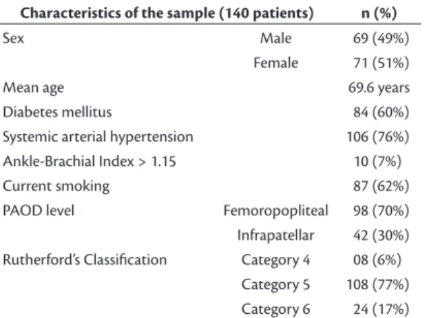
The entire sample comprised 140 patients, 60% of whom were diabetic, 76% hypertensive and 62% smokers. Ninety-eight (70%) had occlusive disease
involving the femoropopliteal territory, with absent popliteal pulses on physical examination, and the
remaining 42 patients (30%) had infrapatellar disease, with normal popliteal pulses. Mean age was 69.6 years. With regard to Rutherford’s Classiication,1 77% of
limbs were classiied as category 5, 17% as category 6 and 6% as category 4. The characteristics of the
sample are shown in Table 1.
Nine diabetic patients (11%) and one (01) non-diabetic patient (2%) had ABI values greater than 1.15 (p = 0.02) and were excluded. After exclusion of the 10 patients with ABI > 1.15, all the remaining 130 patients in the sample were stratiied by Rutherford’s Classiication.
Mean ABI measured at the posterior tibial artery for
the 130 patient with CLI was 0.26 ± 0.05 for patients in Rutherford category 4; 0.30 ± 0.08 for category 5; and 0.15 ± 0.04 for category 6. Mean ABI measured at the dorsalis pedis artery was 0.37 ± 0.06 for category 4; 0.27 ± 0.08 for category 5; and 0.31 ± 0.1 for category 6.
Comparative analysis of mean ABI values calculated for the dorsalis pedis and posterior tibial arteries for both groups of patients with critical ischemia (diabetic and non-diabetic) revealed that mean ABI
for the posterior tibial artery was 0.26 ± 0.07 for diabetics and 0.28 ± 0.08 for non-diabetic patients (p = 0.6). Mean ABI for the dorsalis pedis artery was 0.32 ± 0.07 for diabetic patients and 0.23 ± 0.08 for non-diabetic patients (p = 0.06).
Comparison of diabetic patients with non-diabetic
patients stratiied by the three Rutherford classiication categories (4, 5 and 6) only detected a signiicant difference between patients in category 6, for whom
mean ABI values for the dorsalis pedis artery were higher
for diabetic patients (0.44 ± 0.09) than non-diabetic patients (0.16 ± 0.08) (p = 0.03). Tables 2 and 3 show the results of the comparative analyses of mean ABI values for diabetic and non-diabetic patients calculated for the dorsalis pedis and posterior tibial arteries and
stratiied by the three Rutherford CLI classiication
categories.
DISCUSSION
According to the literature, calciication of the
arterial tunica media is more prevalent among diabetics,4,5 which often leads to discussions of the Table 1. Characteristics of the 140 patients with diagnoses
of critical limb ischemia due to peripheral arterial occlusive disease (PAOD).
Characteristics of the sample (140 patients) n (%)
Sex Male 69 (49%)
Female 71 (51%)
Mean age 69.6 years
Diabetes mellitus 84 (60%)
Systemic arterial hypertension 106 (76%)
Ankle-Brachial Index > 1.15 10 (7%)
Current smoking 87 (62%)
PAOD level Femoropopliteal 98 (70%)
Infrapatellar 42 (30%)
Rutherford’s Classiication Category 4 08 (6%)
Category 5 108 (77%)
Category 6 24 (17%)
Table 2. Comparative analyses of mean dorsalis pedis artery Ankle-Brachial Index (ABI) of diabetic and non-diabetic patients with critical limb ischemia, stratiied by Rutherford Classiication (n = 130).
Rutherford Classification
Mean Ankle-Brachial Index
p
Category 4
Diabetic 0.50 ± 0.01 0.2
Non-diabetic 0.29 ± 0.07
Category 5
Diabetic 0.29 ± 0.07 0.4
Non-diabetic 0.24 ± 0.08
Category 6
Diabetic 0.44 ± 0.09 0.03
applicability of the ABI for diagnosis of PAOD cases in these patients and assessment of their severity. The majority of studies of the prevalence of PAOD and ABI in diabetic and non-diabetic patients do not specify the values found in relation to cases of critical limb ischemia.10-12 A study of the prevalence of
peripheral arterial disease that assessed 2,375 people aged ≥ 40 years found that 4.5% of the non-diabetic patients and 9.5% of the diabetics had ABI ≤ 0.9,10 the majority of whom were asymptomatic. Among diabetic patients, Thavitharam et al. found an overall
mean ABI of 1.03 and detected a difference between those with PAOD (mean of 0.81) and those without PAOD (mean of 1.05).11 Jirkovská et al.12 reported
on around 300 diabetic patients screened for risk of
ulceration and stated that the mean ABI of patients
with ulcers was 0.82 ± 0.42, while for those without ulcers it was 0.92 ± 0.26.
In Brazil, Makdisse et al.13 reported a 36.4%
prevalence of PAOD, diagnosed using a cutoff of
ABI ≤ 0.9, in elderly subjects over 75, the majority of whom (64.2%) had some type of abnormal pulse inding and 34.7% of whom reported pain or discomfort in lower limbs.Another study, with 201 chronic renal failure patients found a 14% prevalence
of PAOD diagnosed on the basis of ABI.14 A study
conducted in a university hospital with 248 patients diagnosed with PAOD found that 79 (32%) exhibited an ABI < 0.5, compatible with severe ischemia, and reported a mean ABI of 0.57 for symptomatic patients and 0.7 for asymptomatic patients.15
However, the majority of studies do not highlight differences between diabetic and non-diabetic patients diagnosed with critical ischemia by providing a break-down of their ABI values. In this study we compared diabetic and non-diabetic patients with
critical ischemia caused by PAOD and, after exclusion of patients with falsely elevated ABI, we did not detect differences in mean ABI for the majority of patients. The prevalence of falsely elevated ABI
(> 1.15) among our patients with advanced ischemia was 10%. The only patient in our sample who had
excessively high ABI but did not have diabetes had chronic renal failure, which is also a disease that is
linked with medial arterial calciication.
The great majority of patients in both groups had ABI values below the limits of normality, which
does not only conirm peripheral vascular disease,
but also indicates advanced disease, with mean ABI
below 0.5. The majority of our patients already had gangrene and tissue loss, conirming the severity of
their ischemic states and explaining the low mean ABIs observed in both groups.
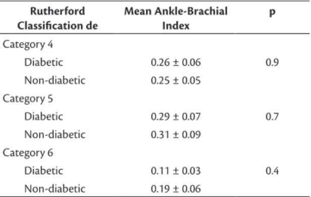
Some arteriographic studies have detected differences between diabetics and non-diabetics, reporting that diabetics are more likely to have atherosclerotic disease below the knee.16,17 However, histological studies show that atherosclerotic lesions of the lower limbs appear to have indistinguishable morphology and distribution in both groups.4,18 Our study did not ind differences between diabetic patients and non-diabetic patients in mean ABI at the posterior tibial artery in any of
the Rutherford’s Classiication categories.
For dorsalis pedis artery ABI there were only
difference between category 6 patients, i.e. patients
with extensive tissue loss, and diabetic patients had a
signiicantly higher mean ABI, which is probably the result of medial arterial calciication of the dorsalis pedis artery in this patients. During the 1960s,
Strandness et al.19 found that diabetics exhibited greater involvement of the posterior tibial, anterior
tibial and ibular arteries than non-diabetic patients,
but when they studied amputated lower limbs from both groups they found that little more than half of them had a patent dorsalis pedis artery. Histological studies have found similarities in the pattern of atherosclerotic disease in diabetic and non-diabetic patients.4,20-22 We believe that since there is a similar pattern of atherosclerotic disease, but medial arterial
calciication is more prevalent among diabetics, this
was probably the cause of the difference in mean ABI
among the category 6 patients with advanced ischemia.
It should be stressed that the mean ABI values among
these diabetics in category 6 were neither normal nor
falsely elevated. The ABI values among diabetics in
category 6 of the Rutherford Classiication were still, on average, below 0.5, despite being statistically higher
than the values observed among the non-diabetics. Table 3. Comparative analyses of mean posterior tibial artery
Ankle-Brachial Index (ABI) of diabetic and non-diabetic patients with critical limb ischemia stratiied by Rutherford Classiication (n = 130).
Rutherford Classification de
Mean Ankle-Brachial Index
p
Category 4
Diabetic 0.26 ± 0.06 0.9
Non-diabetic 0.25 ± 0.05
Category 5
Diabetic 0.29 ± 0.07 0.7
Non-diabetic 0.31 ± 0.09
Category 6
Diabetic 0.11 ± 0.03 0.4
Our study has the limitations inherent to a retrospective study. Notwithstanding, it offers a detailed analysis of the behavior of ABI values in diabetic and non-diabetic patients with severe critical limb ischemia (CLI), reporting the parameters observed in these patients. We very often see the claim that ABI is not a reliable method in diabetics. Our study shows that a small percentage of diabetic patients with CLI have a falsely elevated ABI and that in the majority
of these patients with CLI ABI relects the advanced
arterial obstruction in leg arteries. Further studies are needed to evaluate the correlation between ABI values and prognosis in the cases of critical limb ischemia.
CONCLUSIONS
In our study, diabetic patients with critical limb ischemia had a higher prevalence of falsely elevated ABI. In general, excluding these patients with a falsely elevated ABI, diabetics and non-diabetics with CLI were not different in terms of mean ABI measured using the dorsalis pedis and posterior tibial arteries.
However, when stratiied by degree of ischemia, it was observed that diabetics in category 6 of the Rutherford Classiication had higher mean ABI for the dorsalis
pedis artery than non-diabetics in the same category.
REFERENCES
1. Norgren L, Hiatt WR, Dormandy JÁ, Nehler MR, Harris KA, Fowkes FG. Inter-society consensus for the management of peripheral arterial disease (TASC II). J Vasc Surg. 2007;45(1, Suppl S):S5-67. http://dx.doi.org/10.1016/j.jvs.2006.12.037. PMid:17223489. 2. Williams DT, Harding KG, Price P. An evaluation of the efficacy
of methods used in screening for lower-limb arterial disease in diabetes. Diabetes Care. 2005;28(9):2206-10. http://dx.doi. org/10.2337/diacare.28.9.2206. PMid:16123491.
3. Resnick HE, Lindsay RS, McDermott MMG, et al. Relationship of high and low ankle brachial index to all-cause and cardiovascular disease mortality: the strong heart study. Circulation. 2004;109(6):733-9. http://dx.doi.org/10.1161/01.CIR.0000112642.63927.54. PMid:14970108.
4. Santos VP, Caffaro RA, Pozzan G, Saieg MA, Castelli V Jr. Comparative histological study of atherosclerotic lesions and microvascular changes in amputated lower limbs of diabetic and non-diabetic patients. Arq Bras Endocrinol Metabol. 2008;52(7):1115-23. http:// dx.doi.org/10.1590/S0004-27302008000700007. PMid:19082299. 5. Everhart JE, Pettitt DJ, Knowler WC, Rose FA, Bennett PH.
Medial arterial calcification and its association with mortality and complications of diabetes. Diabetologia. 1988;31(1):16-23. PMid:3350219.
6. Ramsey DE, Manke DA, Sumner DS. Toe blood pressure: a valuable adjunct to ankle pressure measurement for assessing peripheral arterial disease. J Cardiovasc Surg (Torino). 1983;24(1):43-8. PMid:6833352.
7. Takolander R, Rauwerda JA. The use of non-invasive vascular assessment in diabetic patients with foot lesions. Diabet Med. 1996;13(Suppl 1):S39-42. PMid:8741828.
8. Smith FB, Lee AJ, Price JF, van Wijk MC, Fowkes FG. Changes in ankle brachial index in symptomatic and asymptomatic subjects in the general population. J Vasc Surg. 2003;38(6):1323-30. http:// dx.doi.org/10.1016/S0741-5214(03)01021-8. PMid:14681636. 9. Reich LM, Heiss G, Boland LL, Hirsch AT, Wu K, Folsom AR. Ankle–
brachial index and hemostatic markers in the Atherosclerosis Risk in Communities (ARIC) study cohort. Vasc Med. 2007;12(4):267-73. http://dx.doi.org/10.1177/1358863X07082767. PMid:18048462. 10. Gregg EW, Sorlie P, Paulose-Ram R, et al. Prevalence of lower-extremity disease in the US adult population > 40 years of age with and without diabetes. Diabetes Care. 2004;27(7):1591-7. http://dx.doi.org/10.2337/diacare.27.7.1591. PMid:15220233. 11. Tavintharam S, Cheung N, Lim SC, et al. Prevalence and risk
factors for peripheral artery disease in an Asian population with diabetes mellitus. Diab Vasc Dis Res. 2009;6(2):80-6. http://dx.doi. org/10.1177/1479164109336043. PMid:20368197.
12. Jirkovská A, Boucek P, Wosková V, Bartos V, Skibová J. Identification of patients at risk for diabetic foot: a comparison of standardized noninvasive testing with routine practice at community diabetes clinics. J Diabetes Complications. 2001;15(2):63-8. http://dx.doi. org/10.1016/S1056-8727(00)00141-0. PMid:11274901. 13. Makdisse M, Ramos LR, Moreira F, et al. Escore para rastrear
idosos (≥75 anos) de alto risco para doença arterial periférica. Arq Bras Cardiol. 2007;88(6):630-6. http://dx.doi.org/10.1590/ S0066-782X2007000600002. PMid:17664989.
14. Aragão JA, Reis FP, Borges Neto RR, Aragão MECS, Nunes MAP, Feitosa VLC. Prevalência de doença arterial obstrutiva periférica em doentes com insuficiência renal crônica. J Vasc Bras. 2009;8(4):301-6. http://dx.doi.org/10.1590/S1677-54492009000400004. 15. Panico MDB, Spichler ES, Neves MF, Pinto LW, Spichler D.
Prevalência e fatores de risco da doença arterial periférica sintomática e assintomática em hospital terciário, Rio de Janeiro, Brasil. J Vasc Bras. 2009;8(2):125-32. http://dx.doi.org/10.1590/ S1677-54492009005000009.
16. Jude EB, Oyibo SO, Chalmers N, Boulton AJ. Peripheral arterial disease in diabetic and nondiabetic patients. Diabetes Care. 2001;24(8):1433-7. http://dx.doi.org/10.2337/diacare.24.8.1433. PMid:11473082.
17. Rueda CA, Nehler MR, Perry DJ, et al. Patterns or artery disease in 450 patients undergoing revascularization for critical limb ischemia: implications for clinical Trial design. J Vasc Surg. 2008;47(5):995-1000, discussion 999-1000. http://dx.doi.org/10.1016/j.jvs.2007.11.055. PMid:18372151.
18. Colwell JA, Lopes-Virella M, Halushka PV. Pathogenesis of atherosclerosis in diabetes mellitus. Diabetes Care. 1981;4(1):121-33. http://dx.doi.org/10.2337/diacare.4.1.121. PMid:7009108. 19. Strandness DE Jr, Priest RE, Gibbons GE. Combined clinical and
pathologic study of diabetic and nondiabetic peripheral arterial disease. Diabetes. 1964;13(4):366-72. http://dx.doi.org/10.2337/ diab.13.4.366. PMid:14210680.
20. Goldenberg S, Alex M, Joshi RA, Blumenthal HT. Nonatheromatous periferal vascular disease of the lower extremity in diabetes mellitus. Diabetes. 1959;8(4):261-73. http://dx.doi.org/10.2337/ diab.8.4.261. PMid:13663710.
21. Conard MC. Large and small artery occlusion in diabetics and nondiabetics with severe vascular disease. Circulation. 1967;36(1):83-91. http://dx.doi.org/10.1161/01.CIR.36.1.83. PMid:6027218. 22. Ferrier TM. Comparative study of arterial disease in amputated
*
Correspondence
Vanessa Prado dos Santos Universidade Federal da Bahia – UFBA Rua Barão de Jeremoabo, s/n - Ondina CEP 40170-115 – Salvador (BA), Brazil Tel.: +55 (71) 3283-6799 E-mail: [email protected]
Author information
VPS - MSc and PhD in Surgery from Faculdade de Ciências Médicas da Santa Casa de São Paulo; Professor at Instituto de Humanidades, Artes e Ciências, Universidade Federal da Bahia (UFBA); Supervisor of the Medical Residence Program in Vascular Surgery at Hospital Universitário Professor Edgard Santos (UFBA). CASA - Chief of the Vascular Surgery Service at Hospital Universitário Professor Edgard Santos, Universidade Federal da Bahia (UFBA); Preceptor of the Medical Residence Program in Vascular Surgery at Hospital Universitário Professor Edgard Santos, UFBA. RJRF - PhD in Surgery from Universidade de São Paulo (USP); Preceptor of the Medical Residence Program in Vascular Surgery at Hospital Universitário Professor Edgard Santos, Universidade Federal da Bahia (UFBA); President of Sociedade Brasileira de Angiologia e Cirurgia Vascular in Bahia (SBACV-BA). CF - MSc in Medicine from Faculdade de Medicina da Bahia (FMB); Professor at FMB, Universidade Federal da Bahia (UFBA); Preceptor of the Medical Residence Program in Vascular Surgery at Hospital Universitário Professor Edgard Santos, UFBA. JSAF - Professor at Faculdade de Medicina da Bahia (UFBA); Preceptor of the Medical Residence Program in Vascular Surgery at Hospital Universitário Professor Edgard Santos, UFBA.
Author contributions
Conception and design: VPS Analysis and interpretation: VPS, CASA, RJRF Data collection: VPS, RJRF Writing the article: VPS, CASA, JSAF Critical revision of the article: VPS, CASA, CF, JSAF Final approval of the article*: VPS, CASA, RJRF, CF, JSAF Statistical analysis: VPS, RJRF Overall responsibility: VPS, CASA, RJRF, CF, JSAF