resistance exercise: a pilot study
Respostas da frequência cardíaca fetal durante exercício
de força materno: um estudo piloto
Abstract
PURPOSE: To determine fetal heart rate (FHR) responses to maternal resistance exercise for the upper and lower body at two different volumes, and after 25 minutes post-exercise. METHODS: Ten pregnant women (22–24 weeks gestation, 25.2±4.4 years of age, 69.8±9.5 kg, 161.6±5.2 cm tall) performed, at 22–24, 28–32 and 34–36 weeks, the following experimental sessions: Session 1 was a familiarization with the equipment and the determination of one estimated maximum repetition. For sessions 2, 3, 4 and 5,FHR was determined during the execution of resistance exercise on bilateral leg extension and pec-deck ly machines, with 1 and 3 sets of 15 repetitions; 50% of the weight load and an estimated repetition maximum. FHR was assessed with a portable digital cardiotocograph. Results were analyzed using Student’s t test, ANOVA with repeated measures and Bonferroni (α=0.05; SPSS 17.0). RESULTS: FHR showed no signiicant differences between the exercises at 22–24 weeks (bilateral leg extension=143.8±9.4 bpm, pec-deck ly=140.2±10.2 bpm, p=0.34), 28–30 weeks (bilateral leg extension=138.4±12.2 bpm, pec-deck ly=137.6±14.0 bpm,
p=0.75) and 34–36 weeks (bilateral leg extension=135.7±5.8 bpm, pec-deck ly=139.7±13.3 bpm, p=0.38),
between the volumes(bilateral leg extension at 22–24 weeks: p=0.36, at 28–30 weeks: p=0.19 and at 34–36 weeks: p=0.87; pec-deck ly at 22–24 weeks: p=0.43, at 28–30 weeks: p=0.61 and at 34–36 weeks: p=0.49) and after 25 minutes post-exercise. CONCLUSION: Results of this pilot study would suggest that maternal resistance exercise is safe for the fetus.
Resumo
OBJETIVO: O objetivo do presente estudo foi determinar a frequência cardíaca fetal (FCF) enquanto gestantes realizavam exercícios de força para os membros superiores e inferiores, com dois volumes diferentes, e 25 minutos pós-exercício.
MÉTODOS: Dez gestantes (22–24 semanas, 25,2±4,4 anos, 69,8±9,5 kg, 161,6±5,2 cm) realizaram as seguintes sessões experimentais com 22–24, 28–32 e 34–36 semanas de gestação: A Sessão 1 foi a familiarização com os equipamentos e determinação de uma repetição máxima estimada. Para as Sessões 2, 3, 4 e 5, foi determinada a FCF durante a execução do exercício de força nos equipamentos extensão de joelhos bilateral e voador, com 1 e 3 séries de 15 repetições e carga de 50% de uma repetição máxima estimada. A FCF foi avaliada com um cardiotocógrafo digital portátil. Os resultados foram analisados com teste t de Student, ANOVA com medidas repetidas e Bonferroni (α=0,05; SPSS 17.0). RESULTADOS: A FCF não demonstrou diferença signiicativa entre os exercícios com 22–24 semanas (extensão de joelhos bilateral=143,8±9,4 bpm, voador=140,2±10,2 bpm, p=0,34), 28–30 semanas (extensão de
joelhos bilateral=138,4±12,2 bpm, voador=137,6±14,0 bpm, p=0,75) e 34–36 semanas (extensão de joelhos
bilateral=135,7±5,8 bpm, voador=139,7±13,3 bpm, p=0,38), entre os volumes (extensão de joelhos bilateral com 22–24 semanas: p=0,36, 28–30 semanas: p=0,19 e 34–36 semanas: p=0,87; voador com 22–24 semanas: p=0,43, 28–30 semanas: p=0,61 e 34–36 semanas: p=0,49) e 25 minutos pós-exercícios. CONCLUSÃO: Os resultados deste estudo piloto podem sugerir que o exercício de força materno é seguro para o feto.
Laboratory of Exercise Studies of School of Physical Education of Universidade Federal do Rio Grande do Sul – UFRGS – Porto Alegre (RS),Brazil.
1Graduate Program in Medicine, Medical Sciences, School of Medicine, Universidade Federal do Rio Grande do Sul – UFRGS –
Porto Alegre (RS),Brazil.
2School of Physical Education, Universidade Federal do Rio Grande do Sul – UFRGS – Porto Alegre (RS),Brazil.
Conlict of interests: none.
Keywords
Pregnancy Fetus Heart rate, fetal/physiology Exercise/physiology
Palavras-chave
Gestação Feto Frequência cardíaca fetal/isiologia Exercício/isiologia
Correspondence
Roberta Bgeginski Rua Felizardo, 750, room 208 – Jardim Botânico Zip code: 90690-200 Porto Alegre (RS), Brazil
Received
08/26/2014
Accepted with modiications
01/07/2015
Introduction
Regular participation of pregnant women in a physical exercise program is encouraged, as it can control weight gain without compromising fetal development and can facilitate postpartum recovery1. Several types of exercise have been recommended for pregnant women2-8. However, there is insuficient data to counsel pregnant women on resistance exercise, particularly those women who wish to continue training during pregnancy, and also insuf-icient data regarding the corresponding fetal response9. The analysis of fetal heart rate (FHR) during maternal exercise provides the assessment of fetal well-being as a safe and non-invasive method. There is little information regard-ing resistance exercise and FHR, because of methodological dificulty in analyzing this variable during maternal exercise, and potential measuring artifact. Therefore, reports of fetal well-being during maternal exercise are scarce.
Maternal aerobic exercise may lead to fetal brady-cardia and tachybrady-cardia due to blood redistribution, the circulation of catecholamines and increased maternal body temperature1,10,11. However, there is a lack in the litera-ture about the fetal responses during maternal resistance exercise. Nevertheless, pregnant women were tested while performing three different resistance exercises and fetal well-being was not compromised12.
Results of the present pilot study may provide professionals with new information that will inform a safer training prescription for pregnant women. It was hypothesized that maternal resistance exercise would not affect FHR when performed in different gestational trimesters. In this context, the aim of the present study was to determine FHR responses while pregnant women perform resistance exercise for the upper and lower body at two different volumes, at three gestational ages.
Methods
The experimental design was previously approved by the Ethics Committee of Universidade Federal do Rio Grande do Sul (2008185) and by the Ethics Committee of the Municipal Health Department of Porto Alegre (log 449; process 001.000397.10.5). All participants were informed about the study purpose and protocol and signed a written informed consent.
Participants
Ten pregnant women from Porto Alegre, Rio Grande do Sul, Brazil, volunteered. The inclusion criteria included: age over 18 years, gestational age between 22nd and 24th weeks, conirmed by ultrasound, not practicing regular exercise in the last three months, without orthopedic limi-tations or medical constraints (consented by a physician)13,
and not taking any medication that could alter heart rate. Each pregnant woman performed the exercises at 22–24, 28–32 and 34–36 weeks gestational age.
Body height and weight were measured using a stadiometer and a scale at 0.5 cm and 100 g, respectively (Filizola, São Paulo, SP, Brazil). Uterine height was measured with an anthropometric tape at 0.1 mm (Gulick model, Mabbis, Curitiba, PR, Brazil) by ixing the tape from the middle of the upper margin of the pubic symphysis to the height of the fundus of the uterus.
Experimental procedure
Each pregnant woman performed one estimated rep-etition maximum test (1-RM) randomized for lower and upper limbs, using a bilateral leg extension and a pec-deck ly exercise machines, respectively (Taurus, Porto Alegre, RS, Brazil). The exercise protocol has been published previously14. The muscles mainly activated during the exercise in the bilateral leg extension are the quadriceps femoris, and during the exercise in the pec-deck ly, the pectoralis major and anterior deltoid muscles are activated. Women were seated with their back supported for both exercises, and when performing the pec-deck ly exercise, they also had a foot hold support.
The women were familiarized with the procedures, followed by a speciic warm-up (10 repetitions with a low load) and by a 1-RM test. The 1-RM test was estimated from the highest possible number of repetitions in the re-sistance exercise, up to a maximum of ten. The maximum load for each woman was determined with no more than ive attempts, with a ive minute recovery between each attempt and 40-minute recovery between exercises to avoid the inluence of fatigue in the 1-RM values. Performance time for each contraction (concentric and eccentric) was two seconds, controlled by an electronic metronome (Korg, New York, USA). Apnea was avoided. After this proce-dure, the load was scaled using the coeficients proposed by Lombardi15, to estimate the corresponding 1-RM value. From the values of 1-RM for each exercise, a value corre-sponding to 50% of this load was calculated16 and this value was used to perform 15 repetitions of the following stage.
The tests consisted of performing two resistance exer-cises preceded by a rest period, followed by 25 minutes of recovery. Such exercise protocols were selected respecting the guidelines and recommendations published in relation to resistance training in pregnancy1,13,17,18. The exercises chosen were pec-deck ly for the upper body and bilateral leg extension for the lower body, since it represents major muscle mass.
a minimum interval of 48 hours between each visit, to complete four randomized resistance exercise protocols: 1 set of 15 repetitions of the pec-deck ly, 1 set of 15 repeti-tions of bilateral leg extension, 3 sets of 15 repetirepeti-tions of the pec-deck ly, 3 sets of 15 repetitions of bilateral leg extension. A passive interval of two minutes between sets was allowed and a relative load of 50% of 1-RM was used.
Prior to exercise, participants were positioned in a semi-recumbent position (45°) with legs extended on a gurney for 25 minutes and FHR at rest was recorded every minute. The FHR evaluation was performed with a portable digital cardiotocograph (model MD 700D; MICROEM, Ribeirão Preto, Brazil) and the maternal heart rate was monitored (model T61TM; POLAR, Kempele, Finland) for certiication that the beats recorded in the cardiotocograph were fetal. The resistance exercise was performed and FHR was recorded on the 7th and 15th repetitions of each set and maternal heart rate was recorded every ten seconds until the ifteenth repetition. The post-exercise recovery lasted 25 minutes, with the woman positioned as in the pre-exercise rest described above. FHR and maternal heart rate were recorded once each minute during recovery and an average of every ive minutes was reported.
Statistical analysis
Normal distribution was veriied with Shapiro-Wilk’s test. Results were reported as mean±SD. Sample
charac-terization between the three gestational ages, FHR at rest, post-exercise and between the three sets were compared via repeated measures analysis of variance (ANOVA) with Bonferroni (α=0.05). Paired t test was used to compare FHR
between the two exercises at 1 set of 15 repetitions at each gestational age (α=0.05). Statistical analysis was performed
with the Statistical Package for Social Sciences software (ver-sion 17.0 for Windows; SPSS Inc, Chicago, Illinois, USA).
Results
The maternal characteristics are shown in Table 1. Birth weight was 3192.00±403.65 g, birth length was
48.80±2.05 cm, 1-minute APGAR score was 8.40±1.52
(median 9, range 610), and 5-minutes APGAR score was 9.40±0.55 (median 9, range 910).
FHR was measured 25 minutes prior to exercise with no signiicant difference at rest during 22–24 weeks (n=7); Session 1: 144.3±11.0 bpm; Session 2: 145.9±8.1 bpm;
Session 3: 144.3±10.4 bpm; Session 4: 144.0±9.1 bpm
p=0.80), 28–30 weeks (n=5); Session 1: 141.9±10.0 bpm;
Session 2: 142.8±11.8 bpm; Session 3: 138.0±14.7 bpm;
Session 4: 142.8±13.3 bpm; p=0.44) inally at 34–36 weeks
gestation (n=4); Session 1: 140.2±10.2 bpm; Session 2:
138.4±13.1 bpm; Session 3: 140.2±10.8 bpm; Session 4:
135.8±9.1 bpm, p=0.53).
FHR was compared while performing the bilateral leg extension and pec-deck ly exercise in 1 set of 15 repetitions and 3 sets of 15 repetitions. No signiicant differences were found between the exercises at 22–24 weeks (n=5; Bilateral leg extension=143.8±9.4 bpm; Pec-deck ly=140.2±10.2
bpm, p=0.3) at 28–30 weeks (n=5; Bilateral leg exten-sion=138.4±12.2 bpm; Pec-deck ly=137.6±14.0 bpm, p=0.75)
at 34–36 weeks (n=4; Bilateral leg extension=135.7±5.8 bpm;
Pec-deck ly=139.7±13.3 bpm, p=0.38) respecting the volume
(1 or 3 sets of 15 repetitions) performed during bilateral leg extension at 22–24 weeks (n=8, p=0.3); at 28–30 weeks (n=6, p=0.1) and at 34–36 weeks (n=4, p=0.8) about pec-deck ly exercise at 22–24 weeks (n=9, p=0.4); at 28–30 weeks (n=6, p=0.6) and 34–36 weeks (n=3, p=0.4) (Figure 1).
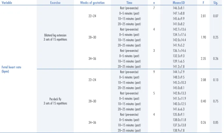
The post-exercise analysis showed no signiicant difference between resting 0–5, 10–15, 20–25 minutes after performing 1 set of 15 repetitions (Table 2) and 3 sets of 15 repetitions (Table 3) for both bilateral leg extension and pec-deck ly exercises.
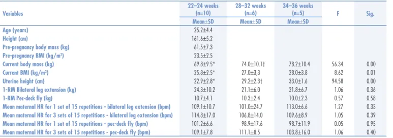
SD: standard deviation; BMI: body mass index; 1-RM:one estimated maximum repetition; HR: heart hate; *signiicantly different from 2832 weeks and 3436 weeks, p≤0.05;
†signiicantly different from 3436 weeks, p≤0.05 Table 1. Maternal characteristics
Variables
22–24 weeks (n=10)
28–32 weeks (n=6)
34–36 weeks
(n=5) F Sig.
Mean±SD Mean±SD Mean±SD
Age (years) 25.2±4.4
Height (cm) 161.6±5.2
Pre-pregnancy body mass (kg) 61.5±7.3
Pre-pregnancy BMI (kg/m2) 23.5±2.5
Current body mass (kg) 69.8±9.5* 74.0±10.1† 78.2±10.4 56.34 0.00
Current BMI (kg/m2) 25.8±2.5* 27.0±3,3 28.0±3.8 8.62 0.01
Uterine height (cm) 22.9±2.8* 29.2±2.3† 33.0±1.6 94.58 0.00
1-RM Bilateral leg extension (kg) 24.3±10.2 21.1±6.0 21.8±6.7 1.06 0.36
1-RM Pec-deck ly (kg) 10.7±4.1 10.3±2.4 10.0±2.3 0.57 0.58
Mean maternal HR for 1 set of 15 repetitions - bilateral leg extension (bpm) 109.1±10.7 101.0±24.7 113.0±6.6 1.27 0.33
Mean maternal HR for 3 sets of 15 repetitions - bilateral leg extension (bpm) 114.8±17.0 106.8±14.0 109.6±8.9 1.05 0.39
Mean maternal HR for 1 set of 15 repetitions - pec-deck ly (bpm) 101.2±6.6 98.9±17.6 98.7±11.9 0.05 0.95
Figure 1. Comparison of fetal heart rate (bpm) at set 1 (15 repetitions), set 2 (15 repetitions) and set 3 (15 repetitions) of leg extension and pec-deck ly exercises for speciic gestational ages. Data are presented as mean±standard deviation (p<0.05).
170
160
150
140
130
120
110
100
Set 1
22–24 weeks 28–30 weeks Exercise – 3 sets / 15 repetitions
Bilateralleg extension
Exercise – 3 sets / 15 repetitions
34–36 weeks
Set 2 Set 3 Set 1 Set 2 Set 3
170
160
150
140
130
120
110
100
Fetal heart rate (bpm) Fetal heart rate (bpm)
Table 2. Mean±standard deviation for fetal heart rate at rest pre-exercise and 0–5, 10–15 and 20–25 minutes post bilateral leg extension and pec-deck ly exercise (1 set of 15 repetitions) (p≤0.05)
Variable Exercise Weeks of gestation Time n Mean±SD F Sig.
Fetal heart rate (bpm)
Bilateral leg extension 1 set of 15 repetitions
22–24
Rest (pre-exercise) 7 141.8±8.3
0.95 0.43
0–5 minutes (post) 144.2±7.2
10–15 minutes (post) 142.9±7.9 20–25 minutes (post) 141.1±7.9
28–30
Rest (pre-exercise) 6 141.0±9.13
0.02 0.99
0–5 minutes (post) 141.6±12.7
10–15 minutes (post) 141.1±8.0 20–25 minutes (post) 141.3±10.2
34–36
Rest (pre-exercise) 5 138.8±9.4
1.50 0.26
0–5 minutes (post) 135.7±9.4
10–15 minutes (post) 137.4±8.8 20–25 minutes (post) 136.6±8.3
Pec–deck ly 1 set of 15 repetitions
22–24
Rest (pre-exercise) 8 143.7±8.9
0.92 0.44
0–5 minutes (post) 143.9±8.8
10–15 minutes (post) 141.6±9.9 20–25 minutes (post) 144.0±5.8
28–30
Rest (pre-exercise) 6 138.5±13.2
1.16 0.35
0–5 minutes (post) 136.1±8.8
10–15 minutes (post) 137.1±8.6 20–25 minutes (post) 141.2±5.6
34–36
Rest (pre-exercise) 4 140.2±10.8
0.73 0.55
0–5 minutes (post) 141.9±12.7
Discussion
The purpose of the present study was to determine the response of FHR at three gestational ages to maternal resis-tance exercises for the upper and lower body at two different volumes. As originally hypothesized, the current study dem-onstrated that FHR was not altered by maternal resistance exercise and during 25 minutes of post-exercise recovery. All fetuses showed a normal response, between 120–160 bpm, indicating that the fetuses did not experience hypoxia and their well-being was not compromised throughout the exercise or during recovery12,19. In the present study, one protocol of a single type of exercise was assessed each day the women visited the laboratory, which may indicate that the duration of the muscle contraction was small and did not relect in the FHR response. Even with the accumulation of sets and the increase in total exercise time, no FHR alterations were found. These results are similar to those of Avery et al.12,who tested (n=12, gestational age 31±1 weeks) the performance of bilateral leg
extension exercise with 3 sets of 10 repetitions at 50, 70 and 90% of 10-RM and found no signiicant difference in FHR between all intensities measured.
The biggest concern regarding the safety of maternal resistance exercise is the possibility of decrease in uterine blood low to increase blood perfusion to the muscles
required for physical exertion20. However, no alterations were observed in FHR in any of the fetuses examined. The protocol design was selected in agreement with the literature, which recommends resistance training for non-pregnant individuals and beginners, with emphasis on improving muscular endurance, with the performance of 1 to 3 sets with 10–15 repetitions and a relative load ranging from 50 to 70% of 1-RM21, which are similar recommendations for women during pregnancy17,18.
The literature reports cases of fetal bradycardia and tachycardia during maternal aerobic exercise1,10,11 due to blood redistribution, the circulation of catecholamines and increased maternal body temperature. Webb et al.11 reported one event of fetal bradycardia while pregnant women (n=22) exercised for 15 minutes on a cycle ergometer (intensity of 145 bpm) and two events during exercise recovery, but FHR quickly returned to the normal baseline and none was associated with adverse fetal responses. The investigators also found that FHR was similar in trained and untrained subjects. Fetal bradycardia and tachycardia did not occur in the present study, probably due to the intermittent characteristic of the resistance exercise, in addition to the small amount of time these women exercised, since each set lasted 1 minute (15 repetitions with 2 seconds for each phase of movement), adding a maximum of 3 minutes of effort when the woman performed 3 sets.
Table 3. Mean±standard deviation for fetal heart rate at pre-exercise rest and at 0–5, 10–15 and 20–25 minutes post-exercise for bilateral leg extension and pec-deck ly exercises (3 sets of 15 repetitions) (p≤0.05)
Variable Exercise Weeks of gestation Time n Mean±SD F Sig.
Fetal heart rate (bpm)
Bilateral leg extension 3 sets of 15 repetitions
22–24
Rest (pre-exercise) 7 146.3±8.1
2.81 0.07
0–5 minutes (post) 147.1±8.8
10–15 minutes (post) 145.6±9.9 20–25 minutes (post) 141.0±8.2
28–30
Rest (pre-exercise) 4 142.7±13.6
1.90 0.25
0–5 minutes (post) 134.1±17.6
10–15 minutes (post) 142.0±14.4 20–25 minutes (post) 141.9±5.2
34–36
Rest (pre-exercise) 3 136.1±14.6
2.35 0.26
0–5 minutes (post) 132.3±9.3
10–15 minutes (post) 139.1±6.5 20–25 minutes (post) 141.2±7.8
Pec-deck ly 3 sets of 15 repetitions
22–24
Rest (pre-exercise) 9 144.1±7.9
2.08 0.13
0–5 minutes (post) 148.2±9.5
10–15 minutes (post) 145.2±10.3 20–25 minutes (post) 145.0±8.1
28–30
Rest (pre-exercise) 5 142.8±13.3
0.40 0.75
0–5 minutes (post) 141.5±11.9
10–15 minutes (post) 140.3±12.5 20–25 minutes (post) 141.6±6.3
34–36
Rest (pre-exercise) 4 135.8±9.1
0.26 0.85
0–5 minutes (post) 138.0±11.8
1. Artal R, O’Toole M, White S. Guidelines of the American College of Obstetricians and Gynecologists for exercise during pregnancy and the postpartum period. Br J Sports Med. 2003;37(1):6-12. 2. Mottola MF, Campbell MK. Activity patterns during pregnancy.
Can J Appl Physiol. 2003;28(4):642-53.
3. Finkelstein I, Bgeginski R, Tartaruga MP, Alberton CL, Kruel LFM. Heart rate and blood pressure behavior throughout pregnancy, with training in water medium. Rev Bras Med Esporte. 2006;12(6):376-80. 4. Chuntharapat S,Petpichetchian W,Hatthakit U. Yoga during
pregnancy: effects on maternal comfort, labor pain and birth outcomes. Complement Ther Clin Pract. 2008;14(2):105-15. 5. Yeo S, Davidge S, Ronis DL, Antonakos CL, Hayashi R, O’Leary S.
A comparison of walking versus stretching exercises to reduce the incidence of preeclampsia: a randomized clinical trial. Hypertens Pregnancy. 2008;27(2):113-30.
6. Bgeginski R, Finkelstein I, Alberton CL, Tartaruga MP, Kruel LFM. Effects of water-gymnastics training on hemodynamic variables in pregnant women at rest. IJARE. 2009;3(2):151-61.
7. Finkelstein I, Bgeginski R, Figueiredo PAP, Alberton CL, Stein R, Kruel LFM. [Cardiorespiratory responses of pregnant women during different types of exercise on land and in water]. Rev Bras Med. 2009;66(6):174-7. Portuguese.
8. Finkelstein I, Figueiredo PA, Alberton CL, Bgeginski R, Stein R, Kruel LFM. Cardiorespiratory responses during and after water exercise in pregnant and non-pregnant women. Rev Bras Ginecol Obstet. 2011;33(12):388-94.
9. Szymanski LM, Satin AJ. Strenuous exercise during pregnancy: is there a limit? Am J Obstet Gynecol. 2012;207(3):179.e1-6. 10. Stevenson L. Exercise in pregnancy. Part 1: Update on pathophysiology.
Can Fam Physician. 1997;43:97-104.
11. Webb KA, Wolfe LA, McGrath MJ. Effects of acute and chronic maternal exercise on fetal heart rate. J Appl Physiol. 1994;77(5):2207-13. 12. Avery ND, Stocking KD, Tranmer JE, Davies GA, Wolfe LA. Fetal
responses to maternal strength conditioning exercises in late gestation. Can J Appl Physiol. 1999;24(4):362-76.
13. American College of Obstetricians and Gynecologists. Exercise during pregnancy and the postpartum period. Clin Obstet Gynecol. 2003;46(2):496-9.
14. Bgeginski R, Almada BP, Kruel LFM. Cardiorespiratory responses of pregnant and non-pregnant women during resistance exercise. J Strength Cond Res.2014 Sep 15. [Epub ahead of print]. 15. Lombardi VP. Beginning weight training: the safe and effective
way. Dubuque: Brown & Benchmark; 1989.
References
The current literature cannot afirm yet whether the selective blood redistribution during regular or prolonged exercise in pregnancy interferes with the transplacental transport of oxygen, carbon dioxide and nutrients, and if such interference can cause any lasting effect. Even though there is indirect evidence that there are no lasting effects, Barakat et al.22-24 tested a training period for previously sedentary pregnant women (12 to 39 gestational weeks) with 12 different resistance exercises in each session, using elastic bands and free weights (never more than 3 kg). The authors did not assess maternal hemodynamic responses throughout the training; nevertheless, it was concluded that the training did not negatively affect the fetus, and did not show a negative impact on body size and general health of the newborn. Recently, another study assessed pregnancy outcomes after an intervention with supervised resistance exercises twice a week, performed at an activity level equivalent to moderate-to-vigorous exercise (n=51, 14 to 25 gestational weeks), compared to the control group (n=41), who received a general-ized exercise recommendation, a home-based training program and a telephone follow-up. Even though the newborns delivered by women who underwent resistant exercise during pregnancy were signiicantly heavier than those born to control women; 3,561 g (±452)
versus 3,251 g (±437) (p=0.02), the supervised
resis-tance training appeared to be an appropriate form of exercise in healthy pregnancies25.
It is important to note the limitations of this study. First, a larger sample size may have increased the statis-tical power. Since this is a pilot study, there was a very small sample number. However, even though there were only 10 subjects, the resistance exercise was safe for FHR and fetal well-being. Second, the sample loss during the protocol, due to the methodological dificulty in analyz-ing FHR, made the sample size even smaller for some analyzes. Finally, only tested two resistance exercises and two volumes were tested, thus FHR results cannot be generalized to different resistance exercises, mainly if they are performed with multiple joints or in a sequence, like in a resistance training session, for example.
Results from the present pilot study suggest that FHR does not seem to be affected by maternal resistance exercise or during 25 minutes post-exercise, and these activities seem to be safe for the fetus. These preliminary indings support the need for a large study that tests other resistance exercise protocols, such as the effects of selec-tive acselec-tive muscle mass, as well as studies regarding FHR responses to a maternal workout with at least 10 differ-ent resistance exercises in sequence, for longer duration.
Acknowledgements
16. Baechle TR, Earle RW. Essential of strength training and conditioning. Champaign: Human Kinetics; 2000.
17. Pivarnik JM, Mudd L. Oh Baby! Exercise during pregnancy and the postpartum period. ACSMs Health Fit J. 2009;13(3):8-13. 18. Evenson KR, Barakat R, Brown WJ, Dargent-Molina P, Haruna
M, Mikkelsen EM, et al. Guidelines for physical activity during pregnancy:comparisons from around the world. Am J Lifestyle Med. 2014;8(2):102-21.
19. ACOG practice bulletin. Antepartum fetal surveillance. Number 9, October 1999 (replaces Technical Bulletin Number 188, January 1994). Clinical management guidelines for obstetrician-gynecologists. Int J Gynaecol Obstet. 2000;68(2):175-85. 20. Wolfe LA, Brenner IKM, Mottola MF. Maternal exercise, fetal
well-being and pregnancy outcome. Exerc Sport Sci Rev. 1994;22(1):145-94.
21. Kraemer WJ, Adams K, Cafarelli E, Dudley GA, Dooly C, Feigenbaum MS, Fleck SJ, Franklin B, Fry AC, Hoffman JR, Newton RU, Potteiger J,
Stone MH, Ratamess NA, Triplett-McBride T; American College of Sports Medicine. American College of Sports Medicine position stand. Progression models in resistance training for healthy adults. Med Sci Sports Exerc. 2002;34(2):364-80.
22. Barakat R, Stirling JR, Lucia A. Does exercise training during pregnancy affect gestational age? A randomised controlled trial. Br J Sports Med. 2008;42(8):674-8.
23. Barakat R, Lucia A, Ruiz JR. Resistance exercise training during pregnancy and newborn’s birth size: a randomised controlled trial. Int J Obes (Lond). 2009;33(9):1048-57.
24. Barakat R, Ruiz JR, Stirling JR, Zakynthinaki M, Lucia A. Type of delivery is not affected by light resistance and toning exercise training during pregnancy: a randomized controlled trial. Am J Obstet Gynecol. 2009;201(6):590.e1-6.