205
Radiol Bras. 2012 Jul/Ago;45(4):205–209
Magnetic resonance imaging findings of disc-related epidural
cysts in nonsurgical and postoperative microdiscectomy
patients
*
Achados de ressonância magnética em cistos epidurais de origem discal em pacientes não operados e após microdiscectomia
Marcelo Novelino Simão1, Clyde A. Helms2, William J. Richardson3
Objective: To demonstrate five discal cysts with detailed magnetic resonance imaging findings in nonsurgical and following postoperative microdiscectomy. Materials and Methods: Five discal cysts in four patients who underwent magnetic resonance imaging were found through a search in our database and referral from a single orthopedic spine surgeon. Computed tomography in two cases and computed tomography discography in one case were also performed. Results:
Five discal cysts were present in four patients. Three patients had no history of previous lumbar surgery and the other patient presented with two discal cysts and recurrent symptoms after partial laminectomy and microdiscectomy. All were oval shaped and seated in the anterior epidural space. Four were ventrolateral, and the other one was centrally positioned in the anterior spinal canal. One showed continuity with the central disc following discography. Three were surgically removed.
Conclusion: Magnetic resonance imaging can easily depict an epidural cyst and the diagnosis of a discal cyst should be raised when an homogeneous ventrolateral epidural cyst contiguous to a mild degenerated disc is identified.
Keywords: Magnetic resonance imaging; Lumbar spine; Discal cyst; Extradural cyst.
Objetivo: Demonstrar os achados de imagem em cinco casos de cisto discal em pacientes sem cirurgia prévia e após microdiscectomia. Materiais e Métodos: Cinco cistos discais em quatro pacientes submetidos a exames de resso-nância magnética foram identificados após procura em nossos sistemas de dados e por referência de um cirurgião ortopédico especialista em coluna. Exames de tomografia computadorizada também estavam disponíveis em dois casos e discografia por tomografia computadorizada em um caso. Resultados: Três pacientes não tinham história prévia de cirurgia lombar e o outro paciente, que tinha dois cistos discais, apresentava recorrência dos sintomas após laminec-tomia parcial e microdisceclaminec-tomia. Todos os cistos mostravam aspecto ovalado e estavam localizados no espaço epi-dural anterior, sendo quatro ventrolaterais, e o outro estava posicionado na região central do espaço epiepi-dural anterior. A discografia por tomografia computadorizada, disponível em um caso, demonstrou continuidade do cisto com o disco. Três foram submetidos a ressecção cirúrgica. Conclusão: A ressonância magnética pode facilmente identificar um cisto epidural e o diagnóstico de cisto discal deve ser considerado quando uma imagem cística homogênea, localizada no espaço epidural anterior, ventrolateral, e em contato com um disco parcialmente degenerado for identificada.
Unitermos: Ressonância magnética; Coluna lombar; Cisto discal; Cisto epidural.
Abstract
Resumo
* Study developed at Central de Diagnóstico Ribeirão Preto (Cedirp), Ribeirão Preto, SP, Brazil, and Duke University Medical Center, Durham, NC, USA.
1. PhD, MD, Physician at Central de Diagnóstico Ribeirão Preto (Cedirp), Ribeirão Preto, SP, Brazil.
2. MD, Professor of Radiology, Chairman of Musculoskeletal Section, Duke University Medical Center, Durham, NC, USA.
3. MD, Professor of Orthopedic Surgery, Chief of Spine Sur-gery Section, Duke University Medical Center, Durham, NC, USA.
Simão MN, Helms CA, Richardson WJ. Magnetic resonance imaging findings of disc-related epidural cysts in nonsurgical and postopera-tive microdiscectomy patients. Radiol Bras. 2012 Jul/Ago;45(4):205–209.
scribed by Kono et al. in 1999(5). A more
complete description was made by Chiba et al. in 2001(6). The pathogenesis of discal
cysts is unclear, with several suggested hypotheses including a mechanism similar to that of meniscal cyst of the knee and synovial cyst of the facet(5); hematoma
re-sorption(6) or that such cysts are ganglion
cysts that derive from the annulus fibrosus(7)
or the posterior longitudinal ligament; find-ings of residual disc tissue in the cyst wall(1)
and discal cyst followed by a herniated disc(8) supporting the theory of focal
degen-eration. Cyst-like fluid collections commu-associated with radiculopathy, among them
epidural cysts(1,2). The most common
loca-tions are the facet joint and the ligamentum flavum(3). A case of a ganglion cyst of the
lumbar spine annulus fibrosus compressing a nerve root was reported by Kornberg in 1995(4). A cyst that communicates with the
intervertebral disc is called a “discal cyst” and is very rarely found, being first de-INTRODUCTION
Lumbar radiculopathy is a very com-mon condition usually related to degenera-tive disease of the spine and herniated discs. Several other conditions have been
Corresponding author: Dr. Marcelo N. Simão. Rua Manoel Achê, 921, ap. 172, Jardim Irajá. Ribeirão Preto, SP, Brazil, 14020-590. E-mail: [email protected]
nicating with the annulus fibrosus have also been described as a rare complication of microdiscectomy in patients with recur-rence of radicular pain(9,10).
The authors’ purpose is to demonstrate five discal cysts with detailed magnetic resonance imaging (MRI) findings, includ-ing two cases of cysts which appeared fol-lowing microdiscectomy.
MATERIALS AND METHODS
The subjects of our study consisted of five discal cysts found at MRI in four pa-tients identified after a search in the au-thors’ institution database and referral from a single orthopedic spine surgeon. All im-ages were obtained with a 1.5 T scanner (Signa Excite or HDx; GE Healthcare, Mil-waukee, WI, USA) and the protocol in-cluded sagittal T1-weighted sequences (TR 550/TE 14) and T2-weighted sequences (TR 4000/TE 60), 26 × 26 cm field of view, 4.0 mm slice thickness with 1.0 mm sec-tion gap, matrix 256 × 256 and axial T2-weighted sequences (TR 4000/TE 70–90), 16 × 16 cm field of view and 4.0 mm slice thickness with 0.4–1.0 mm section gap continuous or angulated in discal spaces and matrix 256 × 256. Also, computed to-mography (CT) was performed in two cases and CT discography (CTD) in one case. A two-year imaging follow-up was available for one patient with clinical fol-low-up. Surgical report was available for three of the cases.
RESULTS
A summary of clinical and imaging findings is shown on Table 1.
The present study includes data regard-ing five discal cysts in four patients (three men and one woman, mean age 29.7 years, age range 21–36 years). Three patients had no previous history of lumbar surgery at the level of the discal cyst and presented with clinical symptoms of lumbar pain and radiculopathy. The other patient presented with two discal cysts and recurrent symp-toms after partial laminectomy and micro-discectomy performed at both levels twelve weeks before symptoms recurrence.
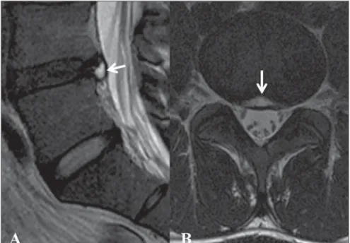
There were three cysts at L4-L5 level and two at L5-S1 level. All cysts presented
homogeneous, low-to-moderate signal in-tensity on T1-weighted images and high signal intensity on T2-weighted images, and were oval shaped, being seated in the anterior epidural space. Four cysts were ventrolaterally located, one to the right, two to the left, and one with bilateral extension. The other one was centrally positioned in the anterior spinal canal. No MRI sign of intracystic hemorrhage or bone erosion was
observed. The adjacent disc presented mild signs of degeneration in all cases.
A communication with the annulus fibrosus was suggested in all cases based on the MRI T2-weighted images (Figure 1) and in one case was confirmed by CT dis-cography (Figure 2). In two cases, CT im-ages were also available and showed epi-dural tissue slightly hypodense to the disc (Figure 3).
Table 1 Summary of patients’ clinical and imaging findings.
Age (years) Sex Lumbar pain/ radiculopathy Level Disc degeneration Adjacent disc protrusion Communication annulus fibrosus Signal intensity Epidural position Previous surgery at level Evolution Case 1 28 Male Yes L4-L5 Mild Yes Yes (MRI) High T2 Low T1 Central No Clinical follow-up Case 2 34 Male Yes L5-S1 Mild No Yes (MRI/CTD) High T2 Low T1 Left ventrolateral No Unknown Case 3 36 Female Yes L4-L5 Mild Yes Yes (MRI) High T2 Low T1 Right ventrolateral No Surgery Case 4 21 Male Yes L4-L5 Mild Yes Yes (MRI) High T2 Low T1 Left ventrolateral Yes Surgery Case 4 21 Male Yes L5-S1 Mild Yes Yes (MRI) High T2 Low T1 Bilateral ventrolateral Yes Surgery
CTD, computed tomography discography; MRI, magnetic resonance imaging.
One patient (case 1) had no available follow-up data. Case 3 was conservatively treated and had unchanged cyst findings at one-year MRI follow-up, and two years of unchanged tissue with epidural mass effect since the first CT examination.
Surgery was performed in patient 2 and findings included a discal cyst in the extra-dural space with a distinct fibrous capsule and containing slightly bloody serous fluid (Figure 3).
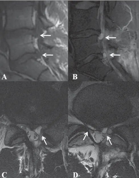
Patient 4 had previously undergone lumbar microdiscectomy at L4-L5 and L5-S1 levels, and 12 weeks later presented re-current lumbar pain related to postsurgical pseudocysts at both levels (Figure 4), and was surgically treated in another institution.
DISCUSSION
Lumbar pain and radiculopathy are very common clinical conditions and MRI is performed to rule out the presence of a herniated disc which is probably the most common reason for lumbar spine investi-gations. Epidural cysts may be clinically indistinguishable from a herniated disc, but constitute a less common cause of radiculo-pathy(6). Differential diagnoses include
discal cysts, perineural cysts, facet synovial cysts, ganglion cysts of the posterior lon-gitudinal ligament or ligamentum flavum, cystic nerve sheath tumors, extradural arach-noid cysts, enteric or dermoid cysts or other cyst-like conditions such as epidural va-rices and premembranous hematomas(5–7).
In a review of uncommon presentations of degenerative disc disease at MRI in eight patients, Eerens et al.(11) have found one
case of discal cyst.
The ventrolateral epidural space could also be occupied by an extruded disc frag-ment, just like a discal cyst. Usually, a disc fragment is easily differentiated from a discal cyst at MRI due to signal character-istics and enhancement pattern. A disc frag-ment has low signal intensity on T1- and T2-weighted images, and a cystic mass is homogeneous and isointense to cerebrospi-nal fluid, although it may have some het-erogeneous components due to protein contents. A discal fragment rarely presents myxoid degeneration of disc material with diffuse apoptotic change and may cause troubles in the diagnosis due to the high Figure 2. Sagittal MRI T2-weighted and reformatted CTD images (A,B), axial MRI T2-weighted and CTD
images (C,D) of case 1 showing left-sided, ventrolateral cyst in epidural space communicating with the disc and filled by contrast medium on CTD image (arrows). Courtesy from Dr. Xavier Stump, São Paulo, Brazil.
signal intensity on T2-weighted images(12).
Although not essential and not available in any case of our series, post-contrast images features include a nonenhancing disc frag-ment surrounded by thick granulation tis-sue in extruded hernia(13), while
contrast-enhanced MRI delineates a thin rim en-hancement in discal cyst(5), which
contrib-utes to elucidate the differential diagnosis.. The diagnosis of discal cyst may be achieved with basis on MRI findings of a ventrolateral homogeneous epidural cyst hypointense on T1- and hyperintense on T2-weighted images. Occasionally, in the presence of intracystic hemorrhage, cysts appear as hyperintense images on T1- and
T2-weighted sequences(13). The finding of
continuity to the annulus fibrosus in a cyst contiguous to the disc may not be seen at MRI. In this case, discography or CTD may be useful to identify such communica-tion(1,5,6,8,12,14,15). Marhsam et al.(7) suggest
that the finding of a communication at dis-cography could be predominantly iatro-genic, the result of a tear in the annulus fibrosus opened by forced injection. In cases of minimal apparent disruption of the annulus fibrosus during probing at surgery in young individuals, discal cyst may have a more optimistic long-term prognosis, as opposed to an intervertebral disc hernia-tion(16).
A direct continuity between the cyst and the disc on sagittal T2-weighted images could be defined in all of the present nonoperative cases, likewise previously reported in the literature(17). Atypical
pre-sentations also include osteolytic change of the vertebral body adjacent to the cyst(5,18)
and gas-containing cysts(19). The
identifica-tion of communicaidentifica-tion of epidural gas col-lection implicated as a possible cause of radicular symptoms and the disc tissue may also be made by CTD. The vacuum phe-nomenon has been proposed as the cause of the gas formation(20).
Histological findings of discal cysts in-clude a capsule consisting of dense fibrous connective tissue with no disc material in-side(5–7). This finding in association with lack of definitive communication between cyst and disc on MRI T2-weighted se-quences is indicative that discal cyst is probably synonymous with ganglion cysts of either posterior longitudinal ligament or annulus fibrosus, and may represent a de-velopmental or degenerative anomaly, with no particular advantage in distinguishing them(21,22). On the other hand, the identifi-cation of residual herniated tissue in the cyst wall(1), disc material with myxoid
de-generation and apoptosis of chondrocytes in the herniated disc material(12), and discal
cyst followed by intervertebral disc hernia-tion(8) could be associated with the
patho-genesis of the discal cyst, suggesting the hypothesis of focal degeneration.
Postsurgical pseudocysts have been de-scribed after laminectomy and lumbar discectomy, and suggested causes include arachnoid hernia through a fissure in the dura mater induced by surgical trauma and persistent spinal fluid or blood leakage leading to the development of pseudome-ningocele. A ventral pseudocyst may ossify but usually it contains serous-hemosiderin fluid(2). Postoperative annular pseudocyst
is a rare complication of microdiscectomy. Its pathogenesis is probably related to the granulation tissue “pseudocapsule” sur-rounding the herniated disc that was not disrupted during surgery, creating a poten-tial space where fluid might accumulate by diffusion through the disrupted annulus fibrosus. The increased disc pressure caused by repetitive loading and enlarge-ment of this pseudocyst could cause symp-Figure 4. Sagittal MRI T1- and T2-weighted images (A,B), axial MRI T2-weighted image at L4-L5 level
toms(10). The first description of a
postsur-gical intervertebral disc cyst was made by Grabel et al. in 1988(9) in a patient with clinical symptoms of failed microdiscec-tomy. It was interpreted as most likely rep-resenting blood breakdown products result-ing from surgery. The blood was prevented from escaping into the epidural space prob-ably because the L5 nerve root had tam-ponated the incised segment of the poste-rior longitudinal ligament. To the authors’ knowledge, cases of double postsurgical annular pseudocysts have never been re-ported.
Several treatments have been proposed for discal cysts, including surgical resec-tion(5,6), fluoroscopy- or CT-guided
percu-taneous steroid injection(23,24), CT-guided percutaneous aspiration(25) or by
percutane-ous endoscopic transforaminal ap-proach(26). A case of spontaneous regres-sion of a discal cyst was reported by Chou et al.(27), although the patient has received
epidural injection and S1 selective nerve block, both injections containing anesthetic and steroid agents. The authors have felt that is was not clear whether the drugs con-tributed to the cyst regression, but the ste-roids could certainly have diffused toward the cyst and caused regression.
CONCLUSION
Based on the above described cases and on the literature review, it is clear that MRI can easily depict an epidural cyst and that the diagnosis of a discal cyst should be considered in cases where homogeneous ventrolateral epidural cyst contiguous to a partially degenerated disc is identified even if a definitive connection with the annulus fibrosus is not possible in nonsurgical pa-tients or as microdiscectomy complication.
Acknowledgments
The authors thank Dr. Xavier M. G. R. G. Stump, São Paulo, Brazil, for providing some images for the present study.
REFERENCES
1. Kobayashi S, Takeno K, Uchida K, et al. Patho-genesis of the discal cysts communicating with an adjacent herniated disc. Histological and ul-trastructural studies of two cases. Joint Bone Spine. 2010;77:184–6.
2. Jeong GK, Bendo JA. Lumbar intervertebral disc cyst as a cause of radiculopathy. Spine J. 2003;3: 242–6.
3. Nabeta M, Yoshimoto H, Sato S, et al. Discal cyst of the lumbar spine. Report of five cases. J Neurosurg Spine. 2007;6:85–9.
4. Kornberg M. Nerve root compression by a gan-glion cyst of the lumbar annulus fibrosus. A case report. Spine. 1995;20:1633–5.
5. Kono K, Nakamura H, Inoue Y, et al. Intraspinal extradural cysts communicating with adjacent herniated disks: imaging characteristics and pos-sible pathogenesis. AJNR Am J Neuroradiol. 1999;20:1373–7.
6. Chiba K, Toyama Y, Matsumoto M, et al. Intraspi-nal cyst communicating with the intervertebral disc in the lumbar spine: discal cyst. Spine. 2001; 26:2112–8.
7. Marshman LAG, Benjamin JC, David KM, et al. “Disc cysts” and “posterior longitudinal ligament ganglion cysts”: synonymous entities? Report of three cases and literature review. Neurosurgery. 2005;57:E818.
8. Tokunaga M, Aizawa T, Hyodo H, et al. Lumbar discal cyst followed by intervertebral disc hernia-tion: MRI findings of two cases. J Orthop Sci. 2006;11:81–4.
9. Grabel JC, Davis R, Zappulla R. Intervertebral disc space cyst simulating a recurrent herniated nucleus pulposus. Case report. J Neurosurg. 1988; 69:137–9.
10. Young PM, Fenton DS, Czervionke LF. Postop-erative annular pseudocyst: report of two cases with an unusual complication after microdiscec-tomy, and successful treatment by percutaneous aspiration and steroid injection. Spine J. 2009;9: e9–e15.
11. Eerens I, Demaerel P, Haven F, et al. Imaging characteristics of noncontained migrating disc fragment and cyst. Eur Radiol. 2001;11:854–7. 12. Okada K, Saito H, Nishida J, et al. Discal cyst
associated with myxoid change and apoptosis of
herniated disc materials: a case report. Ups J Med Sci. 2007;112:39–47.
13. Lee HK, Lee DH, Choi CG, et al. Discal cyst of the lumbar spine: MR imaging features. Clin Im-aging. 2006;30:326–30.
14. Murata K, Ikenaga M, Tanaka C, et al. Discal cysts of the lumbar spine: a case report. J Orthop Surg. 2007;15:376–9.
15. Norman ER, Beall DP, Kitley CA, et al. Interver-tebral disk cyst: a case report. J Comput Assist Tomogr. 2006;30:313–5.
16. Coscia MF, Broshears JR. Lumbar spine intra-canalicular discal cysts: two case reports. J Spi-nal Disord Tech. 2002;15:431–5.
17. Kishen TJ, Shetty AP, Rajasekaran S. Variant of lumbar disc cyst in a 13-year-old girl: a case re-port. J Orthop Surg (Hong Kong). 2006;14:184– 6.
18. Marushima A, Uemura K, Sato N, et al. Osteolytic lumbar discal cyst: case report. Neurol Med Chir (Tokyo). 2008;48:363–6.
19. Kakitsubata Y, Theodorou SJ, Theodorou DJ, et al. Symptomatic epidural gas cyst associated with discal vacuum phenomenon. Spine. 2009;34: E784–9.
20. Pierpaolo L, Luciano M, Fabrizio P, et al. Gas-containing lumbar disc herniation. A case report and review of the literature. Spine. 1993;18: 2533–6.
21. Marshman LAG. Disc cyst. [Letter to the editor]. J Neurosurg Spine. 2007;7:113–4.
22. Marshman LAG. Discal cysts. [Letter to the edi-tor]. J Neurosurg Spine. 2007;7:575–7. 23. Dumay-Levesque T, Souteyrand AC, Michel JL.
Steroid injection performed with fluoroscopy for treatment of a discal cyst. [Letter to the editor]. J Rheumatol. 2009;36:1841–3.
24. Koga H, Yone K, Yamamoto T, et al. Percutane-ous CT-guided puncture and steroid injection for the treatment of lumbar discal cyst: a case report. Spine. 2003;28:E212–6.
25. Kang H, Liu WC, Lee SH, et al. Midterm results of percutaneous CT-guided aspiration of symp-tomatic lumbar discal cysts. AJR Am J Roent-genol. 2008;190:W310–4.
26. Kim JS, Choi G, Lee CD, et al. Removal of discal cyst using percutaneous working channel endo-scope via transforaminal route. Eur Spine J. 2009;18 Suppl 2:201–5.