Cop
yright
© ABE&M t
odos os dir
eit
os r
eser
vados
.
1 Department of Pediatric
Endocrinology, Instituto Nacional de Câncer (Inca), Rio de Janeiro, RJ, Brazil
2 Division of Pathology, Inca,
Rio de Janeiro, RJ, Brazil
3 Department of Head and
Neck Surgery, Inca, Rio de Janeiro, RJ, Brazil
4 Department of Pediatric Oncology,
Inca, Rio de Janeiro, RJ, Brazil
Correspondence to: Maria Alice Neves Bordallo Praia de Botafogo 132, ap. 501 22250-040 − Rio de Janeiro, RJ, Brazil
Received on Apr/13/2010 Accepted on Aug/21/2010
Thyroid spindle epithelial tumor
with thymus-like differentiation
(SETTLE): case report and review
Tumor epitelial de células fusiformes com diferenciação
thymus-like da tireoide (SETTLE): relato de caso e revisão da literatura
Luiz Antônio Magnata Filho1,2, Maria Alice Neves Bordallo1, Cencita H. C. N. Pessoa1, Rossana Corbo1, Daniel Alves Bulzico1,
Fernando Luiz Dias3, Avelino L. Machado4, Andréia B. Soares2, Sima Ferman4
SUMMARY
Spindle epithelial tumor with thymus-like element (SETTLE) is a rare malignant neoplasm of the thyroid, occurring predominantly in children, adolescents, and young adults. SETTLE usually presents itself as a thyroid mass, without metastases at diagnosis. It is believed to derive from branchial pouch or thymic remnant tissueshowing primitive thymic differentiation. This article reports the clinical, cytological, histological and immunohistochemical features of a SETTLE in a 3-year-old girl. Microscopic exam revealed a nodular, highly cellular neoplasm displayed in the classic biphasic pattern, with mixture of prominent spindle cell component and a minor glandular component lined by mucinous or respiratory-type epithelium. The immunohistochemical study showed strong and diffuse positivity for pan-CK, vimentin and smooth muscle actin. The present case is the irst SETTLE case reported in Brazil. To date, the patient described remains without evidence of recurrence or metastasis 5 years after surgery. Arq Bras Endocrinol Metab. 2010;54(7):657-62
SUMÁRIO
O tumor epitelial de células fusiformes com elemento thymus-likeé uma rara neoplasia ma-ligna da tireoide, ocorrendo predominantemente em crianças, adolescentes e adultos jovens. Habitualmente, esse tumor se apresenta como massa tireoideana, sem metástases ao diag-nóstico. Acredita-se derivar de arco branquial ou tecido remanescente tímico, apresentando diferenciação tímica primitiva. Este artigo descreve os aspectos clínicos, citológicos, histológi-cos e imuno-histoquímihistológi-cos de um caso de SETTLE diagnosticado em uma menina de 3 anos de idade. O aspecto microscópico encontrado no tumor foi de uma lesão nodular, hipercelu-lar, disposta em aspecto bifásico clássico, com componente de células fusiformes, e de tecido glandular acompanhado por epitélio mucinoso e respiratório. O estudo imuno-histoquímico foi positivo para pan-CK, vimentina e actina de músculo liso. Esse caso é o primeiro relato de SETTLE no Brasil. A paciente descrita permanece sem evidência de doença em atividade cinco anos após o tratamento cirúrgico. Arq Bras Endocrinol Metab. 2010;54(7):657-62
INTRODUCTION
T
he spindle epithelial tumor with thymus-like element (SETTLE) is a very rare neoplasm related to the thy-roid of young individuals. According to the US National Center for Biotechnology Research (PubMed database), the term SETTLE has been reported only 26 times in the English literature until July of 2010. In 1991, Chan and Rosai (1) uniied the concept of SETTLE when they described 8 neoplasms situated in the neck and thyroidCop
yright
© ABE&M t
odos os dir
eit
os r
eser
vados
.
carcinoma with thymus-like element (CASTLE) (3). This tumor is composed predominantly of spindle and epithelioid cells with glandular or ductular structures lined by a mucinous or respiratory epithelium (4). In spite of indolent growth, SETTLE may give metastases many years after the diagnosis. Therefore, a long-term follow-up is required. We present one case, the irst re-port from Brazil, of a patient who has benign clinical course and tumoral features suggestive of a myoepithe-lial differentiation. The clinicopathologic features of the case reports in the literature are also reviewed.
CASE REPORT
A 3.5 year-old girl was referred to the Brazilian Natio-nal Cancer Institute with a mass in the right lobe of the thyroid. Laboratory exams showed normal free-T3 free; free-T4 and TSH levels. Neck ultrasonography re-vealed a 2.3 x 1.8 x 1.4 cm heterogeneous solid mass which occupied the entire right lobe of the thyroid. There was also bilateral enlargement of lymph nodes at the second cervical level. The thyroid scintigraphy sho-wed decrease of radioactive iodine. Chest tomography and abdominal ultrasonography were normal.
The ine needle aspiration (FNA) cytological study performed was inconclusive. Therefore, resection of the mass was suggested and partial thyroidectomy perfor-med 2 months later. The inal diagnosis was SETTLE. Despite no other therapy having been performed, at the present moment the child is alive without evidence of recurrence or metastasis 5 years after the initial treatment.
MATERIAL AND METHODS
The FNA material was received in physiological solution, processed routinely and stained with Papanicolaou. The surgical specimen was received in 10% buffered formalin and processed routinely. An immunohistochemical analy-sis was performed using formalin-ixed, parafin-embe-dded tissue sections and the standardized streptavidin-biotin peroxidase complex with DAB as a chromogen.
RESULTS
Cytological indings
The biopsy preparation obtained from the FNA reve-aled a highly cellular aspirate consisting of cohesive or isolated sheets of spindle cells with clusters of polygonal
cells with benign features in a background of cyanophi-lic material which resembles mucin. The spindle cells showed elongated or oval nuclei, inely dispersed chro-matin, inconspicuous nucleoli, distinct nuclear mem-brane and a high nuclear-cytoplasm ratio. There was minimal cellular pleomorphism and neither necrosis nor mitotic igures was observed.
Pathological indings
Macroscopic features
The gross examination of the specimen showed a bos-selated, well-circumscribed and irm mass, measuring 2.5 x 2.0 x 1.8 cm. The cut surface was solid, grayish-brown, slightly lobular with residual thyroid at the peri-phery. There were focal cysts and mucoid areas. Necro-sis, calciications and haemorrhage were not observed.
Microscopic features
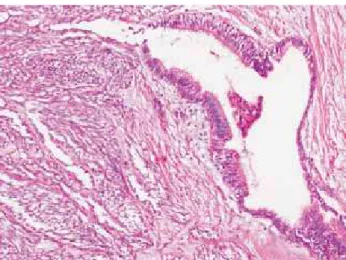
Microscopically, the tumor was circumscribed with collagen bands incompletely dividing the tumor in no-dules (Figure 1). There was a biphasic pattern com-posed of a mixture of a predominant component of spindle cells and a minor glandular component with mucinous and respiratory-type epithelium (Figure 2). The mitotic index was low and there were no areas with necrosis or haemorrhage.
The tumor was highly cellular and formed predo-minantly by reticulated to compact fascicles of spindle cells with a slightly storiform pattern (Figure 3). The spindle cells displayed scanty eosinophilic cytoplasm and elongated to plump nuclei. The nuclei were mini-mally pleomorphic with both pointed and blunt ends, distinct nuclear membranes, delicate chromatin and in-conspicuous nucleoli.
The second component of the tumor consisted main-ly of glandular and ductular structures lined by mucinous epithelium and ciliated pseudo-stratiied epithelium with occasional goblet cells. These structures varied from lar-ge cystic spaces to smaller structures and cell clusters, merging imperceptibly with the spindle cells. These cells displayed a columnar to cuboidal nuclei.
Focal areas with squamous epithelium arising abruptly from the spindle cells were observed. There were interstitial mucine pools, intercellular edema and scant lymphocytes.
Cop
yright
© ABE&M t
odos os dir
eit
os r
eser
vados
.
and smooth muscle actin in both spindle and glandular components. Focal positivity for EMA, mostly in the glandular component, was observed. Desmin, CD5, thyroglobulin, calcitonin, chromogranin, synaptophy-sin and CEA had negative immunoreactivity.
DISCUSSION
Up to July, 2010, from the available data in the indexed literature, there were 30 case reports of SETTLE. This rare tumor occurs in young individuals with a mean age of 17.9 years at presentation (ranging from 2 to 59 ye-ars), with a male-to-female ratio of 1.1.In this case, the patient was 3.5 years old at the time of the diagnosis, the irst report from Brazil.
The generally initial presentation described is a painless mass, within or around the thyroid. In the lite-rature, there are at least 4 patients who had the thyroid mass noticed for four or more years, which suggest a slow tumoral growth (5-8).
In the majority of the reported cases, disease wi-despread occurred years after surgical resection of the primary lesion. In spite of the disease progression with metastasis development, low mortality rates are obser-ved, demonstrating the indolent course of the tumor. SETTLE may not produce metastasis until many years after the diagnosis (mean of 11 years) and according to case reports with follow-up information, the ove-rall rate of metastasis is of about 20% (2,8-10,12-15). Otherwise, when the analysis is made only on patients with more than 5 years of follow-up, the incidence increases to 50% (8,12-14)with 2 patients develo-ping metastasis more than 20 years after the diagnosis (13,14). Metastatic spread is reported to lung, medias-tinum, local lymph nodes and kidney. The principal site of metastasis is the lung, which represents more than 60% of the metastasis sites (2,8,10,12,14). The gap be-tween diagnosis and detection of metastasis varies from a few months after the primary tumor manifestation to 25 years (14,15).
All descriptions of FNA show a highly cellular aspi-rate composed mainly of cohesive or isolated sheets of spindle cells. The cells show an elongated to oval nuclei with well deined nuclear membrane, ine chromatin and inconspicuous nucleoli. Nuclear pleomorphism is not found and mitotic igures are rare. Well differen-tiated polygonal cells and mucin deposits can be found in the background intermingled with the spindle cells (2,6,16).
Figure 1. Collagen bands incompletely dividing the tumor in nodules.
Figure 2. Biphasic pattern composed of a mixture of a predominant
component of spindle cells and a glandular component.
Figure 3. Compact fascicles of spindle cells with a slightly storiform
pattern.
Immunohistochemistry
Cop
yright
© ABE&M t
odos os dir
eit
os r
eser
vados
.
Grossly, SETTLE is a well circumscribed tumor, va-rying in size from 1.8 to 12 cm. It is usually a lobular mass with, sometimes, cysts and mucous focal areas (8).
The external surface of the tumor is frequently bos-selated, with irm, gritty, white-gray to brown, slightly lobular and sometimes with cysts, appearance at the cut surface. The residual thyroid is often identiied at the periphery of the tumor (17). Haemorrhage, necrosis and infarct areas are generally not observed.
Microscopically the tumor can be circumscribed or iniltrative, with sclerotic bands that incompletely sepa-rate the tumor in lobules or nodules. The hallmark fe-ature is a biphasic pattern composed of a predominant component of spindle cells and a minor glandular mu-cous component, occasionally cystic (1,3,9).The colla-gen stripes can produce an eosinophilic hyaline mass si-milar to amyloid, but stain negative on Congo red (2). The tumor has a high cellularity represented by im-perceptibly reticulated to compact fascicles of spindle cells, which are intermixed with epithelial structures. The spindle cells have a mesenquimal appearance, with scanty to moderate slightly eosinophilic cytoplasm. The nuclei are elongated or oval, with little pleomor-phism, pointed ends, ine chromatin, well deined nu-clear membranes and inconspicuous nucleoli. These cells present with a relatively monomorphic aspect, and sometimes with plump features and evident epithelial differentiation (1,6,9,10).
The cells with evident epithelial differentiation may be arranged in cords, narrow complex tubules, short pseudo-papillae, trabecular islands or solid sheets. In many cases, there are cystic branched glands lined by ciliated pseu-do-stratiied epithelium with goblet cells. There may be squamous or mucinous epithelium with cuboidal to co-lumnar nuclei and calciication foci (17-21).
Interstitial mucin pools and intercellular edema are seen frequently (17). Vascular invasion can be present (6,17). Lymphocytes are rare or absent (19).
Mitotic igures are uncommon. A high mitotic in-dex is rare and necrosis is unusual (19). Kirby and cols. (16) observed a high cellularity at the periphery of the tumor near the resection margin, and that the mitotic index increased from the center (1 to 2 mitosis per 10 high-power ields) to the periphery of the tumor (10 to 20 mitosis per 10 high-power ields) (16).
In general, the distant metastasis of SETTLE retains the same morphology of the original tumor (8).
By deinition, SETTLE has an epithelial phenotype of both spindle and clearly epithelial components, as
demonstrated by strong and diffuse immunostaining for cytokeratins (CK), mostly pan-CK.
Vimentin can be expressed in both components of SETTLE, with positivity varying from only 20% of the spindle cells to diffuse for both components (2,18,21). The expression of EMA is also variable, but with focal predominance of the epithelial component (8,2,18,20).
The evident epithelial component does not show, generally, positivity for muscular or neuroendocrine di-fferentiation antibodies. Kirby and cols. (16)reported minimal positivity for smooth muscle actin and NSE in the evident epithelial component (16). Otherwise, Su and Xu observed diffuse immunostaining for smooth muscle actin in the spindle cell component (6,21). We also found expression of smooth muscle actin, which was strong and diffuse in both components, suggesting a myoepithelial differentiation, as found in the other case reports.
The two components of SETTLE are always ne-gative for thyroglobulin, calcitonin, chromogranin A, synaptophysin, CEA, desmin and S-100 protein (2,6,8,16,18,21). Special care is required in cases with trapping of normal thyroid follicles, because when stai-ned with thyroglobulin, the tumoral cells can stain po-sitive due to overlow of the antibody (16).
Xu and Cheuk reported negativity for CD5 (which is expressed in high number of thymic carcinoma, in-cluding CASTLE) and for CD20 (which is expressed in high number of the spindle cells of spindle thymoma) (21,8). Cheuk and cols.(8)still showed negativity for terminal deoxynucleotide transferase (TdT), which is expressed in immature T lymphocytes that accompany the thymomas (8).
Both immunohistochemical and ultrastructural fe-atures conirm the epithelial phenotype of SETTLE. The inding of tonoilaments and desmosomes in all ca-ses studied, in both spindle and clear epithelial cells, de-monstrates the epithelial differentiation of the spindle cells (2,6). Additionally, Su and cols. (6) found the pre-sence of intracytoplasmic thin ilaments with fusiform dense bodies, which suggested, together with immu-noreactivity for smooth muscle actin, a myoepithelial differentiation (6).
Cop
yright
© ABE&M t
odos os dir
eit
os r
eser
vados
.
The differential diagnosis includes many epithelial and biphasic spindle cell tumors of the head and neck which needs a correlation with clinical, radiological, pa-thological and immunohistochemical indings (6).
Among the tumors derived from branchial or thy-mic remnants, “ectopic hamartomatous thymoma” is a benign tumor, compounded by an admixture of plump spindle cells, delicate ibroblast-like spindled cells, ma-ture adipose tissue, and epithelial elements with squa-mous, glandular or indeterminate morphology. This tumor, which has recently been proposed to be called branchial anlage mixed tumor by Fetsch and cols., ge-nerally affects males (male-to-female ratio > 10:1), with a median age of 42.5 years, and can be found in the lo-wer neck, sternoclavicular and presternal regions (22). In addition to the age of the patients (older adults) and the site of the tumor (never affecting the thyroid gland), the presence of mature adipose tissue (present in all cases), rules out this tumor. Ectopic cervical thy-moma is a benign tumor, with a jigsaw puzzle appea-rance, comprised of plump spindle or epithelial cells, which often presents with lymphocytes and occurs in females, with a mean age of 42.7 years. The inding of lymphocytes admixed with the cells in most cases, besides the older age group, helps to differentiate from SETTLE. The third lesion that comprises this group is CASTLE. This tumor does not effectively make diffe-rential diagnosis with SETTLE, since this is clearly a malignant neoplasm, which resembles thymic carcino-ma with lymphoepitheliocarcino-ma-like pattern and occurs in older patients (mean age of 48.5 years) (6,19,20).
In general, thymomas, and mostly the spindle cell variant, present with the characteristic jigsaw puzzle-li-ke lobulation, in addition to CD20 and TdT immunos-taining, ultrastructurally interdigitating cell processes, and lack of glands, as found in SETTLE (8).
Teratomas of the thyroid gland are differentiated from SETTLE based on the inding of mature ele-ments, originated from the 3 germ cell layers (6).
The spindle cell variant of medullar thyroid carci-noma (MTC) can occur in the same age group as SET-TLE, although the inding of prominent ibrovascular septa, amyloid, occasional to frequent mitotic igures and lack of glands, suggests MTC. Furthermore, the immunostaining for calcitonin, chromogranin and CEA conirms the diagnosis of MTC (6).
The spindle cell variant of anaplastic thyroid carci-noma is easily differentiated based on the clinical data, which includes older patients (mean age between 60
and 65 years), and history of rapid tumor growth as-sociated with hoarseness, dysphagia, and dyspnea. The pathologic features resemble a high-grade sarcoma, with prominent cellular pleomorphism, necrosis, high mitotic index, vascular invasion, prominent vascularity, giant cells, and wide extrathyroid extension. The im-munohistochemistry may show positivity for cytokera-tins and CEA, with invariably negative thyroglobulin (6,16).
Synovial sarcoma (SS) is the most dificult differen-tial diagnosis. Clinically, symptoms are often present and there is a rapid tumor growth. The spindle cells are usually monomorphic, hyperchromatic, but present a higher mitotic index. The glandular component is well differentiated, but does not display mucinous or goblet cells. The immunohistochemical study seems to be the best way to distinguish between SS and SETTLE. It reveals only patchy immunopositivity for cytokeratins, (which is strongest in the epithelial cells), strong posi-tivity for vimentin and EMA staining (reliable marker of SS). In addition, there is usually strong and diffuse immunostaining for CD99 and BCL-2 protein. The ul-trastructural lack of tonoilaments, as well as the speci-ic translocation t(x; 18) (p11.2; q11.2), involving the gene SS18 (SYT or SSXT) in chromosome 18, and the genes SSX1, SSX2 and SSX4 in chromosome X, sup-port this distinguishing (2,3,16,18,23).
In the literature, all cases (1,2,3,5-16,18,21,24-33) of SETTLE were treated with surgical resection of the tumor. Adjuvant chemo and radiotherapy were perfor-med in some cases, although the role of the best sche-me is not clear. Both chemo and radiotherapy adminis-tered after surgery led to complete metastatic disease remission in one case (29).
Cop
yright
© ABE&M t
odos os dir
eit
os r
eser
vados
.
treatment, despite the addition of radiotherapy and/or chemotherapy.
Acknowledgements: According to Ethics Committee’s recom-mendations the authors had the patient’s legal representative’s consent and the patient’s assent to report this case.
Disclosure: no potential conlict of interest relevant to this article was reported.
REFERENCES
1. Chan JKC, Rosai J. Tumors of the neck showing thymic or related
branchial pouch differentiation: a unifying concept. Hum Pathol. 1991;22:349-67.
2. Saw D, Wu D, Chess Q, Shemen L. Spindle epithelial tumor with thymus-like element (SETTLE), a primary thyroid tumor. Int J Surg Pathol. 1997;4:169-74.
3. Bradford CR, Devaney KO, Lee JI. Spindle epithelial tumor with thymus-like differentiation: a case report and review of the litera-ture. Otolaryngol Head Neck Surg. 1999;120:603-6.
4. Hadju SI, Hadju EO. Malignant teratoma of the neck. Arch Pathol Lab Med. 1967;83:567-70.
5. Weigensberg C, Daisley H, Asa SL, Bedard Y, Mullen JBM. Thyroid thymoma in childhood. Endocr Pathol. 1990;1:123-7.
6. Su L, Beals T, Bernacki EG, Giordano TJ. Spindle epithelial tumor with thymus-like differentiation: a case report with cytologic, histologic, immunohistologic, and ultrastructural indings. Mod Pathol. 1997;10:510-4.
7. Murao T, Nakanishi M, Toda K, Konishi H. Malignant teratoma
of the thyroid gland in na adolescent female. Acta Pathol Jap. 1979;29:109-17.
8. Cheuk W, Jacobson AA, Chan JK. Spindle epithelial tumor with thymus-like differentiation (SETTLE): a distinctive malignant thyroid neoplasm with signiicant metastatic potential. Mod Pa-thol. 2000;13:1150-5.
9. Erickson ML, Tapia B, Moreno ER, McKee MA, Kowalski DP, Reyes-Múgica M. Early metastasizing spindle epithelial tumor with thymus-like differentiation (SETTLE) of the thyroid. Pediatr Dev Pathol. 2005;8(5):599-606.
10. Kloboves-Prevodnik V, Jazbec J, Us-Krasovec M, Lamovec J. Thyroid spindle epithelial tumor with thymus-like differentiaion (SETTLE): is cytopathological diagnosis possible? Diagn Cytopa-thol. 2000;26:314-9.
11. Abrosimov AY, LiVolsi VA. Spindle epithelial tumor with thymus-like differentiation (SETTLE) neck lymph node metastasis: a case report. Endocr Pathol. 2005;16(2):139-43.
12. Levey M. An unusual thyroid tumor in a child. Laryngoscope. 1976; 86:1864-8.
13. Kingsley DPE, Elton A, Bennett MH. Malignant teratoma if the thyroid: case report and a review of the literarure. Br J Cancer. 1968;22:7-11.
14. Williams ED. Presented at the World Health Organization meeting for the classiication of thyroid tumors; Zurich, Switzerland; 1986. 15. Trabelsi A, Stita W, Zakhama A, Mokni M. An enlarged thyroid in a
20-year-old woman. Arch Pathol Lab Med. 2006;130:405-6.
16. Kirby PA, Ellison WA, Thomas PA. Spindle epithelial tumor with thy-mus-like differentiation (SETTLE) of the thyroid with prominent mi-totic activity and focal necrosis. Am J Surg Pathol. 1999;23:712-6. 17. Rosai J, Carcangiu ML, DeLellis RA. Miscellaneous tumors. In:
Tumors of the thyroid gland. Washington, D.C.: Armed Forces Ins-titute of Pathology; 1990. p. 282-5.
18. Chetty R, Goetsch S, Nayler S, Cooper K. Spindle epithelial tumor with thymus-like element (SETTLE): the predominantly mono-phasic variant. Histopathology. 1998;33:71-4.
19. Fletcher CDM. Diagnostic histopathology of tumors. 2nd ed. Lon-don: Churchill Livingstone; 2002.
20. Rosai J. Rosai and Ackerman’s Surgical Pathology. 9th ed. Phila-delphia: Mosby; 2004.
21. Xu B, Hirokawa M, Yoshimoto K, Miki H, Takahashi M, Kuma S, et al. Spindle epithelial tumor with thymus-like differentiation of the thyroid: a case report with pathological and molecular genetics study. Hum Pathol. 2003;34(2):190-3.
22. Fetsch JF, Laskin WB, Michal M, Remotti F, Heffner D, Ellis G, et al. Ectopic hamartomatous thymoma: a clinicopathologic and immunohistochemical analysis of 21 cases with data supporting reclassiication as a branchial anlage mixed tumor. Am J Surg Pathol. 2004;28:1360-70.
23. Fisher C, De Bruijn DRH, Van Kessel AG. Synovial sarcoma. In: Fletcher DM, Unni KK, Mertens F, editors. Tumors of soft tissue and bone. Lion: OMS; 2002.
24. Hofman P, Mainguene C, Michiels JF, Pages A, Thyss A. Thyroid spindle epithelial tumor with thymus-like differentiation (the “SETTLE” tumor): an immunohistochemical and electron micros-copic study. Euro Arch Otorhinolaryngol. 1995;252:316-20. 25. Iwasa K, Imai MA, Noguchi M, Tanaka S, Sasaki T, Katsuda S, et al.
Spindle epithelial tumor with thymus-like differentiation (SET-TLE) of the thyroid. Head Neck. 2002;24:888-93.
26. Satoh S, Toda S, Narikawa K, Watanabe K, Kuratomi Y, Sugihara H, et al. Spindleepithelial tumor with thymus-like differentiation (SET-TLE): patient. Pathol Int. 2006;56(9):563-7.
27. Tong GX, Hamele-Bena D, Wei XJ, Toole K. Fine-needle aspiration biopsy of monophasic variant of spindle epithelial thymus-like differentiaion of the thyroid: report of one case and review of the literature. Diagn Cytophathol. 2007;35(2):113-9.
28. Haberal AN, Aydin H, Turan E, Demirhan B. Unusual spindle cell tumor of thyroid (SETTLE). Thyroid. 2008;18(1):85-7.
29. Raffel A, Cupisti K, Rees M, Janig U, Bernbeck B, Jazbec J, et al. Spindle epithelial tumor with thymus-like differentiation (SET-TLE) of the thyroid gland with widespread metastasis in a 13-ye-ar-old girl. Clin Oncol. 2003;15:490-5.
30. Murao T, Nakanishi M, Toda K, Konishi H. Malignant teratoma of the thyroid gland in na adolescent female. Acta Pathol Jap. 1979;29:109-17.
31. Lee FY, Wen MC, Jan YJ, Wang J. The predominant monophasic variant of spindle epithelial tumour with thymus-like differentia-tion (SETTLE) of neck soft tissue with late pulmonary metastasis. Pathology. 2010;42(2):188-90.
32. Amji S, Trimeche S, Nouira M, Sfar R, Trabelsi A, Essabbah H. Spindle epithelial tumor with thymus-like differentiation of the thyroid. Clin Nucl Med. 2008;33(12):887-8.