Acute Corneal Hydrops in Children with
Primary Infantile Glaucoma: A Report of 31
Cases over 23 Years at the LVPEI
Anil K. Mandal*
Jasti V Ramanamma Children’s Eye Care Centre, L V Prasad Eye Institute, Hyderabad, India
Abstract
Purpose
Relatively little data exist regarding the outcomes of children with primary infantile glaucoma presenting with acute corneal hydrops. The aim of our study was to determine the surgical outcome of children of infantile glaucoma who presented with acute corneal hydrops.
Methods
In total, 38 eyes of 31 consecutive children of infantile glaucoma presented with acute cor-neal hydrops who underwent primary combined trabeculotomy-trabeculectomy (CTT) by a single surgeon from January 1990 to December 2012 at the LV Prasad Eye Institute (LVPEI), a tertiary eye care centre in Southern India were enrolled in this retrospective study. Primary outcome measures were intraocular pressure (IOP) control (IOP16 mmHg under anaesthesia or IOP21 mmHg without anaesthesia) and clearance of cor-neal edema. Secondary outcome measures were visual acuity (VA), corcor-neal diameter, bleb appearance, intraoperative and postoperative complications.
Results
Mean age at presentation was 6.4 months (range, 2–11 months) and seven eyes (23%) had bilateral affliction. At presentation, all eyes (100%) had moderate to severe degree of cor-neal edema with a mean preoperative IOP of 25.6±5.1 mmHg. Postoperatively, the IOP reduced to 12.0±3.8 mmHg (difference = -13.6, 95% CI = -15.7 to -11.5, t = -13.18, p<0.0001), and the percentage reduction in IOP was 53.05%. Preoperatively 83% of the eyes were on antiglaucoma medication, and postoperatively 2 eyes (5.3%) required 1 anti-glaucoma medication for control of IOP. Preoperatively, corneal edema was present in all eyes and postoperatively it cleared in all of them. Significant myopic astigmatism was pres-ent in 28 eyes (74%), the commonest being compound myopic astigmatism (75%) followed by simple myopic astigmatism (21%). Normal VA (best-corrected VA; BCVA20/60) was achieved in 44.4% of the eyes and 22.2% eyes had low vision (BCVA,<20/60 to 20/400). Complete success (IOP control and clearance of corneal oedema) was obtained in 94.7%
a11111
OPEN ACCESS
Citation:Mandal AK (2016) Acute Corneal Hydrops in Children with Primary Infantile Glaucoma: A Report of 31 Cases over 23 Years at the LVPEI. PLoS ONE 11(6): e0156108. doi:10.1371/journal.pone.0156108
Editor:James Fielding Hejtmancik, National Eye Institute, UNITED STATES
Received:March 21, 2016
Accepted:May 9, 2016
Published:June 1, 2016
Copyright:© 2016 Anil K. Mandal. This is an open access article distributed under the terms of the Creative Commons Attribution License, which permits unrestricted use, distribution, and reproduction in any medium, provided the original author and source are credited.
Data Availability Statement:Institutional Review Board (IRB) restrictions make data unsuitable for public deposition. An anonymized data set will however be made available for researchers who meet the criteria for access to confidential data upon request to the corresponding author or IRB.
Funding:This study was supported by Hyderabad Eye Research Foundation. The funders had no role in study design, data collection and analysis, decision to publish, or preparation of the manuscript.
eyes. There were no significant intraoperative or postoperative complications. Two thirds of the patients showed low, elevated functional filtering bleb. No patient had any bleb leak, ble-bitis or bleb related endophthalmitis. The median follow-up was 36 months (range 2–228 months).
Conclusions
Primary CTT is safe and effective in controlling IOP, resulting in complete clearance of cor-neal edema with modest visual improvement in children of infantile glaucoma presenting with acute corneal hydrops. The outcome of the study will have a positive impact on counsel-ing the parents preoperatively.
Introduction
Infantile glaucoma is a form of developmental glaucoma that manifest after one month of birth until 3 years of age.[1] It manifests with megalocornea associated with increase in intraocular pressure (IOP) but it may remain undetected as the parents believe that the megalocornea may be a manifestation of“beautiful eye”. Consequently, parents of such children may not seek needful attention for medical advice. However some of the children with infantile glaucoma may go onto develop sudden corneal clouding because of acute corneal hydrops.[2–4] This is a medical emergency that prompts the parents to seek consultation with an ophthalmologist or a pediatrician. A detailed ophthalmic evaluation is mandatory to rule out the other causes of acute corneal hydrops.
Once the diagnosis is confirmed, early surgical intervention is required to control the IOP. Although there are various surgical options available in the armamentarium of glaucoma sur-geons, goniotomy is technically difficult because of corneal edema.[5–7] External trabeculot-omy is a suitable alternative as it is possible to perform this procedure even with severe degrees of corneal edema [8–11]; however, the long-term results of trabeculotomy are not encouraging in Indian patient population.[12,13] Another surgical option in the form of primary combined trabeculotomy–trabeculectomy (CTT) is the best alternative as has been reported in various ethnic populations from different parts of the world.[14–18] Encouraged by our results of pri-mary CTT in other forms of developmental glaucoma, we performed the same surgical proce-dure in infantile glaucoma.[12,19–21]
Although acute corneal hydrops is a well recognized but rare manifestation of infantile glau-coma, extensive literature search failed to reveal any series reporting its management outcome. Therefore, the aim of the present study was to report the surgical and visual outcomes of chil-dren with infantile glaucoma who presented with acute corneal hydrops at the L V Prasad Eye Institute (LVPEI) [22], a tertiary eye care centre, in Southern India over a period of 23 years i.e.: from January 1990 to December 2012.
Patients and Methods
(horizontal corneal diameter using calipers, clarity, presence or absence of Haab’s striae and its location) and intraocular pressure (IOP) using Perkin’s hand-held tonometer, In addition, a detailed examination of the lacrimal sac and nasolacrimal duct was performed to rule out the possibility of sac infection. Upon completion of the examination under anesthesia (EUA), the findings were interpreted to establish the diagnosis and decision was taken for surgical inter-vention. Procedures such as refraction, gonioscopy and fundus evaluation were not possible preoperatively given the presence of corneal edema in all cases.
The following information was collected for each patient: age at presentation (in months), gender, age at surgery, pre-and postoperative (at last follow-up visit) corneal diameter and clar-ity, and diameter, pre-and postoperative IOP, visual acuclar-ity, refractive error, number of anti-glaucoma medications at last visit, bleb characteristics, complications, if any. The primary outcome was surgical success as observed at the last follow-up visit. Complete success was clearance of corneal edema and IOP of16 mmHg in patients examined under general anaes-thesia or<21 mmHg in patients who were old enough to be examined with the slit—lamp.
However, refractive error, gonioanomaly, disc evaluation were not considered in the definition of success given that corneal oedema prevented their assessment in the preoperative period. Consequently, comparison of these parameters was not possible between preoperative and postoperative visits Likewise, visual acuity was not considered in the definition of surgical suc-cess. Qualified success was defined when such IOP was maintained with one antiglaucoma medication (AGM). Failure was defined when such IOP could not be achieved even with the addition of one AGM; persistent corneal edema; reduction of vision to no light perception, dev-astating complications; additional glaucoma procedures (including cyclodestructive proce-dures). Devastating complications included endophthalmitis, retinal detachment or chronic hypotony.
Primary CTT was the surgical technique performed in all cases. The methods undertaken for primary CTT have been described previously.[12,13,19–21] Briefly, the Schlemm’s canal was dissected under a partial thickness limbal-based triangular sclera flap and trabeculotomy ab externo was performed on the sides of the radial incision trabeculectomy was then per-formed in the usual manner. In cases of bilateral affliction, after completion of surgery on the first eye, the second eye was operated using a similar technique but with new a set instruments, drapes, gown, gloves etc., simulating a surgical procedure on a different patient.
Ethics Statement
The study was approved by the Ethic Committee of LVPEI. All methods adhered to the tenets of the Declaration of Helsinki. All patient records/information was anonymized and de-identi-fied prior to analysis. The individual in this manuscript has given written informed consent (as outlined in PLOS consent form) to publish these case details.
Postoperative Regimen
Visual acuity (VA) was measured using age-appropriate methods. These included grating acu-ity assessment using Teller Acuacu-ity Cards [23] (TAC) (Vistech Consultants Inc., Dayton, OH, USA) for infants and toddlers and the grating acuity was, however, converted to Snellen equiv-alent for purposes of analysis and comparison based on the norms provided by Mayer et al. [24]. Other assessment procedures included Lea symbols [25] for preschoolers and the log-MAR chart for school-aged children and adolescents. Retinoscopy was performed manually using a streak retinoscope (Heine, Beta 2000) under cycloplegia (1% cyclopentolate hydrochlo-ride eye drops) in the postoperative period.
Statistical Analysis
Descriptive statistics were employed to summarize demographic and outcome characteristics of the participants. Paired t test was used to compare the pre-and postoperative IOP. A P value less than 0.05 was considered statistically significant. All data analyses were performed using SPSS Statistics for Windows version 19.0 (IBM Corp. Armonk, NY).
Results
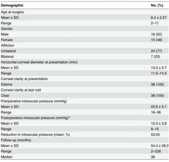
A total of 31 patients (38 eyes) were identified from our retrospective chart review. The demo-graphic characteristics of the participants are summarized inTable 1. The mean age at surgery was 6.4 months (range, 2–11 months). The majority had unilateral affliction (77%) while 7 (23%) had bilateral affliction. The mean follow-up was 54.4 ± 58.2 months (range, 2–228 months; median, 36 months). There was significant lowering of the mean IOP from preopera-tive (25.6 ± 5.1 mmHg) to the postoperapreopera-tive visit (12.0 ± 3.8 mmHg; difference = -13.6, 95% CI = -15.7 to -11.5, t = -13.18, p<0.0001), and this reduction was by a little over 50%. At the last
visit, two eyes (5.3%) required one antiglaucoma medication (Timolol Maleate 0.5%) for con-trol of IOP.
Corneal diameter and clarity
Mean preoperative horizontal corneal diameter was 13.2 ± 0.7mm (Range 11.5–14.5 mm). Although preoperative corneal edema was present in all the eyes, it cleared in all of them (100%) postoperatively.Fig 1A and 1Bshow the preoperative and 1-year postoperative appear-ance of both the eyes of a child who underwent simultaneous bilateral CTT at the age of 6 months. Similarly,Fig 1C and 1Dshow the preoperative and 1-year postoperative appearance of the right and left cornea with Haab’s striae of the same child.Fig 1E and 1Fshow the 1-year postoperative bleb appearance of the right and left eyes respectively.
Refractive error
Data about refractive error was available for all eyes. One eye was emmetropic, 9 eyes had simple myopia and 28 eyes (74%) had astigmatism—the commonest being compound myopic astigma-tism (75%) followed by simple myopic astigmaastigma-tism (21%) and mixed astigmaastigma-tism (4%).
Visual acuity
Success rate
Complete success as defined in the present study was obtained in 36 eyes (94.7%). Two eyes (5.3%) were classified as failures.
Surgical complications
Intraoperative hyphema occurred in 1 eye and was well managed. Postoperative persistent shallow anterior chamber was noted in 1 eye which was surgically reformed with air bubble injection into the anterior chamber on the third postoperative day.
Repeat surgery
Repeat trabeculectomy with mitomycin-C (MMC) was performed in one eye for uncontrolled IOP despite 3 AGMs. Trans-scleral cyclophotocoagulation was done in one eye of a patient with advanced disc damage and poor visual potential.
Table 1. Participant characteristics of 38 eyes of 31 patients with acute hydrops secondary to Infantile Glaucoma.
Demographic No. (%)
Age at surgery
Mean±SD 6.4±2.57
Range 2–11
Gender
Male 16 (52)
Female 15 (48)
Affliction
Unilateral 24 (77)
Bilateral 7 (23)
Horizontal corneal diameter at presentation (mm)
Mean±SD 13.2±0.7
Range 11.5–14.5
Corneal clarity at presentation
Edema 38 (100)
Corneal clarity at last visit
Clear 38 (100)
Preoperative intraocular pressure (mmHg)
Mean±SD 25.6±5.1
Range 16–36
Postoperative intraocular pressure (mmHg)*
Mean±SD 12.0±3.8
Range 8–15
Reduction in intraocular pressure (mean, %) 53.05
Follow-up (months)
Mean±SD 54.4±58.2
Range 2–228
Median 36
SD- Standard deviation;
*intraocular pressure recorded at last follow-up visit, P<0.0001.
Post-operative complications
No patient had any blebitis, bleb leak or bleb-related endophthalmitis.
Discussion
Our study evaluated the results of primary combined CTT in infantile glaucoma presenting with acute corneal hydrops. We found good IOP control (16 mmHg) without AGM in 95% eyes and normal corneal clarity was achieved in all eyes. The results of visual outcome in our series are comparable to that reported by other investigators in the literature.[26,27]
Fig 1. Clinical photograph of a child with bilateral acute corneal hydrops secondary to infantile glaucoma.A) Preoperative appearance showing corneal edema which is more severe in the left eye. B) One-year postoperative appearance of the child seen in figure A showing clear cornea in the right eye with linear horizontal corneal scar and Haab’s striae in the left eye. C) Photograph of the right eye in higher magnification showing Haab’s striae as curvilinear parallel lines in the temporal side (white arrows). D) Fundus retroillumination picture of the left eye showing Haab’s striae as illuminated parallel curved lines crossing the pupillary area. E) Clinical photograph of the right eye showing bleb appearance after 1 year of surgery (asterisk). F) Clinical photograph of the left eye showing the bleb appearance after 1 year of surgery (asterisk).
Nonetheless, visual outcome was sub-optimal in little over 50% of the eyes, with a final best-corrected visual acuity of<20/60 in our study. Such a visual outcome is the consequence of
high corneal astigmatism (up to 7 diopters of cylinder) from Haab’s striae running across the papillary area in most of the eyes.
The development of acute hydrops in our patient series is because of breaks in the desce-met’s membrane under the influence of high IOP in an eye with pre-existing megalocornea. The aqueous humor gains access into the corneal stroma leading to sudden corneal edema.[2] This sudden corneal clouding prompted the parents of children with infantile glaucoma to seek urgent medical attention and consequently early glaucoma surgery.[3] We believe that in the absence of acute hydrops these children with silent infantile glaucoma would have remained undetected and it is plausible that further glaucomatous damage may have occurred.
Mean age at presentation of our patients was 6.4 months (range, 2–11 months) and most of the initial visual development is complete within the first 6 months of life.[28,29] Naturally one would have expected excellent visual outcome in these children following CTT. However, we did not obtain such excellent results in the present series. We offer a couple of reasons for this. Firstly, the Haab’s striae seen in these children crossed the papillary area in most of the cases resulting in photophobia and high irregular corneal astigmatism, and this has the poten-tial to produce meridional amblyopia. Secondly, there were corneal scars resulting from Haab’s striae—a permanent sequel and this may act as an impediment to good visual recovery.[30] In the present series, three-quarters of the eyes had high compound myopic astigmatism with cylindrical component as high as 7 diopters and despite aggressive amblyopia therapy, the visual outcome remained sub-optimal in about 50% of the eyes. Pragmatically, prompt surgical intervention at an early age as in our series should have reduced the risk of deprivation ambly-opia, but our data have not borne this out. Similar results of amblyogenic factors resulting in visual impairment have been reported in the literature. [31] Nonetheless, the results of visual outcome in our series should be interpreted with caution given that there is also a concurrent maturation of the visual system with increasing age of the child that may have contributed to the improvement of VA and changed the refractive status.
Primary CTT is a promising surgical technique for the management of different forms of developmental glaucoma in the Indian context.[12,13,19–21] The alternative surgical tech-nique of goniotomy is technically impossible given the presence of significant corneal edema from acute hydrops.[12,20,21] In the present series significant corneal edema was present in all the eyes and trabeculotomy abexterno may have been a viable option, but long term results of such a procedure is not encouraging in our patient population.[12] Primary CTT offers advantages of the dual mechanism of IOP control (trabeculectomy and trabecultomy) and good long term results have been reported by us previously in various forms of developmental glaucoma. [12,13,19–21] In our series, 84% of the eyes were using AGM at the time of presen-tation to us. By comparison, only 2 eyes (5%) required only a single AGM postoperatively. Two-thirds of the eyes showed mild to moderate filtering bleb which was low to moderately elevated with microcystic changes. These blebs are not prone for infection and we did not have any bleb-related infection or endophthalmitis in the present series.
Children with infantile glaucoma presenting with acute corneal hydrops require prompt surgical intervention. It has been established in the literature that the success rates are highest with the first surgical procedure.[6,11,18,33] Furthermore, there is always a search for the best surgical technique that should be employed in a given patient.[34–37] Recently, 360° tra-beculotomy has emerged as an attractive choice which can be performed with 6’O Proline suture or with the help of an illuminated microcatheter.[17,38–41] Several encouraging reports have emerged in the literature favoring circumferential trabeculotomy with illuminated microcatheter. [17,39–41] Steep learning curve and the cost of therapy using disposable illumi-nated microcatheter is a limiting factor especially in our scenario of a developing country. In a comparative study from India illuminated microcatheter assisted circumferential trabeculot-omy achieved comparable results to primary CTT with mittrabeculot-omycin-C.[17] In the present study, we performed CTT without antifibrotic therapy, and achieved excellent IOP control. However, the long-term results remain unknown.
In the present series, none of the patients required second antiglaucoma surgery. If needed, MMC-augmented trabeculectomy or glaucoma drainage device are viable options as second surgical procedures.[42–44] The benefits of good IOP control in the long term with a single surgical procedure as obtained in the present study are encouraging for the treating ophthalmologist and we believe that such results will have a positive impact while counseling the parents preoperatively. However, the risk of long term drifts in IOP control is well known [42,43] and the life-long follow-up of these children is mandatory. It is also possible that perhaps in the future, with advances in corneal treatment methods, these patients may be able to undergo corneal treatment of their high astigmatism as well as Haab's striae after the IOPs have stabilized and corneal edema cleared to help rehabilitate their vision and prevent amblyopia.
Limitations of this study include the flaws inherent in a retrospective study including the nonrandomization of patients. This study analyzed the surgical experience of a single glaucoma surgeon. Moreover, the mean follow-up was 54.4 ± 58.2 months (range, 2–228 months; median, 36 months) so the longer-term outcomes of primary CTT in a similar patient population remain to be determined.
In conclusion, primary CTT showed promise as an initial surgical procedure in the manage-ment of children with infantile glaucoma with acute corneal hydrops. The IOP control is excel-lent with acceptable bleb appearance with no bleb-related complications and moderate visual recovery in our patient population. Hence, same surgical procedure may be a viable option in other ethnic populations.
Author Contributions
Conceived and designed the experiments: AKM. Performed the experiments: AKM. Analyzed the data: AKM. Contributed reagents/materials/analysis tools: AKM. Wrote the paper: AKM.
-References
1. Mandal AK, Netland PA. Terminology and classification of developmental glaucomas. In: Mandal AK, Netland PA, editors. The Pediatric Glaucomas. Philadelphia, PA: Elsevier Butterworth Heinemann; 2006.
2. Biglan AW. Glaucoma in children: are we making progress? J AAPOS. 2006; 10(1):7–21. Epub 2006/ 03/11. S1091-8531(05)00296-X [pii] doi:10.1016/j.jaapos.2005.10.001PMID:16527674.
3. Mandal AK, Gothwal VK. Images in clinical medicine. Sudden unilateral corneal clouding in an infant. N Engl J Med. 2013; 369(1):74. Epub 2013/07/05. doi:10.1056/NEJMicm1213817PMID:23822779. 4. Zacharia PT, Harrison DA, Wheeler DT. Penetrating keratoplasty with a valved glaucoma drainage
5. deLuise VP, Anderson DR. Primary infantile glaucoma (congenital glaucoma). Surv Ophthalmol. 1983; 28(1):1–19. Epub 1983/07/01. 0039-6257(83)90174-1 [pii]. PMID:6353647.
6. Haas J. Principles and problems of therapy in congenital glaucoma. Invest Ophthalmol. 1968; 7 (2):140–6. Epub 1968/04/01. PMID:5641564.
7. Hoskins HD Jr, Shaffer RN, Hetherington J. Goniotomy vs trabeculotomy. J Pediatr Ophthalmol Stra-bismus. 1984; 21(4):153–8. Epub 1984/07/01. PMID:6470912.
8. Luntz MH. The advantages of trabeculotomy over goniotomy. J Pediatr Ophthalmol Strabismus. 1984; 21(4):150–3. Epub 1984/07/01. PMID:6470911.
9. McPherson SD Jr, Berry DP. Goniotomy vs external trabeculotomy for developmental glaucoma. Am J Ophthalmol. 1983; 95(4):427–31. Epub 1983/04/01. PMID:6837685.
10. McPherson SD Jr. Results of external trabeculotomy. Am J Ophthalmol. 1973; 76(6):918–20. Epub 1973/12/01. PMID:4759851.
11. McPherson SD Jr, McFarland D. External trabeculotomy for developmental glaucoma. Ophthalmology. 1980; 87(4):302–5. Epub 1980/04/01. PMID:7393536.
12. Mandal AK. Current concepts in the diagnosis and management of developmental glaucomas. Indian J Ophthalmol. 1993; 41(2):51–70. Epub 1993/07/01. PMID:8262604.
13. Mandal AK, Naduvilath TJ, Jayagandan A. Surgical results of combined trabeculotomy-trabeculectomy for developmental glaucoma. Ophthalmology. 1998; 105(6):974–82. Epub 1998/06/17. S0161-6420 (98)96022-5 [pii] doi:10.1016/S0161-6420(98)96022-5PMID:9627644.
14. Elder MJ. Combined trabeculotomy-trabeculectomy compared with primary trabeculectomy for congen-ital glaucoma. Br J Ophthalmol. 1994; 78(10):745–8. Epub 1994/10/01. PMID:7803349; PubMed Cen-tral PMCID: PMC504926.
15. Hazmi-AL A, Awad A, Zwaan J, Mesfer-AL SA, Jadaan-AL A, Mohammed-AL A. Correlation between surgical success rate and severity of congenital glaucoma. Br J Ophthalmol. 2005; 89:449–53. PMID: 15774922
16. Mullaney PB, Selleck C, Al-Awad A, Al-Mesfer S, Zwaan J. Combined trabeculotomy and trabeculect-omy as an initial procedure in uncomplicated congenital glaucoma. Arch Ophthalmol. 1999; 117 (4):457–60. Epub 1999/04/17. PMID:10206572.
17. Temkar S, Gupta S, Sihota R, Sharma R, Angmo D, Pujari A, et al. Illuminated microcatheter circumfer-ential trabeculotomy versus combined trabeculotomy-trabeculectomy for primary congenital glaucoma: a randomized controlled trial. Am J Ophthalmol. 2015; 159(3):490–7 e2. Epub 2014/12/09. doi:10. 1016/j.ajo.2014.12.001S0002-9394(14)00776-4 [pii]. PMID:25486542.
18. O'Connor G. Combined trabeculotomy-trabeculectomy for congenital glaucoma. Br J Ophthalmol. 1994; 78(10):735. Epub 1994/10/01. PMID:7803346; PubMed Central PMCID: PMC504923. 19. Mandal AK, Bhatia PG, Bhaskar A, Nutheti R. Long-term surgical and visual outcomes in Indian
chil-dren with developmental glaucoma operated on within 6 months of birth. Ophthalmology. 2004; 111 (2):283–90. Epub 2004/03/17. doi:10.1016/j.ophtha.2003.05.027S0161-6420(03)01189-8 [pii]. PMID: 15019376.
20. Mandal AK, Gothwal VK, Nutheti R. Surgical outcome of primary developmental glaucoma: a single surgeon's long-term experience from a tertiary eye care centre in India. Eye (Lond). 2007; 21(6):764–
74. Epub 2006/04/01. 6702324 [pii]doi:10.1038/sj.eye.6702324PMID:16575414.
21. Mandal AK, Matalia JH, Nutheti R, Krishnaiah S. Combined trabeculotomy and trabeculectomy in advanced primary developmental glaucoma with corneal diameter of 14 mm or more. Eye (Lond). 2006; 20(2):135–43. Epub 2005/04/09. 6701817 [pii] doi:10.1038/sj.eye.6701817PMID:15818392. 22. Rao GN. The Barrie Jones Lecture-Eye care for the neglected population: challenges and solutions.
Eye (Lond). 2015; 29(1):30–45. Epub 2015/01/09. doi:10.1038/eye.2014.239eye2014239 [pii]. PMID: 25567375; PubMed Central PMCID: PMC4289831.
23. Teller DY, McDonald MA, Preston K, Sebris SL, Dobson V. Assessment of visual acuity in infants and children: the acuity card procedure. Dev Med Child Neurol. 1986; 28(6):779–89. Epub 1986/12/01. PMID:3817317.
24. Mayer DL, Beiser AS, Warner AF, Pratt EM, Raye KN, Lang JM. Monocular acuity norms for the Teller Acuity Cards between ages one month and four years. Invest Ophthalmol Vis Sci. 1995; 36(3):671–85. Epub 1995/03/01. PMID:7890497.
25. Hyvarinen L, Nasanen R, Laurinen P. New visual acuity test for pre-school children. Acta Ophthalmol (Copenh). 1980; 58(4):507–11. Epub 1980/08/01. PMID:7211248.
27. Richardson KT Jr, Ferguson WJ Jr, Shaffer RN. Long-term functional results in infantile glaucoma. Trans Am Acad Ophthalmol Otolaryngol. 1967; 71(5):833–7. Epub 1967/09/01. PMID:6066201. 28. Daw NW. Critical periods and amblyopia. Arch Ophthalmol. 1998; 116(4):502–5. Epub 1998/05/02.
PMID:9565050.
29. Wright KW. Visual development and amblyopia. In: Wright KW, Strube Y, N J., editors. Pediatric Oph-thalmology and Strabismus. 3rd ed. Oxford, New York: Oxford University Press; 2012. p. 231–47. 30. Patil B, Tandon R, Sharma N, Verma M, Upadhyay AD, Gupta V, et al. Corneal changes in childhood
glaucoma. Ophthalmology. 2015; 122(1):87–92. Epub 2014/09/10. doi:10.1016/j.ophtha.2014.07.029 S0161-6420(14)00638-1 [pii]. PMID:25200398.
31. Khitri MR, Mills MD, Ying GS, Davidson SL, Quinn GE. Visual acuity outcomes in pediatric glaucomas. J AAPOS. 2012; 16(4):376–81. Epub 2012/08/30. doi:10.1016/j.jaapos.2012.05.007S1091-8531(12) 00237-6 [pii]. PMID:22929453.
32. Mandal AK, Bhatia PG, Gothwal VK, Reddy VM, Sriramulu P, Prasad MS, et al. Safety and efficacy of simultaneous bilateral primary combined trabeculotomy-trabeculectomy for developmental glaucoma. Indian J Ophthalmol. 2002; 50(1):13–9. Epub 2002/07/02. PMID:12090080.
33. Zagora SL, Funnell CL, Martin FJ, Smith JE, Hing S, Billson FA, et al. Primary congenital glaucoma out-comes: lessons from 23 years of follow-up. Am J Ophthalmol. 2015; 159(4):788–96. Epub 2015/01/31. doi:10.1016/j.ajo.2015.01.019S0002-9394(15)00048-3 [pii]. PMID:25634533.
34. Khaw PT. What is the best primary surgical treatment for the infantile glaucomas? Br J Ophthalmol. 1996; 80:495–6. PMID:8759255
35. Chen TC, Chen PP, Francis BA, Junk AK, Smith SD, Singh K, et al. Pediatric glaucoma surgery: a report by the American Academy Of Ophthalmology. Ophthalmology. 2014; 121(11):2107–15. Epub 2014/07/30. doi:10.1016/j.ophtha.2014.05.010S0161-6420(14)00430-8 [pii]. PMID:25066765. 36. Ko F, Papadopoulos M, Khaw PT. Primary congenital glaucoma. Prog Brain Res. 2015; 221:177–89.
Epub 2015/11/01. doi:10.1016/bs.pbr.2015.06.005S0079-6123(15)00096-5 [pii]. PMID:26518078. 37. Papadopoulos M, Edmunds B, Fenerty C, Khaw PT. Childhood glaucoma surgery in the 21st century.
Eye (Lond). 2014; 28(8):931–43. Epub 2014/06/14. doi:10.1038/eye.2014.140eye2014140 [pii]. PMID:24924446; PubMed Central PMCID: PMC4135261.
38. Beck AD, Lynch MG. 360 degrees trabeculotomy for primary congenital glaucoma. Arch Ophthalmol. 1995; 113(9):1200–2. Epub 1995/09/01. PMID:7661757.
39. Girkin CA, Rhodes L, McGwin G, Marchase N, Cogen MS. Goniotomy versus circumferential trabecu-lotomy with an illuminated microcatheter in congenital glaucoma. J AAPOS. 2012; 16(5):424–7. Epub 2012/10/23. doi:10.1016/j.jaapos.2012.05.013S1091-8531(12)00291-1 [pii]. PMID:23084377. 40. Sarkisian SR Jr. An illuminated microcatheter for 360-degree trabeculotomy [corrected] in congenital
glaucoma: a retrospective case series. J AAPOS. 2010; 14(5):412–6. Epub 2010/11/03. doi:10.1016/j. jaapos.2010.07.010S1091-8531(10)00396-4 [pii]. PMID:21035067.
41. Lim ME, Neely DE, Wang J, Haider KM, Smith HA, Plager DA. Comparison of 360-degree versus tradi-tional trabeculotomy in pediatric glaucoma. J AAPOS. 2015; 19(2):145–9. Epub 2015/04/22. doi:10. 1016/j.jaapos.2015.01.008S1091-8531(15)00068-3 [pii]. PMID:25892042.
42. Mandal AK, Walton DS, John T, Jayagandan A. Mitomycin C-augmented trabeculectomy in refractory congenital glaucoma. Ophthalmology. 1997; 104(6):996–1001; discussion 2–3. Epub 1997/06/01. PMID:9186441.
43. Mandal AK, Prasad K, Naduvilath TJ. Surgical results and complications of mitomycin C-augmented trabeculectomy in refractory developmental glaucoma. Ophthalmic Surg Lasers. 1999; 30(6):473–80. Epub 1999/07/07. PMID:10392736.