P
ROTEIN
F
OLDING AND
D
ISEASE
T
HE
M
ITOCHONDRIAL
P
ROTEIN
F
RATAXIN
Dissertation presented to obtain a Ph.D. degree in Biochemistry
at Instituto de Tecnologia Química e Biológica,
Universidade Nova de Lisboa
Ana R. Correia
T
HE
M
ITOCHONDRIAL
P
ROTEIN
F
RATAXIN
Ana Raquel Viegas Correia
Dissertation presented to obtain a PhD degree in Biochemistry at Instituto de Tecnologia Química e Biológica,
Universidade Nova de Lisboa
Supervisor
Cláudio Emanuel Moreira Gomes
Opponents
Mark M. Fisher & Ana Margarida Damas
Instituto de Tecnologia Química e Biológica, Universidade Nova de Lisboa
ii
ITQB - Protein Biochemistry Folding and Stability group Instituto de Tecnologia Química e Biológica,
Universidade Nova de Lisboa
Av. da República (EAN), 2785-572 Oeiras, Portugal
iii This dissertation describes the work performed under the supervision of Dr. Cláudio Gomes, at the Protein Biochemistry Folding and Stability Laboratory, at the Instituto de Tecnologia Química e Biológica, from January 2006 to August 2009.
The studies presented here aim to contribute to a better understanding of frataxin conformational and functional properties and, at the same time, also intend to shed some light into the pathological process associated to Friedreich's ataxia (FRDA). Initially, the impact of FRDA-related mutations in human frataxin (FXN) was studied and their potential rescue by chemical chaperones addressed. Still focusing on the mechanisms underlying FRDA pathogenesis the susceptibility and possible role of oxidative stress relations on human frataxin were investigated. Further, the yeast frataxin orthologue (Yfh1) was used to explore the conformational and functional properties of frataxin.
v I would like to express my sincere gratitude to the following people: My supervisor, Cláudio M. Gomes, to whom I am deeply grateful for the opportunity to learn and to grow to be a scientist in an amazing environment. His passion for science, dedication, critical sense, persistence and broad awareness are a source of inspiration. I am thankful for his support, for the freedom to explore different paths and for the opportunities to visit other laboratories.
Annalisa Pastore from the National Institute for Medical research (UK), for accepting me as a visiting student at her laboratory and for scientific training in molecular biology techniques and NMR.
Salvatore Adinolfi and John McCormick for teaching me molecular biology techniques. Chiara Pastore, Veronica Esposito and Goeff Kelly for the NMR experiments.
Paula Chicau and Ana Coelho for the HPLC separations and MS analysis of the peptides from the limited proteolysis experiments.
Peter Bross from the University of Aarhus (DK), for useful discussions and for sharing Lon and ClpXP plasmids.
Mark Fisher and Subhashchandra Naik from the University of Kansas Medical Center (US) for support, sharing of immobilized GroEL beads and for optimizing the conditions for the GroEL-Frataxin system.
vi
for useful discussions, sharing of plasmids and for their functional studies on FRDA yeast model.
Miguel Teixeira from ITQB for useful discussions, support and for performing the EPR measurements.
Joan Valentine, from UCLA (US), is gratefully acknowledged for sharing SOD1 and Apo-SOD1 proteins.
My colleagues and friends at the Protein Biochemistry, Folding and Stability group for the friendly and positive environment, constant support and helpful discussions: Sónia Leal, Hugo Botelho, João Rodrigues, Ema Alves, Vesna Prosinecki and Patrícia Faísca.
Bárbara Henriques, for always being there, for being an amazing listener, for the helpful discussions and for the laughs we shared in and out of the lab.
All my friends, especially Sofia, Ana Paula, Patrícia, Lígia and Vera, with whom I've shared the excitement and also the down moments of these last 4 years.
All my family for the constant support and love, especially my grandfather Viegas who has always listened with unbelievable attention what was going on in the lab in spite of not having the faintest idea of what I was talking about.
vii support, by awarding a PhD fellowship (SFRH/BD/24949/2005)
ix 1. Correia, A. R., Adinolfi, S., Pastore, A., Gomes, C. M. "Conformational stability of human frataxin and effect of Friedreich’s ataxia-related mutations on protein folding"
Biochem. J. 398, 605-611 (2006).
2. Correia, A. R. *, Pastore, C. *, Adinolfi, S., Pastore, A., Gomes, C. M. "Dynamics, stability and iron-binding activity of frataxin clinical mutants" FEBS J. 275, 3680-3690 (2008).
3. Correia, A. R., Ow, S. Y., Wright, P. C., Gomes, C. M. "The conserved Trp-155 in human frataxin as a hotspot for oxidative stress related chemical modifications"
BBRC - Biochemical and Biophysical Research communication, 390(3):1007-11 (2009).
4. Correia, A. R., Wang, T. Craig, E. A., Gomes, C. M. "Iron binding activity in yeast frataxin entails a trade off with stability in the /1 acidic ridge region"
Biochem J. 426(2):197-203 (2010).
Other publications not included in this thesis
5. Amaral. J. D.*, Correia, A. R. *, Steer, C. J., Gomes, C. M., Rodrigues, C. M. P. "No evidence of direct binding between ursodeoxycholic acid and the p53 DNA-binding domain"
Bioscience Reports, in press.
*
xi This dissertation focuses on the study of frataxin, a small mitochondrial protein whose deficiency is associated with the neurodegenerative disease Friedreich's ataxia (FRDA). Aiming at a better understanding of frataxin conformational and functional properties, two lines of research were followed: first, the effect of FRDA-related mutations in human frataxin (FXN) were studied and the role of oxidative stress related modification addressed; second, yeast frataxin (Yfh1) orthologue was used to explore the conformational and functional properties of the protein.
FRDA is the most common recessive ataxia among the Caucasian population with an estimate prevalence of 1-2 cases per 40,000 individuals in Europe. Most patients are homozygous for a GAA triplet expansion in a non-coding region that reduces the transcription of the gene encoding for frataxin, leading to reduced protein levels (5-30% of the normal levels). However, some patients (~4%) are compound heterozygous patients, meaning that they have the expansion in one allele and a deleterious point mutation on the other. The currently known mutations include missense, non-sense and splice mutations. Here we focused on the study and characterisation of four of the indentified missense mutations. Deletion of frataxin gene in eukaryotes is lethal, hence, even if in residual levels, all FRDA patients must express the protein.
xii
xiii chemical chaperones due to their ability to stabilise proteins and to correct their misfolding or folding defects. Thus, the presence of these compounds is particularly relevant in the context of a genetic modification, such as a clinical point mutation, or in adverse environment conditions. The therapeutic potential of these compounds has been explored for many misfolding diseases like Gaucher's or cystic fibrosis and here we have evaluated the potential chaperone-like behaviour of four small compounds on two frataxin FRDA-related variants: FXN-D122Y and FXN-I154F. The wild type was used as a control and 4-phenyl butyrate (small fatty acid), trehalose (sugar), trimethylamine-N-oxide (methylamine) and glycerol (polyol) were tested for their potential chaperone-like effect on frataxin clinical variants.
First, the effect of small compounds on early folding events was tested. Using the E. coli expression system, the mutants were expressed in the presence of different concentrations of the small compounds and the ratio between expressed soluble and insoluble protein allowed to assess the impact of the compounds on the proteins’ folding efficiency. Intriguingly, the effect of the different compounds was mutant dependent. While the FXN-D122Y folding efficiency was only modestly improved, the FXN-I154F folding efficiency improved significantly with the amount of expressed soluble protein increasing up to ~3fold in the presence of glycerol.
xiv
compounds, 4PBA, trehalose and glycerol, showed a positive impact on the FXN-D122Y conformational defects, as evaluated through the GroEL sink assay (GroEL specifically binds exposed hydrophobic regions, thus when conformational defects are present the proteins patition onto GroEL). The effects on the folded FXN-I154F were moderate; stability was only increased up to a Tm of ~2.5ºC. However,
the addition of TMAO (500mM) positively affected this mutant’s conformation, preventing its partitioning onto the molecular chaperone GroEL.
Our results show that the effects induced during the early folding events are distinct from those observed on the folded protein and also, different point mutations are modulated by the same chemical chaperone in a different manner. These differences are likely to result from the conformational variations introduced by each mutation. The results presented here suggest that FXN-D122Y conformational variation might be overcome by the presence of chemical chaperones and in vivo increased concentrations of different chemical chaperones may rescue the pathological phenotype associated to this mutant. On the other hand, the FXN-I154F mutant should only be partially rescued, but the presence of small compounds may attenuate the phenotype observed.
xv presence of oxidative stress related modifications. The protein is susceptible to carbonylation and nitration modifications in residues from the -sheet surface (Tyr143, Tyr174, Tyr205 and Trp155). Frataxin functions are not significantly affected: frataxin-mediated protection against ROS is still observed, as well as iron-binding necessary for the metallochaperone activity. However, the protein is up to 1.0 kcal.mol-1 destabilised, with conformational opening, as shown by the thermal unfolding reactions and the limited proteolysis experiments. Interestingly, the strictly conserved Trp155, whose mutation leads to FRDA, is both susceptible to carbonylation and nitration modifications, suggesting that this residue may be a functional hotspot in frataxin.
The human and yeast frataxin orthologues have a high degree of amino acid and structural identity. In addition, the phenotype caused by frataxin depletion is very similar in human and yeast cells, thus the yeast model system has been proven to be a very useful model to study frataxin function. Here, when addressing frataxin functional and conformational properties we have used the yeast frataxin orthologue (Yfh1). Aiming at a better functional understanding of frataxin structure, we have characterised eight variants involving mutations on two putative functional regions – the acidic ridge and the conserved
xvi
this region. In addition, our study suggests that residues Asp101 and Glu103 are involved in the iron-mediated interaction between Isu and Yfh1, but their alteration does not abrogate the interaction, as evidenced by the rescue of the yfh1 phenotype.
Mutations on the conserved -sheet residues had been shown to severely compromise frataxin function; here we show that these mutations only have a modest impact on the protein stability (Tm
~0-4ºC), which highlights the functional importance of residues 122-124.
xvii copper binding ability. Copper may regulate frataxin iron-chaperone function, and, as shown by copper/iron competition assays, this might be occurring at least to some extent. The presence of copper was shown to be able to reduce Yfh1 affinity to Isu however, even the excess of copper does not abrogate this interaction. Alternatively, it may be suggested that, in addition to its already described iron-chaperone function, frataxin may also act as a copper-iron-chaperone, directing the copper present in the matrix to the appropriate chaperones within the IMM. Thus, we have evaluated frataxin copper-chaperone ability and we have found that frataxin improves SOD1 reconstitution, meaning that this protein may act as a copper chaperone. Frataxin copper binding ability must be further explored: first it has to be assessed whether this feature is also occurring in vivo and if does what is its biological meaning; in addition, a more chemical approach must be followed in order to characterise in detail the binding of copper to this protein.
xix A presente tese foca-se no estudo da frataxina, uma proteína mitocondrial cuja deficiência está associada à doença neurodegenerativa atáxia de Friedreich (FRDA). Com o objectivo de contribuir para uma melhor compreensão das propriedades conformacionais e funcionais da frataxina, foram seguidas duas linhas de investigação: a primeira envolveu o estudo de mutantes clínicos da frataxina humana e também a avaliação do efeito das modificações associadas a condições de stress oxidativo; a segunda focou-se na investigação das propriedades funcionais e conformacionais da proteína, tendo os estudos sido realizados com a frataxina de levedura (Yfh1).
xx
pacientes expressam frataxina.
xxi Em suma, os resultados aqui apresentados sugerem que os efeitos clínicos observados para os pacientes heterozigóticos resultam provavelmente da combinação de várias factores, tais como a redução da capacidade de enrolamento, o aumento da degradação in vivo, o mau enrolamento e a destabilização conformacional, que, em conjunto, contribuem para uma redução dos níveis funcionais de frataxina. Considerando este cenário, a atáxia de Friedreich em pacientes heterozigóticos pode ser considerada uma doença conformacional (misfolding disorder).
Compostos pequenos, como os açúcares e os polióis, são frequentemente designados chaperões químicos uma vez que estabilizam e corrigem alguns defeitos de enrolamento das proteínas. Assim, estes compostos são particularmente relevantes num contexto biológico, quando ocorre uma modificação genética ou em condições adversas como um aumento da temperatura. O potencial terapêutico destes compostos tem sido explorado para muitas patologias conformacionais, como sendo a doença de Gaucher ou a fibrose quística. Assim, avaliámos aqui a potencial utilização de quatro compostos como chaperões químicos de dois mutantes clínicos da frataxina: FXN-D122Y e FXN-I154F. A proteína selvagem foi utilizada como controlo e o 4-phenilbutirato (ácido gordo), a trealose (açúcar), a trimetilamina-N-oxide (metilamina) e o glicerol (poliol) foram testados no sentido de avaliar as suas capacidades como chaperões químicos dos mutantes estudados.
xxii
eficiência do processo de enrolamento proteico. Curiosamente, o efeito dos compostos depende do mutante considerado. Enquanto a eficiência de enrolamento do mutante FXN-D122Y foi apenas marginalmente alterada, a do mutante FXN-I154F aumentou significativamente com a presença dos vários compostos, nomeadamente a adição de glicerol triplicou a quantidade de FXN-I154F solúvel. Posteriormente, o efeito dos compostos na proteína enrolada, mais especificamente o efeito na conformação e estabilidade, foi também avaliado. Considerando a proteína já enrolada, os efeitos no mutante FXN-D122Y são mais significativos. A presença de TMAO resulta num aumento da estabilidade térmica do mutante FXN-D122Y para valores idênticos aos determinados para a proteína selvagem na ausência dos compostos (o aumento máximo do Tm de ~7.8ºC). Os
estudos com a GroEL (GroEL sink assay) permitiram ainda avaliar os defeitos conformacionais dos mutantes e a potencial correcção desses defeitos em consequência da presença dos chaperões químicos. Três dos compostos testados (4PBA, trealose e glicerol) permitiram corrigir pelo menos parte dos defeitos conformacionais associados a mutação D122Y, resultando num atenuamento da ligação da proteína mutante à GroEL. Os efeitos destes compostos no mutante FXN-I154F enrolado são, no entanto, mais modestos: em termos de estabilização da proteína os compostos conseguem apenas induzir pequenas variações (máx. Tm de ~2.5ºC); considerando a conformação deste mutante, o
ensaio com a GroEL revelou que apenas o TMAO (500mM) consegue reverter os defeitos conformacionais associados a este mutante, retardando a sua interacção com o GroEL.
xxiii Provavelmente estas diferenças resultam das variações conformacionais causadas por cada mutação. Os resultados aqui apresentados sugerem que provavelmente a variação conformacional inerente ao mutante FXN-D122Y pode ser ultrapassada, ou parcialmente revertida, pela presença de chaperões químicos, e, in vivo, a presença de chaperões químicos deverá presumivelmente evitar o fenótipo patológico associado à expressão deste mutante. Por outro lado, a presença de chaperões químicos in vivo deve apenas atenuar o fenótipo patológico associado à mutação I154F.
O estudo aqui apresentado abordou ainda o impacto de modificações associadas ao stress oxidativo na frataxina humana. A priori, a frataxina é um potencial alvo de modificações associadas a este tipo de stress, não só devido a sua localização na mitocôndria, em que oxigénio e óxido nítrico estão disponíveis num ambiente redutor, mas também devido a sua função de chaperão de ferro. Assim, a frataxina humana selvagem foi submetida a condições que simulam o aumento de stress oxidativo dentro da mitocondria (H2O2 + Fe(II) ou
xxiv
conservado e cuja mutação resulta em FRDA, é susceptível aos dois tipos de modificação identificadas, o que sugere que este resíduo talvez seja um hotspot funcional.
xxv não é suficiente para abolir a referida interacção, como é evidenciado pela recuperação do fenótipo causado pela yfh1.
É também aqui demonstrado que os resíduos 122-124, cuja mutação compromete de forma drástica a função da frataxina, têm apenas um impacto modesto em termos da estabilidade da proteína (Tm ~0-4ºC) o que evidencia a importância destes resíduos na
interacção com a Isu.
xxvi
esta hipotética função não está associada à oligomerização da proteína. A afinidade para o cobre e para o ferro são idênticas, o que pode sugerir uma função alternativa para a capacidade desta proteína ligar cobre. Este metal pode potencialmente regular a função da frataxina como chaperão de ferro e, de acordo com os ensaios de competição cobre/ferro, isto pode ocorrer parcialmente. A presença de cobre em solução reduz a afinidade da Yfh1 para a Isu, no entanto, mesmo em excesso, a presença de cobre não impede esta interacção. Alternativamente, além de uma função com chaperão de ferro, a frataxina poderá também funcionar como um chaperão de cobre, direccionando o cobre presente na matriz para os chaperões de cobre presentes na membrana interna da mitocôndria. Assim, a potencial capacidade de a frataxina funcionar como um chaperão de cobre foi avaliada através da reconstituição da Apo-SOD1 in vitro. Os resultados aqui apresentados revelam que a presença de frataxina facilita a reconstituição da apo-SOD1, o que sugere que esta proteína poderá efectivamente funcionar como um chaperão de cobre.
A capacidade de a frataxina ligar cobre tem que ser mais explorada: primeiro é necessário estabelecer se, in vivo, a frataxina é também capaz de ligar cobre e, caso isto se verifique, mais estudos serão necessários no sentido de clarificar o significado biológico de a frataxina ligar cobre. Adicionalmente, um estudo mais químico visando a caracterização da ligação do cobre à frataxina terá que ser realizado.
xxvii
4PBA 4-Phenylbutyrate Ksv Stern-Volmer constant
ATP Adenosine triphosphate nucleotide
mtDNA Mitochondrial DNA
CcO Cytochrome c oxidase mFXN Murine frataxin orthologue
CuL Copper -
non-proteinaceous ligand complex
N Native state
CyaY Escherichia coli frataxin orthologue
NOE Nuclear Overhauser Enhancement
D Denatured state OXPH
OS
Oxidative phosphorylation
[D]1/2 Midpoint of the chemical
denaturation curve
PD Parkinson's disease
Dfh1 Drosophila melanogaster frataxin orthologue
QC Quality control
DLS Dynamic light scattering ROS Reactive oxigen species
DMS methylsulfonium solutes SOD Superoxide dismutase
Dox doxycycline T1 Longitudinal relaxation rates
DSF Differential scanning fluorimetry
T2 Transverse relaxation rates
ETF Electron transfer flavoprotein
(TBA)2
-MDA
Tiobarbituric acid- melondialdehyde complex
F Fold TFA Trifluoroacetic acid
FeS Iron-sulphur Tm Midpoint of the thermal
unfolding curve
FRDA Friedreich's Ataxia TMAO Trimethylamine N-oxide
FXN Human frataxin orthologue TS Transition state
GdmCl Guanidinium chloride U Unfolded state
GST Gluthathione-Stransferase UPS ubiquitin-proteasome system GuSCN Guanidine thiocyanate Yfh1 Yeast frataxin orthologue
HD Huntington's disease GH2O Unfolding Gibbs
free energy change in water
Hsp Heat shock protein Hm Enthalphy change for
unfolding at Tm HSQC Heteronuclear single
quantum coherence
G) Difference in unfolding Gibbs free energy change
HTT Huntingtin m Mitochondrial membrane
potential
I Intermediate state c Correlation time
xxix Foreword ... ..iii Acknowledgements ... .v Thesis Publications ... ..ix Dissertation Abstract ... ..xi Resumo da Dissertação...xix Abbreviation...xxvii Figures ...xxxvii Tables ...xlii
C
HAPTER1
P
ROTEIN
M
ISFOLDING AND
D
ISEASE
1.1 The Importance of Folding ... 3
1.1.1. The Protein Folding Mystery ... 4 1.1.2. The Search for a Protein
Folding Mechanism ... 5 1.1.3. Energy Landscapes ... 9 1.1.4. Protein Folding in the Cell ... 13 1.1.5. Protein Evolution ... 17
1.2. When the Folding Process Goes Wrong:
Misfolding and Aggregation ... 20
xxx
Molecules - Chemical Chaperoning ... 27 1.2.3. Protein Quality Control
and Disease Prevention ... 29 1.2.4. Mitochondrial Dysfunction
and Neurodegeneration ... 33
1.3. References ... 41
C
HAPTER2
F
RATAXIN AND
F
RIEDREICH
’
S ATAXIA
2.1 Friedreich’s Ataxia ... 55
2.1.1. Friedreich’s Ataxia:
a Mitochondrial Disease ... 60 2.1.2. Treatments Available ... 62
2.2. Fratain: a Highly Conserved
Mitochondrial Protein ... 64
2.3. Frataxin Function: What is Known so Far ... 71
2.3.1. An Iron Binding Protein ... 71 2.3.2. Iron-Chaperon ... 75 2.3.3. A Ferritin-Like Protein ... 83 2.3.4. Anti-Oxidant Activity ... 85
xxxi
3
FRATAXIN AND EFFECT OF FRIEDREICH
ATAXIA
-
RELATED MUTATIONS ON PROTEIN
’
S
FOLDING
3.1. Summary ... 99
3.2. Introduction ... 99
3.3. Materials and Methods ... 101
3.4. Results ... 103
3.4.1. Frataxin Chemical and Thermal Unfolding ... 103 3.4.2. Conformational Dynamics ... 105 3.4.3. Limited Proteolysis Experiments ... 107 3.4.4. Iron Binding is Impaired
by Protein Destabilisation ... 108 3.4.5. Frataxin Mutants Thermodynamics Stability ... 108
3.4. Discussion ... 109
3.5. Acknowledgements ... 110
3.6. References ... 110
C
HAPTER4
D
A
YNAMICS
CTIVITY OF
,
S
F
RATAXIN
TABILITY AND
C
LINICAL
I
RON
M
UTANTS
B
INDING
xxxii
4.3. Materials and Methods ... 116
4.4. Results ... 120
4.4.1. Mapping Frataxin Mutations on the Structure ... 120 4.4.2. Protein Dynamics of Wild-Type
Human Frataxin ... 121 4.4.3. Conformational Dynamics
of Frataxin Mutants ... 123 4.4.4. Structural Flexibility ... 127 4.4.5. Proteolytic Degradation Kinetics ... 129 4.4.6. Degradation by Lon and ClpXP ... 131 4.4.7. Clinical Mutations Impact
on Stability and Iron Binding ... 132
4.5. Discussion ... 134
4.6. Acknowledgements ... 136
4.7. References ... 136
C
HAPTER5
S
FRDA
MALL
F
M
RATAXIN
OLECULES AS MODULATORS OF
V
ARIANTS
5.1. Summary ... 141
5.2. Introduction ... 142
xxxiii
5.4. Results ... 146
5.4.1. Effect on Early Folding Events ... 146 5.4.2. Probing Conformational Variantions:
GroEL Sink Assay ... 148 5.4.3. Stabilisation of Frataxin’s Native State ... 151
5.4. Discussion ... 153
5.4. Acknowledgements ... 154
5.4. References ... 155
C
HAPTER6
T
HE
C
ONSERVED
T
RP
-155
IN
H
UMAN
F
RATAXIN AS A
H
OTSPOT FOR
O
XIDATIVE
R
ELATED
C
HEMICAL
M
ODIFICATIONS
6.1. Summary ... 159
6.2. Introduction ... 159
6.3. Materials and Methods ... 160
6.4. Results ... 164
6.4.1. Frataxin Carbonylation and Nitration ... 164
6.4.2. Effect on Frataxin Functions ... 167
6.4.3. Impact on Frataxin Conformation and Stability ... 169
6.5. Discussion ... 170
xxxiv
C
HAPTER7
I
RON
B
INDING
A
CTIVITY IN
Y
EAST
F
RATAXIN
E
NTAILS A
T
RADE
O
FF WITH
S
TABILITY IN THE
1/
1
A
CIDIC
R
IDGE
7.1. Summary ... 177
7.2. Introduction ... 177
7.3. Materials and Methods ... 179
7.4. Results ... 183
7.4.1. Frataxin’s Cellular Function ... 183
7.4.2. Iron Binding ... 186
7.4.3. Isu Binding ... 187
7.4.4. Activity-Stability Trade-Off ... 188
7.5. Discussion ... 192
7.6. Acknowledgements ... 192
7.7. References ... 193
C
HAPTER8
F
C
RATAXIN AND ITS
OPPER
M
ETABOLISM
P
UTATIVE
R
OLE ON
8.1. Summary ... 197
xxxv
8.3. Materials and Methods ... 201
8.4. Results ... 203
8.4.1. Copper Binding Capacity and
Stabilisation Effect ... 203
8.4.2. Putative Copper Binding Sites ... 206
8.4.3. Frataxin Can Also Be a Copper Chaperone ... 208
8.5. Discussion ... 209
8.6. Acknowledgements ... 210
8.7. References ... 210
C
HAPTER9
D
ISCUSSION
9.1. Frataxin and FRDA ... 215
9.1.1. FRDA in Heterozygous Patients ... 215 9.1.2. Modulators of FRDA variants ... 220 9.1.3. Oxidative Stress Related Modifications
and FRDA ... 222
9.2. Frataxin Function ... 226
9.2.1. Characterisation of Functional Mutants ... 226 9.2.2. Copper Binding: A New Functional
Feature of Frataxin? ... 228
xxxvii 1.1. Simplified energy diagrams for folding of small
single-domain proteins the differences between
diffusion-collision and nucleation-condensation models. ... 6 1.2. Information redrawn from the -value analysis ... 8 1.3. Three-stage mechanism of folding - Lattice model. ... 10 1.4. Schematic free-energy (F) surface representing features
of the folding of hen lysozyme (a protein comprising an
and a domain with 129 amino acid residues). ... 12
1.5. Model of nascent chain configurations in a
representative polysome. ... 15 1.6. Folding funnels designed to describe the competition
between productive/efficient folding and aggregation. ... 16 1.7. Transition folds trough an evolutionary bridge. ... 19 1.8. The GroEL-ES chaperonin. ... 26 1.9. Protein Quality control. ... 30 1.10. Mitochondrial quality control (QC) mechanisms. ... 37 1.11. Molecular mechanism associated to the development of
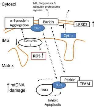
Huntington disease (HD). ... 38 1.12. Molecular mechanisms associated to the development
of Parkinson’s disease (PD). ... 40 2.1. The German pathologist and neurologist, Prof. Nikolaus
xxxviii
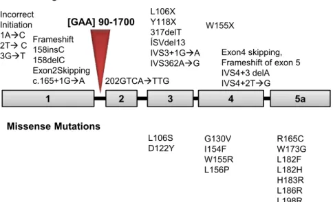
the described mutations. ... 57 2.3. Alignment of eukaryote and bacterial frataxins sequences. .... 65 2.4. Schematic representation of human frataxin processing
by MPP. ... 66 2.5. Molecular surface and ribbon representation of human
frataxin. ... 67 2.6. Ribbon representation of frataxin's orthologues
structure, highlighting the similarities between the orthologues but also the different C-terminal expansion length. ... 69 2.7. Human frataxin putative iron binding sites identified by
NMR spectroscopy. Sites occupied at different iron to protein stoichiometries (1:1, 2:1 and 6:1). ... 72 2.8. A model for FeS protein assembly in Eukaryotes ... 76 2.9. Frataxin dynamic role in the regulation of aconitase
during oxidative stress. ... 77 2.10. Schematic representation of the alternative molecular
mechanism of frataxin as a gate-keeper of FeS clusters assembly. ... 78 2.11. Ribbon representation ofthe structure of human frataxin
and sequence alignment of CyaY, Yfh1 and FXN highlighting the putative iron and ferrochelatase binding sites identified by NMR ... 80 2.12. Model for the regulation of frataxin iron chaperon
xxxix 3.1. Chemical denaturation curve for FXN at pH 7.9. with
GdmCl, GuSCN and urea. ... 105 3.2. Stern–Volmer plots of acrylamide and iodide quenching
of frataxin tryptophan fluorescence. Plots of F0/F against
concentration of acrylamide and iodide for the native wild type Fxn, W155R and I154F. ... 106 3.3. Peptide maps resulting from partial tryptic digestions.
Wild-type and mutant variants (I154F andW155R) after being incubated with trypsin for 100 min at 37◦C. ... 107 4.1. Frataxin mutations involved in Friedreich’s ataxia. ... 121 4.2. Comparison of the HSQC spectra for the four frataxin
mutants. (A)D122Y; (B) G130V; (C) W155R; and (D) I154F. The spectra were recorded at 600 MHz and 25ºC. ... 122 4.3. Effect of frataxin clinical mutations on the protein
aggregation propensity. ... 124 4.4. Representative relaxation parameters of the W155R
mutant. The data were collected at 600 MHz and 25 ºC. ... 126 4.5. Trypsin limited proteolysis of frataxin at pH 8.5. ... 128 4.6. Time course of trypsin limited proteolysis. ... 130 4.7. Degradation rate of wild type and D122Y and I154F
proteins in cells co-overexpressing Lon (A) or ClpXP (B)
proteases at 37ºC. ... 131 4.8. Thermal (A) and chemical (B) denaturation curves at pH
7.0 Comparison between wild type and the four
xl
5.2 Effect of chemical chaperones on early folding events. ... 147 5.3. Variation of the folding efficiency induced by the
presence of chemical chaperones. ... 148 5.4. Partition of frataxin variants onto GroEL beads.
Comparison between wild type and mutant variants (FXN - D122Y and FXN - I154F). ... 149 5.5. Modulating the partitioning GroEL partitioning of
FRDA-related frataxin variants. ... 150 5.6. Stabilisation induced by the presence of chemical
chaperones. ... 151 6.1. Iron Fenton reaction and Tryptophan modification by
ROS. ... 161 6.2. Nitration Reaction of Tyrosine by peroxynitrite ... 162 6.3. Structure of the modified Trp and Tyr identified in
frataxin after MCO and peroxynitrite treatment. Ribbon representation of human frataxin structure highlighting the modified residues. ... 165 6.4. Mass spectra of Human frataxin peptide
153-QIWLSSPSSGPK-164. ... 166 6.5. Effect of oxidative modifications on the ability of frataxin
to attenuate Fenton reactions generating the hydroxyl radical. ... 168 6.6. Effect of oxidative related modification on frataxin
xli accession #2ga5). Mutated residues are represented by
sticks, labelled and highlighted in black.. ... 183 7.2. Yeast growth rescue of (yfh1) cells by yfh1 mutant
variants. Normal frataxin expression (minus doxycycline) and reduced expression (plus doxycycline). ... 184 7.3. Aconitase activity. Measured for mutant mitochondria
represented as a percentage of wild type aconitase activity... 185 7.4. Thermal denaturation curves at pH 7.0 following Trp
emission. Impact of mutations on Yfh1 thermal stability. ... 189 7.5. Time course of trypsin limited proteolysis. Comparison
between wild type and functional mutants. ... 191 8.1. Copper binding (CuI vs CuII) and its stabilising effect. ... 204 8.2. Cu(II) binding evaluated through EPR. ... 205 8.3. Dynamic light scattering (DLS) measurements, in the
presence of excess of copper or iron. ... 206 8.4. Binding of Isu1 to Yfh1. Possible displacement of iron by
the presence of copper. ... 207 8.5. SOD1 activity after anaerobic reconstitution with free
copper or copper bound to Yeast frataxin. ... 208 9.1. Schematic representation of mitochondrial copper
xlii
1.1. Major chaperone and proteases families. ... 22 1.2. Proteins associated to neurodegenerative diseases that
involve mitochondrial dysfunction/decline. ... 35 2.1. Clinical point mutations described in heterozygous
patients. ... 59 2.2. Residues identified as being involved in iron binding by
NMR spectroscopy and the sequence number conversion
between different species for this functional residues. ... 73 2.3. Stoichiometry and dissociation constants determined, by
both flurescence or ITC (isothermal titration calorimetry), for the interaction of frataxin with either ferrous or ferric iron. ... 74 2.4. Stoichiometry and dissociation constants determined, by
both flurescence, ITC or Biacone, for the interaction between frataxin and its protein partners Isu and ferrochelatase. ... 79 2.5. Residues identified as being involved in ferrochelatase
binding by NMR spectroscopy and the sequence number conversion between different species for this functional residues. ... 81 3.1. Parameters for FXN thermal denaturation as a function of
pH. ... 104 3.2. Parameters for the chemical denaturation of wild type
FXN, pH 7.9. ... 104 3.3. Effect of FRDA mutations on quencher accessibility,
Stern-Volmer constants using different quenchers. ... 106 3.4. Parameters for urea denaturation of wild type and mutant
xliii and T2 as well, as steady-state H- N NOE and c, were
measured or frataxin variants. ... 123
4.2. Time course of trypsin limited proteolysis. Kinetic constants observed for all the identified peaks. ... 129 4.3. Parameters for urea and thermal denaturation of wild type
and mutant forms (W155R; I154F, D122Y and G130V). Comparing the differences in stability. ... 133 5.1. Melting temperatures determined by the two state fits to
the unfolding curved obtained by the DSF experiments 152 6.1. Thermodynamic stability from thermal denaturation of
modified frataxins. ... 169 7.1 Iron binding affinity determined by Trp fluorescence for
Yfh1 and its functional mutants. ... 186 7.2 Thermodynamic parameters for thermal denaturation of
yeast frataxin variants. Effect of functional mutations on the protein thermal stability. ... 190 8.1. Metal concentrations within the mitochondria. ... 198 8.2. Examples of Copper-binding and copper homeostasis
1
P
ROTEIN
M
ISFOLDING AND
D
ISEASE
1.1. The Importance of Folding ... 3 1.1.1. The Protein Folding Mystery ... 4 1.1.2. The Search for a Protein Folding Mechanism ... 5 1.1.3. Energy Landscapes ... 9 1.1.4. Protein Folding in the Cell ... 13 1.1.5. Protein Evolution ... 17
1.2. When the Folding Process Goes Wrong: Misfolding and Aggregation ... 20 1.2.1. Molecular Chaperones ... 21 1.2.2. Rescuing of Misfolding Defects by Small
Molecules- Chemical Chaperoning ... 27 1.2.3. Protein Quality Control
and Disease Prevention ... 29 1.2.4. Mitochondrial Dysfunction
and Neurodegeneration ... 33
3
1.1. The Importance of Folding
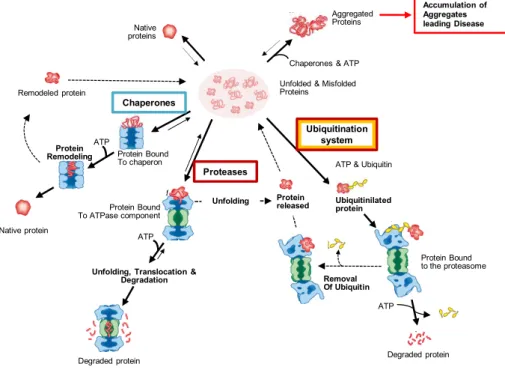
Proteins are remarkable and mysterious molecules. Not only they are the most abundant molecules in biology (besides water) but they are also responsible for every chemical process that makes the existence of living systems possible. Proteins are synthesised as polymeric sequences of amino acid residues but, in order to be functional, most proteins need to acquire a specific three-dimensional structure. The folding process is usually extremely efficient, leading to the formation of highly specific structures that grant proteins selective and diverse functions. However, the folding process is not fail proof and indeed a percentage of all proteins produced in the cell fail to fold correctly. Misfolded proteins are not only a product of inefficient folding but can also be formed as a consequence of stress conditions. Changes in the cellular environment due to ageing or temperature fluctuations, as well as genetic mutations, are known to cause protein misfolding or unfolding. To cope with misfolding and unfolding of proteins, cells have developed a protein quality control system, which comprises both chaperones and proteases and is responsible for limiting this burden. Unfortunately, the capacity of the protein quality control is limited and protein aggregation and misfolding are now recognised as the cause of a large and diverse group of diseases called protein conformational diseases or misfolding diseases.
4
1.1.1. The Protein Folding Mystery
How a protein folds to its functional native state is a fundamental process that has occupied the mind of scientists for almost 50 years. The protein folding process can be divided in two major problems. The first was introduced through the pioneering work of Anfinsen and his co-workers on the folding/refolding of ribonuclease A [1-2]. They have shown that a polypeptide chain can spontaneously fold downhill to its lowest free-energy conformation (predetermined functional native state) without the help of any other biological machinery. This observation encompasses one of the key concepts in protein science, which is that all the information necessary for a protein to fold is encoded in its own amino acid sequence.
The second problem focuses on the kinetics and dynamics of folding, and it was introduced by Levinthal in 1968 [3-4]. He has demonstrated that the folding process could not consist of a systematic search of all the conformational space otherwise it would take an enormous amount of time for a protein to acquire the native state, approx. 1052 sec for a protein with ~100 amino acid residues - Levinthal Paradox.
5
1.1.2. The Search for a Protein Folding Mechanism
During the folding process, amino acid residues are not independent pieces: they interact with each other and these interactions are not limited to consecutive residues. Long-range interactions are also involved in the folding process and if on one hand these interactions allow the folding process to be cooperative, they also increase the complexity of describing the folding process of any given protein [8-9]. In addition, folding kinetics are also puzzling, as folding is generally accomplished in a timescale in the order of milliseconds to seconds. Levinthal postulated the first protein folding model, proposing that the folding pathway occurs through a well defined and sequential path (sequential model) [10]. Since then other models have been postulated aiming at describing the folding process. Here, an overview of those models is presented.
In 1973, Wetlaufer has proposed the classical nucleation model [11]. This model suggests that the folding process is initiated by the formation of native secondary structure through the interaction of consecutive/neighbouring residues. The first elements of secondary structure will then act as seeds for the folding process, which occurs in a stepwise way without the formation of intermediates. During the 1970s and the 1980s, scientists have focused on the study of folding intermediates [12] and the classical nucleation model was discredited.
6
through a unique path or by parallel paths. Folding process dynamics are reduced as they rely on the global properties of each microdomain and its interactions. A few years later, in 1982, and based on experimental evidence, Kim and Baldwin proposed the framework model [12] whose description did not differ significantly from the diffusion-collision.
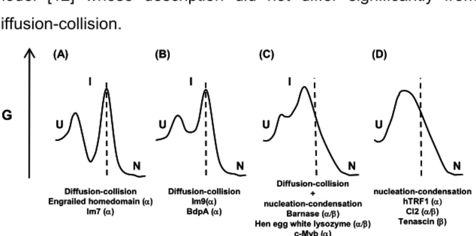
Figure 1.1: Simplified energy diagrams for folding of small single-domain proteins describing the differences between diffusion-collision and nucleation-condensation models. (A) Pure diffusion-collision: first secondary structure is formed leading to the formation of a low energy intermediate and then through diffusion and collision of secondary structure elements the tertiary structure is formed; (B) pure collision as in (A) but a high energy intermediate is formed; (C) mixed diffusion-collision/nucleation-condensation: the formation of a high energy intermediate containing secondary and tertiary structure; (D) pure nucleation-diffusion: no intermediate is formed and at the transition state both secondary and tertiary structures are formed concurrently. As the propensity to form secondary structure increases, the folding mechanism shifts from the nucleation-condensation to the diffusion-collision. Adapted from [17].
The jigsaw-puzzle model was proposed by Harrison and Durbin in 1985 and suggests that folding occurs through different, parallel pathways rather than through a single path [18]. The existence of multiple paths makes folding more robust (kinetically) to mutations that do not disrupt the native structure. According to this model, the selection of the pathways follows the search for structures with increasing stability. Several unfolded states can be considered and the paths are enumerable, meaning that if intermediates do exist they
G
U U U U
N N N N
I I
I
Diffusion-collision Engrailed homedomain ()
Im7 ()
Diffusion-collision Im9() BdpA ()
Diffusion-collision + nucleation-condensation
Barnase () Hen egg white lysozyme ()
c-Myb ()
nucleation-condensation hTRF1 (
Cl2 () Tenascin ()
(A) (B) (C) (D)
G
U U U U
N N N N
I I
I
Diffusion-collision Engrailed homedomain ()
Im7 ()
Diffusion-collision Im9() BdpA ()
Diffusion-collision + nucleation-condensation
Barnase () Hen egg white lysozyme ()
c-Myb ()
nucleation-condensation hTRF1 (
Cl2 () Tenascin ()
7
should comprise a random assortment of substructures of the native protein. Pulsed hydrogen exchange studies have demonstrated that the folding intermediates formed along the folding pathway are specific. These observations contradict what was proposed by the jigsaw-puzzle model and thus the model was abandoned.
The hydrophobic collapse model[19-21], suggests the existence of an initial hydrophobic collapse that restricts and consequently guides the subsequence folding steps, since folding/conformations are only allowed within the confined volume defined by the initial collapse.
Later, folding studies on CI2 [22-23] have demonstrated that folding does not rely on the accumulation of intermediates and that, in fact, folding can occur via a simple two state kinetics. These results, together with the CI2 -value analysis, lead to the formulation of a new folding model: the nucleation-condensation model [24-25]. -value analysis is the only experimental method allowing the evaluation and characterisation of the transition state (Fig. 1.2) [26-29]. It consists on the systematic study of the energetic consequences of introducing mutations throughout the protein. The effect of mutations is measured in both kinetic and equilibrium studies yielding information on the transition states of the folding-unfolding reaction. Mutations can be regarded as structural probes of each residue and the -value is defined by the following equation:
∆∆Φ ∆∆ ∆∆ ∆∆∆∆ (equation 1)
GTS-D and GN-D are the free energies of the transition
8
Figure 1.2: Information redrawn from the -value analysis. After introducing a mutation, if the transition state energetics is the same as in the native state, the protein is immune to that mutation until after the transition state is reached, meaning that both the transition and the native states are equally affected by the mutation (GTS-D = GN-D), 1 (residue a). On the contrary, 0 means that the structure of
the transition state at the site of the mutation is the same as in the denatured stated (residue b). 0 < <1, correspond to structures that are partially folded at the transition state. From [30].
The nucleation-condensation model conciliates the concepts from the framework and the hydrophobic-collapse models. It postulates the existence of a nucleus that constitutes the transition state; if a mutation occurs within the residues of the nucleus, this will not abolish the folding process but it will slow it down. The folding process is guided by an initial collapse around the protein hydrophobic side chains (generating the nucleus), after which the structure rearranges from the restricted conformational space occupied by the intermediate until it acquires the native like structure. According to this model the secondary structure formation is guided by the native-like tertiary interactions formed during the initial collapse (Fig. 1.1).
a b
Native State
Denatured State Transition
9
1.1.3. Energy Landscapes
10
native contacts. The Monte Carlo simulations performed by Karplus and his co-workers on a simplified protein model, a 27-bead self avoiding chain on a cubic lattice, lead to the establishment of a three-stage folding model (Fig. 1.3).
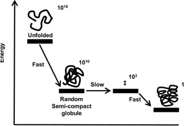
Figure 1.3: Three-stage mechanism of folding proposed by Karplus and co-workers (Lattice model). The number of states was calculated considering a 27-mer.Adapted from [31].
The first step involves a rapid collapse to a semi-compact random globule (with ~60% of the total number of possible contacts), this step reduces the amount of possible conformation from ~1016 to ~1010 [32]. The second step is the rate limiting step and involves the search for the transition states that will successfully and rapidly lead to the native state. The transition states are similar to the native state and comprise 80-95% of the native contacts. In the last step, the native state is rapidly obtained from any of the transition states. As folding proceeds along the three stages, the number of available conformations decreases and this loss of entropy between stages is determinant for the shape of the energy surface. If the contribution to the
free-Unfolded 1016
Fast
Random Semi-compact
globule Slow 1010
Fast ‡
103
En
e
rg
y
Number of native contacts (Q0)
11
energy of the conformational entropy decreases faster than the average energies, a barrier is formed on the free-energy surface, corresponding to the transition states [35]. Also, this decrease in the available conformations as the sequence approaches the native fold leads to the introduction of an important concept in protein folding – the folding funnel [36].
Although all the phenomenological models previously described already contain energetic concepts, the lattice simulation imposed a need to describe the energetic factor biasing the folding process. Also, the Monte Carlo simulations highlighted the fact that a sequence can undergo several folding trajectories at the same time, leading to an ensemble of high energy intermediate states close in structure to the native state that corresponds to the unique transition state described by experimentalists. The pathway view is replaced by the energy landscape description, and the folding process is viewed as an ensemble of conformations sliding down through a unique energy funnel leading to the most energetically stable conformation - the native state. As proteins increase in size, the folding process becomes more complex and barriers to the organisation on the collapse-state are likely to be larger, leading to rougher landscapes and multiple "pathways" [37]. Experiments on lysozyme (129 aa) showed that the folding kinetics of this protein are quite complex and heterogeneous [38-40]. Nonetheless, they can be explained, qualitatively, through a
free-energy surface (Fig. 1.4). At least two folding
12
Figure 1.4: Schematic free-energy (F) surface representing features of the folding of hen lysozyme (a protein comprising an and a domain with 129 amino acid residues). Through the yellow trajectory (fast track) the and the domains are formed concurrently populating the intermediate only briefly. Through the red trajectory (slow track) the polypeptide chain is trapped in a long-lived intermediate with persistent structure involving only the -domain. Further folding from this intermediate involves either a transition over a higher barrier or partially unfolding. Adapted from [35].
13
trajectories seems to be identical and comprises the docking of the two domains [40]. The similarity between the folding landscape obtained experimentally for lysozyme and the one obtained when simulating the folding of a 125-mer with a 5x5x5 cubic native state is remarkable. Therefore, energy landscapes constitute a conceptual framework for understanding both two-state and multi-state folding kinetics [35, 42]. Since the folding time will increase exponentially with the chain length, due to the fact that the number of semi-compact states increases faster than the number of transition states, the three-stage model will not be able to give reasonable folding times for long chains.
The traditional models can, in particular cases, be better phenomenological descriptions of the folding process, but this ‘new view’ of folding provides a unifying mechanistic description for the different proteins regardless of their folding complexity. More importantly, it provides a framework for the interpretation of experimental observations.
1.1.4. Protein Folding in the Cell
14
additional factors will affect the success of folding. The first obvious difference between the test tube and cellular environment is the concentrations of macromolecules. While polypeptide chains in vitro refold in very diluted solutions (<1 mg/ml), the cellular environment is extremely crowded (300-400 mg/ml of proteins and other macromolecules [43]) which significantly increases the affinity between the different species present such as folding intermediates and could lead to aggregation [44]. The crowding effect can indeed increase the association constants between macromolecules, favouring aggregation, but this effect is more significant for small proteins, as larger ones will have a reduced diffusion which limits the encounter rate with other macromolecules. Nevertheless, crowding may also have a positive impact on the folding propensity [45-46]. It promotes the exclusion volume, stabilising the native and compact states. In addition, it may increase molecular chaperones function by promoting their encounter with partially unfolded proteins, facilitating chaperones successive reaction cycles with non-native proteins [44, 47].
15
for the assembly of parallel -sheets and complex antiparallel -sheet topologies since these structure elements involve interactions between residues that are distant in sequence. In addition, translation is relatively slow (~15-75s for a 300 amino-acid protein [52]) leading to the exposure of partially folded nascent chains to cellular environment for a long period of time which represents another challenge for the de novo folding. Individual ribosomes are grouped in polysomes during active protein synthesis.
The 3D organisation of the polysomes revealed that ribosomes are organised along the mRNA through a pseudohelical orientation. The transcript is sequestered inside, while the tRNA entrance sites as well as the polypeptide exit sites face the cytosol (Fig. 1.5). This orientation allows for the maximisation of the distance between nascent chains on adjacent ribosomes, thus also contributing for decreasing the probability of intermolecular interactions that would lead to aggregation.
Considering the complexity inherent to the folding process within the cell, a new schematic energy landscape had to be re-drawn to describe protein folding and the aggregation process under physiological conditions [54].
16
Figure 1.6: Folding funnels designed to describe the competition between productive/efficient folding and aggregation. Left. A higher-dimensional energy landscape contour plot that includes stabilising interactions between two polypeptide chains. In this simplified example, the stable aggregate is formed via intermolecular stabilising interactions that develop between two partially folded conformations with about 50% of the contacts present in the native state. Darker colours represent lower energy conformations. Right. A double funnel depicting the competition between folding and aggregation. The surface shows the ensemble of unfolded conformations funnelling towards the native state via intramolecular contacts formation or towards the formation of aggregated states via intermolecular contacts. The relative population of the different states depend on the kinetics and thermodynamics of the various equilibrium states. The lowest energy state for the aggregate might or might not represent the true global energy minimum regardless of this it is sufficiently kinetically trapped from the native conformation and in living systems, the fate of a given protein molecule is closely related by the protein quality control. Adapted from [54-56].
This new energy landscape formulation was extended to contemplate different scenarios like aggregation, fibril formation and co-translational folding, as well as the action of molecular chaperones and other cellular factors (Fig. 1.6) [57-59]. The surface shows the diversity of the unfolded state as previous descriptions. On the left side of the funnel, intermolecular contacts lead to the formation of non-native structures such as aggregates and amyloid fibrils that are likely to occur due to misfolding and exposure of hydrophobic regions. Intermolecular contacts lead to the deepest minima (more stable), thus the native state is not necessarily the most stable structure. On the
17
funnel right hand side, intramolecular interactions are dominant, and the native state is formed, however, intermediates that might need some refolding to evolve towards the native state are also present.
1.1.5. Protein Evolution
The concept of a funnelled energy landscape has its biological consequences, the first immediate one being that proteins are structurally robust [60]. A mutation may not disrupt the balance between the entropy of the unfolded state and the free energy of the folded state; if this occurs, the funnel landscape model allows for the new native state to be identical in terms of structure and stability to the primordial protein and likely function is also retained [61-62].
18
conformational diversity [65-66]. Natively unfolded proteins constitute the most extreme, yet remarkable, example of protein conformational diversity. These proteins can have two distinct structures: unfolded and folded once they bind to their ligands or proteins partners [67]. The analysis of several proteomes has shown that natively unfolded proteins are not rare events, and in fact eukaryotes have a percentage of natively unfolded proteins between 31-51% [68]. Eukaryote organisms have the higher percentage of natively unfolded proteins which may result from the fact that these organisms have a greater need for protein mediated signalling, regulation and control, functions that are common among natively unfolded proteins. The identification of intrinsically unfolded/disorder proteins has imposed a review of the structure-function paradigm, a defined structure is no longer a prerequisite for function or at least not for all proteins.
19
Figure 1.7: Transition folds through an evolutionary bridge. Two different topologies (mediated by three different disulfide bridges) are found in two naturally occurring cysteine-rich domains (NW1 and Mcol1C) that show almost no sequence identity beyond the conserved cysteines. Conversion between these topologies was demonstrated via one mutation Lys21 → Pro21 (K21P). This mutation leads to an intermediate that equilibrates between the two topologies (evolutionary bridge). A second mutation Gly11→Val11 (G11V) completes the transition [71]. Adapted from [70] .
Protein folds are robust not only as a result of sequence selection, but also due to the presence of chaperones and to the stability thresholds associated to each fold that may lead to the accumulation of damaging mutations [72-74]. By stabilising proteins against misfolding chaperones, they act like buffers preventing the formation of non-native structures extending the neutral networks (no effect results from the introduction of mutations). Due to the chaperones sensitivity and response to environmental stress, they are likely to act like evolutionary aiders.
Considering this scenario of co-evolution of fold and function, and the existence of proteins with structural plasticity, it may be suggested
Lys21Pro Gly11Val
~78% ~22%
20
that the early folds evolved from functional but yet intrinsically unfolded proteins [75].
1.2. When the Folding Process Goes Wrong:
Misfolding and Aggregation
21
interactions that lead to protein aggregation. Amyloid diseases and Huntington's are examples of diseases associated to a 'gain-of-function' [78]. Mutations increasing the protein's hydrophobicity or decreasing its charge favour intramolecular interactions and hence aggregation. Also, alterations to proteins that lead to a deficient processing may result in the accumulation of protein fragments that can act as seeds of the aggregation process [55, 79-80].
1.2.1. Molecular Chaperones
Molecular chaperones are involved in a wide range of cellular processes including de novo folding, refolding of stress-denatured proteins, oligomeric assembly, intracellular protein transport and assistance in proteolytic degradation (Table 1.1) [52]. Chaperones interact, stabilise and guide non-native proteins to fold into the native-state, but do not determine the native fold nor are present in the final functional structure.
22
Table 1.1: Major chaperone and proteases families.
Family E.Coli
Interacting partner
(co-chaperone)
Eukaryotic Homolog
(localization)a Activity
b Phenotype (null mutants) Ref.d AAA+ (Hsp100/ Clp)
Clp A Clp P Clp P (m) AP Phenotype No [76, 84]
Clp B Hsp104 (c), Hsp 78 (m) aggregates AD of
Impaired Thermo-tolerance
[85-89]
Clp X Clp P
Clp X (Humans - m;
plants - ch)
AP No
phenotype [90-94]
Clp Y
(HsIU) Clp Q (HsIV) AP
No
phenotype [95-97]
FtSH Agf3, Rcalp (m) AP Lethal [98-101]
Lon PIM1 (m) AP Mucoid
Growth [102-107]
Hsp 90 Htp G Hsp 82 AC
Reduced growth rate at
44ºC
[108]
Hsp 70 DnaK DnaJ, CbpA, DjlA Hsp70, Hsp72, Bip, etc AC Temperature sensitive growth (39ºC) [108]
Hsc A Hsc B Ssq1p ACc Slow Growth [109]
Hsc 62 (YbeS, YbeY) AC No
phenotype [110-111]
Hsp 60 GroEL GroES CCT, TriC AC Lethal [109]
sHSP IbpA,
IbpB Hsp 25, etc AC
No
phenotype [109]
Hsp 33 Hsp33
Redox-regulated AiC Slightly sensitive towards High temperature and H2O2 [109]
TF Trigger
factor
PPIase, AiC
No
phenotype [109]
a (m) – mitochondria, (c) – cytosol and (ch) – chloroplasts;
b AP – ATP-dependent proteolysis; AD – ATP-dependent disaggregation; AC – ATP-dependent
chaperone; ATP-independent chaperone;
c ATP-dependent chaperone involved in Fe-S cluster formation and protein maturation d References are not all-inclusive. Emphasis on review articles and papers demonstrating the
chaperon/protease activity.
23
that could lead either to aggregation, or to conformational trapping (e.g. Hsp104 and sHSPs). In addition to being able to block deleterious interaction, some chaperones are also able to promote the correct folding (Hsp70 system) [112, 114]. Apart from these two remarkable features, the Hsp60/CCT/Clp/Hsp104 chaperone machinery can also reverse intra-molecular misfolding, by unfolding and disrupting small aggregates [115]. For a growing number of proteins the chaperone activity has to be combined with isomerase activity (PDIs and PPIases) in order for the correct fold to be guaranteed [116].
24
release of the substrate promotes folding and at the same time prevents aggregation [114, 120]. The molecular process by which Hsp70’s induce conformational changes is still not clear, but a mechanism involving entropic pulling is hypothesised [121]. According to this mechanism, when Hsp70 binds to the substrate it locally stabilises it in the unfolded state. Disaggregation is favoured and, once the substrate is released, it can fold into the native state. Co-factors (NEF) and co-chaperones (Hsp40/DnaJ) participate and regulate Hsp70 chaperone activities. While co-factors control the Hsp70 cycles by facilitating the ADP release and rebinding of ATP, co-chaperones control the specificity of Hsp70’s by targeting the substrates to the chaperone [52, 81]. Bag-1, a co-chaperone of this system, binds to the proteosome directing proteins for degradation, and thus has a crucial and regulatory role in protein degradation [122].
25
importance of this chaperone system in the regulation of neuronal susceptibility towards degeneration.
26
Figure 1.8: The GroEL-ES chaperonin. A) Crystal structure of the aymetric GroEL-ES complex (PBD 1AON). B) Working model summarising the conformational changes in a substrate protein upon transfer from Hsp70 system (DnaK, DnaJ and GrpE) to GroEL and during GroEL-GroES-mediated folding (ATP dependent cycles). (1) Substrate protein may be delivered to the GroEl by the Hsp70 system in a non-aggregated but kinetically trapped state. Upon binding to GroELit undergoes local unfolding to an ensemble of expanded and more compact conformations. (2) ATP-dependent domain movement of the apical GroEL domains results in stretching of tightly bound regions of substrate and in release and partial compaction of less stably bond regions. (3) Compaction is completed upon substrate encapsulation by GroES. (4) Folding in the chaperonin cage. (5) Substrate release upon GroES dissociation. (6) Rebinding of incompletely folded states. N, Native state and I, folding intermediate. From [52].
27
that can induce unfolding as it was observed for lysozyme and rhodanese [143-145]. The mechanism of action and the nature of group II binding sites are not yet understood.
1.2.2. Rescuing of Misfolding Defects by Small Molecules-
Chemical Chaperoning
28


![Table 2.2: Residues identified as being involved in iron binding by NMR spectroscopy and the sequence number conversion between different species for this functional residues [89, 91, 113-114]](https://thumb-eu.123doks.com/thumbv2/123dok_br/15769838.641158/117.892.119.642.229.796/residues-identified-involved-spectroscopy-sequence-conversion-different-functional.webp)
