Ana Rita Tomé Ferreira
Perturbation of Calcium Homeostasis, and a Novel Function for
the Ubiquitin Ligase (E4) Ufd2 in Yap8 Stability
Ana Rita Tomé Ferreira
Dissertation presented to obtain the Ph.D degree in Biochemistry
Instituto de Tecnologia Química e Biológica António Xavier |Universidade Nova de LisboaOeiras,
Instituto de Tecnologia Química e Biológica António Xavier (ITQB), Universidade NOVA (UNL)
Av. da República, EAN 2781-901 Oeiras Portugal
President of the Jury:
Dr. Júlia Carvalho Costa (ITQB)
Members of the Jury:
Prof. Dr. Claudina Rodrigues-Pousada, Full Professor, Head of the Genomics and Stress Laboratory, Supervisor
Prof. Dr. Manuela Côrte-Real (CBMA)
Prof. Dr. Miguel Cacho Teixeira (IST)
Dr. Lígia Saraiva Teixeira (ITQB)
Dr. Pedro Neto Domingos (ITQB)
Dr. Regina Andrade Menezes, (ITQB and iBET), Supervisor
(Rita T. Ferreira et al., unpublished)
Financial Support:
FCT and FSE (“no âmbito do III Quadro Comunitário de Apoio”)
Yeast Response to Arsenic’s Threats. Rita T. Ferreira
Contents
Dedication ix
Acknowledgments xi Thesis Publications xiii Foreword xv
Summary xvii Sumário xix
Abbreviations xxi
1 Introduction 1
1.1 Historical Aspects, and Biochemistry of As 1 1.2 Molecular and Cellular Effects of As 2 1.3 As Poisoning to Humans 3
1.4 Applications of As in Medicine 4
1.4.1 Targeted Therapy by ATO: the Current Front-Line Therapy for APL 4
1.5 Budding Yeast as an Eukaryotic Model System 5 1.5.1 The Yeast Genome Sequencing Project 7
1.5.2 Yeast Applications: Biomedical Research, Food and Wine, and Chemical Industries 8
1.5.2.1 Biopharmaceuticals from Health-Care Industries 9 1.5.3 Stress Responses in Yeast 9
Yeast Response to Arsenic’s Threats. Rita T. Ferreira
1.6.1 Yap8, Transcriptional Regulation of ACR genes, and Contribution of Yap1 in the Response to Arsenic 17 1.6.2 As Stress, and Other Affected Signaling Pathways in S.
cerevisiae 18
1.7 Ca2+-Signaling in Eukaryotes 19
1.7.1 Ca2+-Signaling and Transport in Yeast 20
1.8 Protein Modifications by Ub, and Programmed Protein Degradation 22
1.8.1 Ubiquitin-Proteasome System 22 1.8.1.1 The Ubiquitination Cascade 24
1.8.2 Yeast Ubiquitin-Like Proteins (Ulps), and Cognate factors 27 1.8.2.1 SUMO 27
1.8.2.2 Other Proteins Related to Ub 28 1.9 Aims of the Thesis 29
References 31
2 Perturbation of Ca2+ Homeostasis, and Activation of the Calcineurin/Crz1 Pathway by As 39
2.1 Preface 39 2.2 Journal Article 39 2.2.1 Author’s Contribution 40
2.3 References for Chapter’s Preface 40
Full Text of The Article: Arsenic stress elicits cytosolic Ca2+ bursts and Crz1 activation in Saccharomyces cerevisiae 41
3 Regulation of Transcription Factor Yap8, and the E4- Ubiquitin Ligase Ufd2 71
3.1 Preface 71 3.2 Journal Article 72 3.2.1 Author’s Contribution 72
Yeast Response to Arsenic’s Threats. Rita T. Ferreira
Full Text of The Article: E4-Ubiquitin ligase Ufd2 stabilizes Yap8 and modulates arsenic stress responses independent of the U-box motif 73
4 Role of Ubiquitin- and SUMO-Covalent Modification in the Regulation of the Yap8Transcription Factor 107
4.1 Preface 107
4.2 Manuscript in Preparation 108 4.2.1 Author’s Contribution 108
4.3 References for Chapter’s Preface 108
Full Text of The Article in Preparation: Post-translational control of Yap8 transcription factor by ubiquitination and SUMOylation during arsenic stress response in
Saccharomyces cerevisiae 110
5 Discussion 127 5.1 Final Discussion 127
5.1.1 CaN/Crz1 Pathway Activation in Arsenic Stress Response 127
5.1.2 Post-translational Control of Yap8 by Ufd2 129 5.1.3 Deciphering Yap8 Regulation via Ubiquitination, and SUMOylation 131
5.2 Future Research 131 5.3 Final Conclusions 132
References 134
6 Epilogue 135
Appendix: Human Disease Genes Found in Yeast 137 Biopharmaceuticals Produced in
Yeast Response to Arsenic’s Threats. Rita T. Ferreira
Dedication
“You need to be able to present your work for the amount of time you have been allocated, for instance 1 min. We will discuss this at a later stage.”
Yeast Response to Arsenic’s Threats. Rita T. Ferreira
Acknowledgments
I am indebted to many people who contributed with their time, expertise and passion during my doctoral studies.
Foremost, I wish to thank Prof. Dr. Claudina Rodrigues-Pousada—Ph.D Supervisor (by far, the best professor I have ever had the pleasure of studying under) for accepting me in her laboratory, for her bold and memorable leadership, for providing with an inspiring working environment, and for giving me the chance to work on this exciting project. I have no doubt that the many things I learnt under her guidance will benefit me in my future research endeavors.
With great pleasure, I wish to acknowledge Dr. Regina A. Menezes— Ph.D Supervisor, for her very good and accurate handling of my results. I would like to thank Prof. Dr. M. Teixeira and Dr. I. Abreu—Members of the Ph.D. Thesis Committee, for the critical reading of my work, and useful comments and suggestions.
Not to forget the nice contacts of Prof. Dr. E. Melo who introduced me to the use of bioluminescence emission spectra; and Prof. Dr. T. F. Outeiro who helped me to get access to the yeast SUMO strains.
Yeast Response to Arsenic’s Threats. Rita T. Ferreira
Agradeço muito em especial à minha família que me acolhe, que me aconchega, e que me ama, sempre.
December 21st 2015 Rita T. Ferreira
Yeast Response to Arsenic’s Threats. Rita T. Ferreira
Thesis Publications
This thesis is the description of the peer reviewed original research published in the OpenMicrobiology (formerly SGM Open) and Biology Open (BiO) online Open Access journals:
E4-Ubiquitin ligase Ufd2 stabilizes Yap8 and modulates arsenic stress responses independent of the U-box motif.
Rita T. Ferreira, Regina A. Menezes and Claudina Rodrigues-Pousada Biology Open, AOP 27 July 2015.
DOI:10.1242/bio.010405, in press.
Arsenic stress elicits cytosolic Ca2+ bursts and Crz1 activation in Saccharomyces cerevisiae.
Rita T. Ferreira, Ana R. Courelas Silva, Catarina Pimentel, Liliana Batista-Nascimento, Claudina Rodrigues-Pousada and Regina A. Menezes
Yeast Response to Arsenic’s Threats. Rita T. Ferreira
Foreword
Yeast Response to Arsenic’s Threats. Rita T. Ferreira
of Yap8 may be modified by SUMO were collected in a manuscript in preparation that I authored (Chapter 4).
Yeast Response to Arsenic’s Threats. Rita T. Ferreira
Summary
Human exposure to elevated levels of inorganic arsenic (As) poses the greatest threat to global public health. However, arsenic trioxide (ATO) is presently the most active antineoplastic agent in the treatment of acute promyelocytic leukaemia (APL) (Trisenox®, Teva Pharm). Despite the success of this drug, the actual mechanism of action is complex and is not completely understood. We have therefore investigated the mechanisms by which the cell responds to arsenic that perturb homeostasis, and how homeostasis can subsequently be restored, while also focusing on the mechanistic understanding of individual protein regulation important for the cell to thwart the cytotoxic effects of arsenic.
Using S. cerevisiae, also known as baker's yeast—a model system relevant to human biology, we have firstly performed DNA microarrays to obtain novel molecular biological pathways that are mapped by arsenic. The resulting analysis revealed a transcriptional response to arsenic stress of five 'core' components of calcium signaling and calcium transport (CaM, CaN, Crz1, Mid1 and Cch1). Bioluminescence spectroscopy and genetic analyses showed that arsenic induces a transient increase in cytoplasmic calcium ([Ca2+]
cyt), followed by a calcineurin/Crz1-dependent gene expression to
Site-Yeast Response to Arsenic’s Threats. Rita T. Ferreira
directed mutagenesis analyses suggested that all six lysines (K46,47,62,90,96,198) control Yap8 stability, and K62 and K198 as potential SUMOylation sites that control Yap8 activity.
This study contains fundamental biological information that might possibly offer the context for a more reasoned and informed approach to the effects of arsenic toxicity.
Yeast Response to Arsenic’s Threats. Rita T. Ferreira
Sumário
A exposição humana a elevados níveis de arsénio (As), sob a sua forma inorgânica, constitui uma ameaça à saúde pública global. Por outro lado, o trióxido de arsénio (TOA) é, presentemente, o agente antineoplásico mais ativo no tratamento da leucemia promielocítica aguda (LPA) (Trisenox®, Teva Pharm). Apesar da sua eficácia terapêutica estar comprovada, o exato mecanismo de ação é complexo, não sendo completamente conhecido. É preciso, portanto, investigar ao nível da célula os mecanismos de resposta ao arsénio que perturbam a homeostase e como esta pode ser recuperada, e ao mesmo tempo, compreender a regulação individual das proteínas importantes no combate aos efeitos tóxicos do arsénio.
Utilizámos a levedura S. cerevisiae (―baker’s yeast‖), um sistema modelo relevante no campo da biologia humana, e utilizámos a tecnologia de DNA microarrays para obter novas vias e alvos moleculares e biológicos mapeados pelo arsénio. A análise revelou uma resposta, ao nível transcricional, de cinco componentes centrais para a sinalização e o transporte de cálcio (CaM, CaN, Crz1, Mid1 e Cch1). As análises de espectroscopia de bioluminescência e de genética mostraram que o arsénio é responsável por um aumento da concentração de cálcio no citoplasma ([Ca2+]cyt), acompanhado da expressão de genes-alvo do Crz1 e da
calcineurina (CaN). O estudo de interações proteína-proteína baseado no sistema de dois híbridos revelou que, ao nível bioquímico, o enzima E4-Ufd2 ligase de ubiquitina interage com o fator de transcrição Yap8 do tipo AP-1 para desempenhar uma nova função celular, estabilizando o Yap8 e ativando-o sem recorrer ao seu domínio de atividade como ligase,
designado de ―U-box‖. As análises in silico revelaram os locais na proteína
Yeast Response to Arsenic’s Threats. Rita T. Ferreira
com a ubiquitina (SUMO), e uma possivel conexão entre ambos. As análises de mutagénese sítio-dirigida sugeriram que as seis lisinas (K46,47,62,90,96,198) controlam a estabilidade da proteína Yap8, e que as lisinas K62 e K198 como potenciais locais de SUMOilação controlam a sua atividade.
Este estudo contém informação biológica fundamental, podendo vir a facultar um contexto mais fundamentado e informado dos efeitos tóxicos do arsénio.
Yeast Response to Arsenic’s Threats. Rita T. Ferreira
Abbreviations
ABC ATP-binding cassette ACT1 ACTin gene
AD Activation Domain ADP adenosine diphosphate AML Acute Myeloid Leukaemia ALD AdrenoLeukoDystrophy AP-1 Activating Protein-1
APL Acute Promyelocytic Leukaemia AQP Aquaglyceroporin
ARE AP-1 Recognition Element, see AP-1
ARR1-3 ARsenicals Resistance As Arsenic
As(III) Arsenite As(V) Arsenate ATO Arsenic trioxide
ATP Adenosine TriPhosphate BiO Biology Open (Journal) bZIP Basic-region Leucine ZIPper Ca Calcium
[Ca2+]cyt Cytosolic calcium ion concentration
[Ca2+]I intracellular Ca2+ concentration, see Ca
CaM Calmodulin
CaMK Ca2+/CaM-dependent protein kinases, see CaM
CaN Calcineurin
cCRD C-terminal cysteine rich domain cDNA Complementary DNA, see DNA CDRE Calcineurin-dependent response
element
Cmk2 Ca/CaM-dependent protein kinase 2, see Ca, see also CaM CML Chronic Myeloid Leukaemia CN Calcineurin
CFTR Cystic Fibrosis Transmembrane (conductance) Regulator
Cnb1 CaN regulatory subunit B, see CaN
Co-IP Co-immunoprecipitation c.p.s. Counts per second Crz1 CaN-responsive zinc finger
transcription factor, see CaN CsA Cyclosporine A
DABCO 1,4-Diazabicyclo[2.2.2]octane DAPI 4’,6-Diamidino-2-phenylindole DBD DNA binding domain, see DNA DMSO Dimethyl sulfoxide
DNA DeoxyriboNucleic Acid ER Endoplasmic Reticulum EUROSCARF EUROpean
Saccharomyces Cerevisiae ARchive for Functional Analysis
Fps1 Aquaglyceroporin, plasma membrane channel
Fw Forward (oligonucleotide) Gad1 Glutamate Decarboxylase 1 GFP Green Fluorescent Protein GSC2 Glucan Synthase of Cerevisiae GSH Glutathione
GTP Guanosine-5'-triphosphate HA Hemagglutinin
HECT Homologous to E6-carboxy terminus
HBAg Hepatitis B surface antigen Hph1/2 Homologous integral ER
membrane proteins, see ER Hog High osmolarity glycerol HSFs Heat Shock Factors Hxts Hexose transporters IP Immunoprecipitation MAPK Mitogen-Activated Protein
Kinases
Mid1 Mating pheromone-induced death
MYC MyeloCytomatosis (oncogene)
mRNA messenger RNA, see RNA NFATc Nuclear Factor of Activated T
cells
NES Nuclear Export Signal NDB ID Nucleic Acid Database
Identification
NLS Nuclear Localization Signal OATPs Organic Anion Transporting
Polypeptides OD Optical Density ORF Opening reading frame PAGE Polyacrylamide gel
Yeast Response to Arsenic’s Threats. Rita T. Ferreira PBS Phosphate-Buffered Saline
PCR Polymerase Chain Reaction PDB ID Protein Data Bank Identification Pgk1 3-Phosphoglycerate kinase Pho Phosphate transporters Pi inorganic Phosphate Pmc1 Plasma membrane calcium PML Promyelocytic leukaemia
protein
Pmr1 Plasma membrane ATPase-related, see ATP
ppb parts per billion
PTM post-translational modification qPCRs quantitative PCRs, see PCR Rad23 RADiation sensitive
RARα Retinoic Acid Receptor alpha RING ―really interesting new gene‖ RLU Relative Luminescence Units ROS Reactive Oxygen Species r.p.m. Rotations per minute RTG Retrogade
RT-PCR Reverse-Transcriptase PCR, see PCR
Rv Reverse (oligonucleotide) SC Synthetic Complete SCF Skp1, Cullin, F-box SD Synthetic defined/minimal
medium or sd/SD, standard deviation
SDS Sodium Dodecyl Sulfate SEAR South East Asia
SGD Saccharomyces Genome Database
SGM Society for General Microbiology SMT3 Suppressor of Mif Two 3 SPB Spindle Pole Body
STRE STress Responsive Elements SUMO Small ubiquitin-like modifier or
small ubiquitin-related modifier SV-40 Simian Vacuolating virus 40 or
Simian Virus 40 TCA Trichloroacetic acid TFs Transcription factors Ub Ubiquitin
Ubc Ubiquitin-conjugating Ufd2 Ubiquitin fusion degradation 2 Ulp Ubiquitin-like protease UPP Ubquitin-proteasome pathway UPS Ubiquitin-proteasome system UV Ultraviolet
WT Wild-type
Yap Yeast specific AP-1-like activator proteins, see AP-1 Ycf1 Yeast cadmium factor-1 YPD Yeast Peptone Dextrose YRE Yeast Response Element
latin
c.a., circa, approximately cf., confer with, consult
e.g., exempli gratia, for example et al., et alia., and other people i.e., id est, that is to say vs., versus, against, turned
1
Introduction
1.1
Historical Aspects, and Biochemistry of As
Arsenic (As) compounds are extremely toxic with a long history of use as a poison for life. Nevertheless, arsenic has been also used in medicine, and its earliest use goes back to the times of ancient Rome, when Galen and Hippocrates used it to treat ulcers [1]. In the 1800s and early 1900s, arsenic was commonly used to treat diseases, such as psoriasis and syphilis. Through the systematic chemical modification of arsenic derivate, before the ―pre-antibiotic era‖, the discovery of salvarson (arsphenamine) made it the main medicine used against syphilis [2].
As (atomic number, 33; relative atomic mass, 74.92) has chemical and physical properties intermediate between a metal and a nonmetal, and is often referred to as a semi-metal or metalloid. It belongs to Group VA of the Periodic Table, and can occur in four oxidation states (+5,+3,0,-3) [3]. Nevertheless, it is generally found in the environment in two biologically important oxidation states—the pentavalent arsenic [As(V), arsenate] and trivalent arsenic [As(III), arsenite]. Of the inorganic arsenic compounds, arsenic trioxide (ATO, As2O3)—the form of arsenite used in cancer therapy
[3], sodium arsenate (Na3AsO4), and arsenic trichloride are the most
common found in nature [4].
and manganese) and are toxic only in an overdose, whereas others are xenobiotic and highly toxic (e.g., arsenic, cadmium, lead, and mercury) [5]. Thus, all cells and organisms maintain metal homeostasis within physiological or sub-toxic levels, respectively, and utilize metal detoxification mechanisms. (For arsenic detoxification in yeast and mammals, see Section 1.6.)
1.2
Molecular and Cellular Effects of As
In general, the toxicity of a given metal depends on its physicochemical properties and ligand preferences: ‗soft‘ transition metals (like cadmium and mercury) prefer sulfur as their ligand; while ‗hard‘ transition metals (like chromium and manganese) and metalloids (arsenic, antimony and selenium) favor oxygen in their higher oxidation states and sulfur in their lower oxidation states; lead, iron, cobalt, nickel, copper and zinc may use oxygen, sulfur or nitrogen as ligands [6].
At the molecular level, an important feature of As(III) chemistry lies in its high reactivity, forming strong bonds with functional groups, such as thiolates of cysteine residues or the imidazolium nitrogens of histidine residues, and therefore its ability to bind to and affect the activity of many proteins in the cell [7]. Hence, the biochemical effects of the arsenic trivalent form (principally, arsenite and arsenic trioxide) may be mediated by reactions with closely spaced cysteine residues on critical cell proteins [8]. On the other side, As(V)—as a chemical analogue of inorganic phosphate (Pi), can
uncouple the oxidative phosphorylation in mitochondria by displacing phosphate in ATP synthesis [9]. However, As(III) is generally more toxic than As(V), and both of them are more toxic than the organic arsenic compounds [10].
well as to inhibit protein function/activity leading to cell proliferation or cell death [6, 11, 12]. Indeed, arsenic facilitates profound cellular alterations, including induction of apoptosis, inhibition of proliferation, stimulation of differentiation, and inhibition of angiogenesis via numerous pathways [13]. Despite arsenic‘s cytotoxic effects, ATO is currently used as a mitochondria-targeting drug in acute promyelocytic leukaemia (APL) capable of triggering apoptosis in leukaemia cells (see Section 1.4.1), though the mechanisms by which ATO kills cancer cells are not fully understood [14, 15].
1.3
As Poisoning to Humans
Arsenic is a metalloid element, which forms a number of poisonous compounds [16]. As previously referred, the two oxidation states of inorganic arsenic, As(V) and As(III), are most commonly found in nature, namely in rocks and soil. It is widely distributed throughout the earth‘s crust, and is found in groundwater supplies in many countries of the world. South East Asia (SEAR) countries have been identified as the most highly affected geographical areas by arsenic contamination [17]. Furthermore, antrophogenic sources such as industrial and urban pollution are also responsible for introducing this metalloid into the environment. For example Asturias, an autonomous region in Northern Spain with a large industrial area, registers high lung cancer incidence and mortality [18].
Exposure of humans to arsenic occurs by inhalation as well as consumption of contaminated water and food (for a review see Ref. [19]). The environmentally relevant concentrations of arsenic could reach >10 μg/l, as reviewed by Dopp et al. [20].
exposure to arsenic environmental contaminant has been documented to cause genome-wide changes in human newborns [22].
Recently, a journal article review [23] reported that four million people in Argentina are exposed to arsenic contamination from drinking waters of several center-northern provinces, associated with an increased risk of serious chronic diseases, showing the need for adequate and timely actions.
1.4
Applications of As in Medicine
As aforementioned (Section 1.2), arsenic has long been known to act as a carcinogen; paradoxically, it has also been demonstrated that arsenic can have anti-cancer activity in some cases [24, 25]. In traditional Chinese medicine, ATO was recorded in the Compendium of Materia Medica by Li Shi-Zhen. In Western medicine, arsenicous oxide (Fowler's solution) was used as a treatment of choice for chronic myeloid leukaemia (CML) in the 19th century. Osler stated in his textbook of medicine: ‗
There are certain remedies that have an influence upon the disease. Of these, arsenic, given in large doses, is the best. I have repeatedly seen improvement under its use.‘ (1892). Nevertheless, due to toxic side effects of long-term dose of oral arsenic in most patients, and with the advent of modern radiotherapy and chemotherapy, the arsenic treatment for CML was given up in Western medicine [26]. Noticeably, the discovery of the therapeutic effect of As2O3 in
APL has revived this ancient drug [27]. (See section below.)
1.4.1
Targeted Therapy by ATO: the Current Front-line Therapy for APL
As we have seen above, arsenic has been successfully used in the molecularly targeted therapy of APL (the M3 subtype of AML), which was once a lethal disease, yet nowadays the majority of patients with APL can be
APL is characterized by a balanced reciprocal chromosomal translocation
fusing the promyelocytic leukaemia (PML) gene on chromosome 15 with the
retinoic acid receptor α (RARα) gene on chromosome 17 [27]. It has been
found that ATO or all-trans-retinoic acid (ATRA) alone exerts therapeutic
effect on APL patients with the PML-RARα fusion gene, and the combination
of both drugs can synergistically act to further enhance the cure rate of the
patients [28].
In addition, ATO induces apoptosis via changes in the mitochondrial membrane potential; an important aspect of ATO treatment of malignant cells is that elevated intracellular levels of hydrogen peroxide (H2O2) lower
mitochondrial membrane potential, which leads to cytochrome c release, and subsequent activation of the caspase pathway [29].
Trisenox® (Teva Pharm) (www.trisenox.com) injection is indicated for induction of remission and consolidation in patients with APL who are refractory to, or have relapsed from, retinoid and anthracycline chemotherapy, and whose APL is manifested by the presence of the t(15;17) translocation or PML/RARα gene expression [30].
The excellent improvement in the survival rate of APL patients is an
example of the advantage of modern medicine. Nevertheless, despite the
satisfactory advancements regarding the efficacy of ATO-based therapy for APL patients, there is still a large research effort to improve current knowledge of chemotherapy with ATO [31].
1.5
Budding Yeast as an Eukaryotic Model System
The budding yeast Saccharomyces cerevisiae is perhaps the most useful yeast species owing to its use since ancient times in Egyptian methods of baking and brewing [32].
Figure 1.1). As an eukaryote, S. cerevisiae shares the complex internal cell structure of higher eukaryotes, however without the very large fraction of genome containing non-coding DNA–commonly referred to as ―junk DNA‖, that is found in humans [33]. Apart from an even diploid set of chromosomes, yeast cells harbor numerous proteins similar to human cells that make them in particular suitable for recapitulating the fundamental aspects of eukaryotic biology [34].
Small, newly budded S. cerevisiae cells grow until they achieve a critical size, when they can then produce new buds themselves [35]. Furthermore, these cells can be transformed allowing for either the addition of new genes, or the deletion through homologous recombination. In addition, the possibility of working with an haploid state facilitates the study of multiple processes, and allowed the generation of collections of haploid knockout strains [e.g., the collection from European Saccharomyces cerevisiae Archive for Functional Analysis (EUROSCARF)].
Figure 1.1 Budding yeast compartmentalizing metabolic pathways in essential ‗core‘
organelles found in most eukaryotic cells: nucleus, endoplasmic reticulum (ER), mitochondria, Golgiapparatus, peroxisome, and vacuole. (After Ref. [38].)
1.5.1
The Yeast Genome Sequencing Project
transporter protein Pat1 as revealing great functional similarities to the human ALD protein [42, 43]. Almost 200 genes are orthologs to those that cause disease in humans (e.g., the yeast YCF1, and human CFTR); and around 71 human genes complement yeast mutations, which is important in order to unravel the molecular basis of many human diseases ([39, 44-46], cf. also Table 1 in Appendix).
The Saccharomyces Genome Database (SGD) provides scientific high-quality, curate data about the genes and gene products of S. cerevisiae [47].
1.5.2
Yeast Applications: Biomedical Research, Food and Wine, and Chemical Industries
Currently, a large network of scientists takes advantage of the simple yeast assay system that helps them speed up the discovery of more complex biological processes of the eukaryotic biology, and limiting the use of animal models.
Yeast cells—which share the ‗core‘ cell biology of human cells, provide a powerful experimental tool to study the intricacies of eukaryotic biology given its genetic tractability, and remarkable conservation of fundamental cellular processes with higher eukaryotes [48, 49]. They have been extensively used as living test tubes to learn about the underpinnings of human diseases from neurodegenerative, prions, cancer and other age-related diseases [50-54]; to renal disorders [55]; as well as physiological processes such as angiogenesis, cell death or DNA repair [56].
Very recent systematic humanization of yeast genes also determined conserved functions and genetic modularity [49]. In addition, yeast cells allow a global knowledge of toxicological and resistance mechanisms upon exposure to weak acids used as food preservatives, herbicides or in pharmacology [57].
hydrolysates (WSH) [60], but also on food and beverage production processes [61].
1.5.2.1
Biopharmaceuticals from Health-Care Industries
Since the 1980s, development of the pharmaceutical industry has been marked by the rapid expansion of biotherapeutics [62].
The first recombinant protein expressed in S. cerevisiae was human interferon, followed by the production of the first genetically engineered vaccine – hepatitis B surface antigen (HBAg). HBAg has been extracted from yeast cells and sold under various names. The greatest value of S. cerevisiae passes through the production of human insulin, presumably; its production covers nearly half the insulin needed by the diabetic patients worldwide. In addition, the first authorized product for human therapeutics is a commercially available recombinant human albumin, Recombumin® (approved for the production of childhood vaccines for rubella, measles, and mumps) [63]. (see Table 2 in Appendix.)
1.5.3
Stress Responses in Yeast
responding to an array of stress conditions. In the yeast genome, over 186 genes (c.a. 3% of the yeast genome) are potentially regulated in a STRE-dependent manner [74]. Msn2/Msn4 together with Hot1 are required for the induction of subsets of high osmolarity glycerol (HOG) pathway-dependent genes [75].
To summarize, the yeast S. cerevisiae displays a privileged position as a model organism for interpreting the mechanisms of response to chemical stresses with impact in environmental health, pharmacology, and biotechnology, and limiting the use of animal models [76].
1.5.3.1
AP-1 Transcription Factors in Yeast, and Yap Family
The best characterized AP-1 factor in S. cerevisiae is the Gcn4 protein which is involved in the expression of more than 500 genes [77]. In functional terms, Gcn4 is remarkably similar to the oncoproteins Jun and Fos; Gcn4, Jun, and Fos have the same DNA binding specificity, and they are functionally interchangeable for transcriptional activation from AP-1 sites in yeast cells and mammals. Moreover, Gcn4 resembles mammalian AP-1 factors in mediating a protective response against UV which involves the Ras signal transduction pathway, and a translational control mechanism that leads to increased AP-1 transcriptional activity.
Since the release of the yeast genome sequence, Rodrigues-Pousada C. and other scientists have been involved in the functional analysis of the yeast specific AP-1-like activator proteins (Yaps). The Yap family consists of eight transcription factors, named Yap1 to Yap8, belonging to the basic region leucine zipper (bZIP) super-family. Indeed, the bZIP proteins form one of the largest families of transcription factors in eukaryotic cells. Despite relatively high homology between amino acid sequences of bZIP motifs, these proteins recognize diverse DNA sequences [78, 79].
lesser extent to Yap3; Yap4 is most homologous to Yap6; so is Yap5 and Yap7; whereas Yap8 is the most divergent family member. Although they display relatively high homology between the amino acid sequences of their bZIP motifs, these proteins recognize a diversity of DNA sequences [78]. They specifically bind to the Yap recognition element (YRE), and they are implicated in various stresses although with a moderate interplay between the transcriptional regulators [79, 80]. Despite homologues for Yap1 exist in other eukaryotes, none has been described for the remaining family members [81].
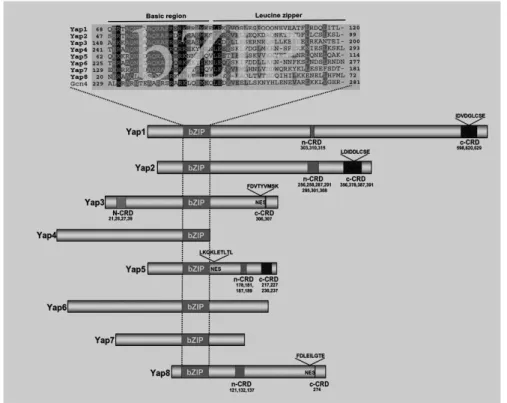
Figure 1.2 Structural features of eight Yap family members of S. cerevisiae. Sequence alignment of Yap basic region and leucine zipper (bZIP) with the true yeast AP-1 protein, Gcn4. Representation of the entire Yaps showing the positioning of cysteine-rich domains (n-CRD and c-CRD), and nuclear export signal (NES). (After Ref [72], adapted.)
Asn31 of the basic region are relevant to Yap8 specificity, with the latter two amino acid residues displaying importance for the stability of the Yap8-DNA-complex. The three amino acid residues at sites 29, 26 and 31 are of highly conserved positions in the other Yap family of transcriptional regulators and Pap1 (of fission yeast). Therefore, a homology model of the complex Yap8bZIP-DNA was built based on the 2.0Å resolution crystal structure of the bZIP motif of Pap1, an AP-1-like transcription factor of fission yeast Schizosaccharomyces pombe (view the NDB ID associated with the structure: PD0180, and PDB ID: 1GD2).
The general picture of functions of Yap members, emerging over the years, is that Yap1 has a central regulatory role of different stress responses, being particularly important for the adaptive response to oxidative stress [83, 84] (for more details, see next Section), and the mediator of stress-induced transcriptional activation of ABC protein genes (together with Yap2) [85]. Yap2 (Cad1) regulates the cadmium stress response, as well as it confers resistance to 1,10-phenantroline, cerulin, and cycloheximide [80, 86], and is involved in pleiotropic drug resistance (together with Yap1) [79]. Yap4 and Yap6 mediate the response to osmotic shock [87]; YAP4 expression is also induced under pro-oxidant conditions [88] regulated by Yap1 and Msn2. Yap5 is involved in remodeling gene expression in response to iron bioavailability [89]. Yap8 (together with Yap1) responds to arsenate and arsenite compounds [90, 91] (see Section 1.6.1). With respect to Yap3 and Yap7, their functions have not been described so far (Yap7 has a paralog, Yap5, which arose from the whole genome duplication) [72, 79, 92]. Members of the Yap family carry out distinct biological functions, however, in some cases overlapping. (e.g., example, mutations in the YAP4 gene affect chromosome stability suppressing the cold-sensitive phenotype of the yap1 mutant, see Ref. [79]).
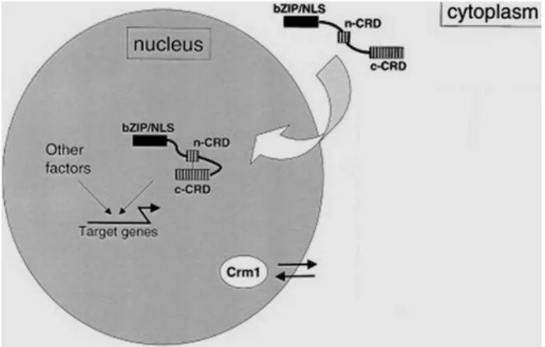
physiological growth [94]. Nonetheless, when cells are treated with various oxidants [95, 96] or with arsenic [97], Yap1 and Yap8 accumulate in the nucleus, respectively.
The molecular mechanisms responsible for Yap1 nuclear accumulation in response to oxidative stress [96] are well understood: the association of Crm1 with the CRD of Yap1 is inhibited by the formation of intramolecular disulfide linkages. In the case of H2O2, these linkages involve cysteine
residues in the N- and C-terminal CRDs, whereas treatment with the thiol oxidant diamide induces disulfide linkages involving only the C-terminal CRD [96]. With respect to Yap8, arsenic is known to directly modify the sulphydril groups of reactive protein cysteines located in the cCRD (C-terminal cysteine responsive domain), and masking the NES preventing recognition by the Crm1 export factor. (In addition, Yap1 accumulates in the nucleus in response to carbon stress in S. cerevisiae [98].)
With respect to Yap8 protein, Cys132, Cys137, and Cys274 are crucial for its Crm1-dependent nuclear export, and arsenite and arsenate compounds directly bind to and modify these cysteine residues and hence the NES [97]. (Attention will be drawn to additional aspects of Yap8 post-translational regulation in the Chapters 3 and 4.)
On the other hand, Yap4 and Yap6 are known nuclear-resident proteins, being their localization unaffected by environmental changes [99]; whereas Yap5 was suggested to constitutively localize to the nucleus [100].
To date, such a characterization in detail of AP-1-like factors has been described only in Saccharomyces cerevisiae, however, a family of Yap proteins also exists in Candida glabrata [79].
1.5.3.1.1
Transcription Factor Yap1 and Multidrug Resistance
Yap1, the first member of the Yap family to be described [101], as well as the best studied one, was initially identified by its ability to bind to and activate the SV-40 AP-1 recognition element (ARE: TGACTAA).
The key function of Yap1 transcription factor consists of the activation of the antioxidant machinery preventing protein damage [91] (see also Section 1.6.1). Yap1 regulates yeast response to H2O2, as well as to several
unrelated chemicals and metals or metalloids. Activation by H2O2 involves
Yap1 Cys303-Cys598 intra-molecular disulfide bond formation directed by the H2O2 sensor Orp1/Gpx3. The electrophile N-ethylmaleimide (NEM)
activates Yap1 through covalent modification of Yap1 C-terminal Cys598, Cys620, and Cys629, in an Orp1 and Yap1-oxidation-independent manner. Also, menadione (a superoxide anion generator and a highly reactive electrophile) operates both modes of Yap1 activation. The reactivity of Yap1 C-terminal domain towards other electrophiles (4-hydroxynonenal, iodoacetamide) and metals (cadmium, selenium) is a common mechanism for sensing thiol reactive chemicals via thiol chemical modification. Within Yap1, two redox centers are relevant for H2O2 and thiol-reactive chemical
signaling: one triggered by reactive oxygen species (ROS) (e.g., hydroperoxides and the superoxide anion); and the other triggered by chemicals with thiol reactivity (e.g., electrophiles and divalent heavy metals cations) [102].
It has to be mentioned that Yap1 was found to be a major determinant of tolerance to polyamine toxicity involving the drug: H+ antiporter Qdr3 and the transcription factor Gcn4 as well; Yap1 is also accumulated in the nucleus of the cells exposed to spermidine-induced stress [104].
Finally, the critical role of Yap1 in cobalt stress response [105] (via the regulation of the high affinity phosphate transporter Pho84), as well as cadmium stress response [106] (involving repression of the low affinity iron transporter gene FET4) has been reported.
1.6
As Uptake, Transport, and Efflux in Yeast and Mammals
Arsenic is highly toxic and can only permeate cells through the transporters that evolved for accumulation of essential ions and nutrients using the molecular mimicry. The sequence of events during arsenic uptake, transport and efflux in yeast and mammals is schematized in Figure 1.4.
As mentioned in Section 1.2, As(V) behaves as a chemical analogue of Pi. Thus, it is imported by phosphate transporters (Pho) (the counterpart in
subfamilies are the major pathways of As(III) extrusion. Both MRP1 and MRP2 are able to transport inorganic and monomethylated forms of As(III) conjugated with glutathione. In addition, MRP2 mediates efflux of seleno-bis(S-glutathionyl) arsenium ion. The exact form of As(III) recognized by MDR1/P-gp is uncertain, but it is not glutathione-S-conjugate [107].
Figure 1.4 Arsenic transport in S. cerevisiae, and mammals. [After Ref. [107] (adapted).]
In addition to eukaryotes, prokaryotic cells have also developed mechanisms to take up and detoxify arsenic compounds [7], being the oxidation state essential for the type of transporter used in this process.
1.6.1
Yap8, Transcriptional Regulation of ACR genes, and Contribution of Yap1 in the Response to Arsenic
the above genes) represent the main pathway of arsenic detoxification. Since Yap8 transcription factor regulates As(III) detoxification, the yap8 mutant strain consequently absorbs increased levels of As(III) [92].
The Yap1 transcription factor also regulates the YCF1 gene expression. In addition, Yap1 overproduction restores arsenite resistance to the ABC transporter deficient mutant ycf1 by activating ACR3 expression [108].
Most importantly, Yap1 promotes cellular antioxidant defenses through the scavenging of ROS that are generated as a side effect of exposure to arsenic [91]. Yap1 and its target genes TRX2, GSH1 and SOD1 are highly induced under these conditions indicating that activation of Yap1 is essential to favour cell adaptation by preventing the ROS accumulation. Genome transcriptional profiling of the wild-type strain stressed with arsenate reveals that genes of the functional categories related with sulphur and methionine metabolism, and with the maintenance of the cell redox homeostasis are highly activated. As a consequence, the yap1yap8 double mutant exhibits a more sensitive phenotype to arsenic stress than single yap1 or yap8 mutant. Nevertheless, YAP8 gene overexpression does not alleviate the yap1 mutant sensitivity to arsenic, as well as YAP1 gene overexpression is not able to rescue the sensitive phenotype of the yap8 mutant either [72]. The work from Menezes et al. [91] has revealed increased levels of intracellular oxidation and lipid peroxidation in the single yap1 and yap8, and the double yap1yap8 mutants; changes in protein carbonylation and the redox equilibrium were also discussed, mostly in the YAP1 deletion strain lacking the antioxidant defenses causing protein damage.
1.6.2
As Stress, and Other Affected Signaling Pathways in Yeasts
Although Yap8 and Yap1 appear as the key responsive transcription factors in controlling arsenic detoxification and redox homeostasis, respectively, additional regulators also contribute to cell tolerance. The transcription factor Rpn4 strongly mediates the yeast adaptation to arsenic stress as revealed by expressing profiling [90]. In addition, the transcriptional regulator Met4 (together with Yap1) controls the assimilation of sulphur into glutathione biosynthesis in order to compensate its continuously oxidation during the detoxification process [110]. The mitogen-activated protein kinase Hog1 is also implicated in arsenic resistance, being required to full activation of ACR3 and YCF1, and to control the As(III) uptake through phosphorylation of the aquaglyceroporin Fps1 [111, 112].
Furthermore, it is now well understood that multiple pathways involved in mitochondrial biogenesis protect S. cerevisiae from arsenic-induced toxicity, as the case of the RTG (retrograde) genes involved in mitochondria-to-nucleus signaling, and the targets of the multifunctional transcription factor Abf1 [113].
Arsenic toxicity in yeast can also be determined through the balance between chronic activation of general stress factors Msn2/Msn4 in combination with lowered/inhibited TORC1 kinase activity [114].
Recent analyses from the laboratory of Rodrigues-Pousada C. [115] have shown that arsenic disrupts the cellular iron homeostasis in yeast by inducing conditions of iron deficiency as consequence of a destabilization of the high-affinity iron uptake system (this perturbation was also observed in mammals).
1.7
Ca2+-Signaling in Eukaryotes
Calcium ions (Ca2+) are ubiquitous, universal, and versatile intracellular
messengers [116]. An increase in the cytosolic Ca2+ concentration activates
many proteins, including the eukaryotic calmodulin (CaM), and Ca2+
In yeast, many downstream transcriptional and translational events are known to be controlled by the Ca2+-mediated activation of CaM. In yeast, as in other organisms, CaM is essential for life [118]. The phosphatase CaN is conserved from yeast to humans (except in plants) [119], and many target proteins of CaN have been identified. However, the most prominent and best-investigated targets are: (1) the nuclear factor of activated T cells (NFAT), which regulates, for example, the cardiac hypertrophic response in mammals [120]; and (2) the yeast zinc-finger transcription factor (Crz1) that activates the transcription of genes whose products promote cell survival during environmental stress conditions [121].
It has been shown that the functions of the signaling of calcineurin-Crz1 (see next section)- ranging from ion homeostasis through cell wall biogenesis to the building of filamentous structures, are conserved in different organisms. Moreover, frequency-modulated gene expression through Crz1 was discovered as a striking mechanism by which the cell can coordinate its response to a signal [119].
The new findings concerning CaN-Crz1 signaling in yeast exposed to arsenic stress will be discussed in Chapter 2.
1.7.1
Ca2+-Signaling and Transport in Yeast
The utilization of Ca2+ to regulate a wide range of cellular processes, in
response to a variety of environmental insults, is a strategy exploited by S. cerevisiae cells exposed to diverse environmental cues [122]. Ca2+ enters the cytoplasm across the plasma membrane complex Cch1/Mid1 or the vacuolar transient receptor potential-like channel Yvc1 (Figure 1.5). Once in the cytosol, Ca2+ is sensed through its receptor, calmodulin; and once activated, calmodulin transduces the Ca2+-signaling to calcineurin.
Dephosphorylation by calcineurin results in nuclear translocation of Crz1/Tcn1, and transcriptional activation of its target genes. Then, Ca2+
homeostasis can be restored via Pmc1 (a Ca2+ ATPase), as well as by the
secretory pathway in the ER and Golgi apparatus by Pmr1 (a plasma membrane ATPase related) [123].
Crz1 phosphorylation responds to calcium, and regulate more than 100 different targets [124], majorly involved in: (i) cell wall, and lipid biosynthesis, (ii) ion, and small molecule transport, and (iii) vesicle trafficking [125].
CaM also participates in the Ca2+-dependent stress response pathways
Figure 1.5 Ca2+ transport in
S. cerevisiae. (After Ref. [122].)
1.8
Protein Modifications by Ub, and Programmed Protein Degradation
1.8.1
Ubiquitin-Proteasome System
The story of protein degradation started with an observation by Aaron Ciechanover in Avram Hershko‘s laboratory (in the late 1970s), followed by studies on the role of ATP in this process- mainly contributed by the laboratory of Irwin Rose [128].
The Ubiquitin-Proteasome System (UPS) is a central proteolytic pathway present in every eukaryotic cells, which regulates vital physiological events. Protein quality control, protein trafficking, cell division and differentiation, and signal transduction- are all controlled processes (to some extent) by the UPS [129].
critical in the protein quality control process (as mentioned above)- which importantly prevents accumulation of aberrant proteins. For those proteins in which refolding is no longer possible, the cells count on proteolytic systems to eliminate unstable proteins, and to recycle their amino acids [130]. Furthermore, also the properly folded proteins- whose levels must be down-regulated, such as cell cycle regulators [131], or transcription factors [132] represent substrates of the proteasome. An important aspect is that, regulation of transcription controls cell development, differentiation, and maintains homeostasis- which supports the importance of the proteasome. The Ubiquitin-Proteasome Pathway (UPP) was initially recognized as a selective degradation pathway [133], and a large number of studies were devoted to characterize protein ubiquitination (or ubiquitylation) as a signal for protein degradation. However, it is increasingly realized that Ubiquitin conjugation to proteins can be used for many other purposes. Furthermore, in every virtually eukaryotic cells, there are many Ubiquitin (Ub) and Ubiquitin-like proteins (Ulps) [134] that control the activities of many proteins. (For Ulps, see Section 1.8.2.).
A large portion of the proteasomes localizes to the nucleus, therefore protein degradation can occur in the nucleus; and proteasome-dependent protein degradation of nuclear proteins has proven to be instrumental in nuclear function [135]. Degradation processes both in the cytoplasm and the nucleus can occur through two pathways: (1) an N terminus-dependent pathway [136], (2) and a lysine-dependent pathway. These pathways are respectively characterized by the site of initial ubiquitination of the protein- the N terminus, or an internal lysine [137]. Nevertheless, a study has been characterized that the lysine-dependent pathway is the more active pathway within the cytoplasm, whereas in the nucleus the two pathways are both active in protein degradation.[138].
1.8.1.1
The Ubiquitination Cascade
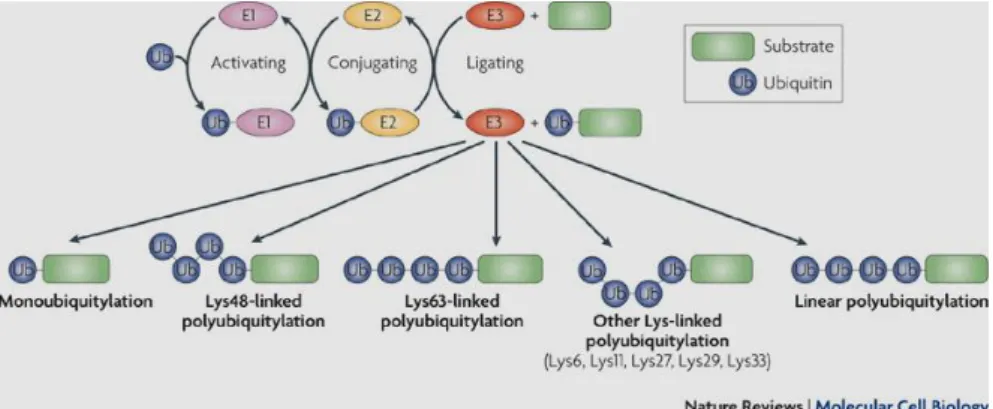
Ubiquitination of substrates recognized by the 26S proteasome, a multisubunit enzyme complex (molecular mass ~ 2000 kDa), is catalyzed by three classes of enzymes: Ub-Activating enzymes (E1), Ub-Conjugating (E2) or Ubc enzymes, and Ub-Ligating (E3) enzymes (Figure 1.6, Ref. [140]). Ubiquitin-binding domains (UBDs) are a collection of modular protein domains that bind to the Ub, in a non-covalent fashion. The preferences of UBDs for ubiquitin chains, of specific length and linkage (see once again Figure 1.6), originate from multimeric interactions, and UBDs synergistically bind multiple Ub molecules, and from contacts with regions that link Ub molecules into a polymer. It was reported that the sequence context of UBDs, and the conformational changes that follow their binding to Ub greatly contribute to ubiquitin signaling in vivo [140].
Figure 1.6 The activity of three enzymes (E2, E2, and E3) required for ubiquitylation.
(After Ref.[140]).
Like Ub-Conjugating enzymes, there are multiple classes of Ub-Ligating enzymes. These classes include the HECT (homologous to E6-associated protein C-terminus)-type, RING (really interesting new gene)-type, and U-box (a modified RING motif without the full complement of Zn2+-binding
ligands)-type, all named after the domain in the E3 enzyme that interacts with the E2 enzyme [141]. In addition, E3s can be (i) simple- one protein chain recruits and interacts with both the E2 and the substrate, or (ii) complex- multiple proteins are required to facilitate the ligase function. E3s can facilitate their function either through one protein chain, or as part of a protein complex. Some E3 ligases contain a substrate recruitment domain, as well as a respective E2 binding domain, and are functional as a single entity [144]. However, in other cases, additional proteins are necessary to form a complex where one subunit will recruit the substrate, and another will bind the E2. The primary example of complex E3s is the SCF (Skp1, Cullin, F-box) family [145]– discovered through genetic requirements for cell cycle progression in budding yeast, which requires at least four different protein subunits for ligase function.
or salt bridges provide structural stabilization to the 70 residues of the domain [147]. Although HECT and RING domain E3 ligases have been studied extensively, little is known about the box proteins. To date, the U-box family of E3 enzymes is the smallest class of ligases (with relatively few E3s have been characterized). Nevertheless, it is notable that the U-box containing E3 ligases have been shown to play diverse roles in RNA splicing [148], collaborating with molecular chaperones [149], as well as the ER-associated protein degradation [150].
It has to be mentioned that the two E2 enzymes- Ubc4 and Ubc5, were shown to be extremely necessary for most of the Ub-dependent protein degradation processes in yeast [151].
In some cases, selective protein degradation by the UPS requires multiubiquitylation, which can subsequently require the additional activity of certain Ub-chain elongation factors- the E4s, that indeed represent a distinct and novel class of enzymes [152]. Yeast Ufd2 (ubiquitin fusion degradation protein-2), was the first E4 enzyme to be described [153]- that binds to the Ub moieties of preformed conjugates, and catalyses Ub-chain elongation, and in association with E1, E2, and E3 enzymes. Ufd2 belongs to the UFD (Ub fusion degradation) pathway in S. cerevisiae (like Ufd1, Ufd3, Ufd4, and Ufd5) [154]. Together with an orchestra of Ub-binding factors, the E4-Ufd2 enzyme co-operates with in an escort pathway to transfer and deliver polyubiquitinated substrates to the 26S proteasome [155]. Surprisingly, Ufd2 can have other functions beyond degradation; one example is the Ufd2-mediated stabilization of the Yap8 transcription factor in the arsenic stress response of S. cerevisiae. These findings will be discussed in the Chapter 3.
1.8.2
Yeast Ubiquitin-Like Proteins (Ulps), and Cognate factors
1.8.2.1 SUMO
Several ubiquitin-like protein (Ulp) modifiers have been identified for yeast, vertebrates, and plants. One of the most challenging classes are the ‗small ubiquitin-like modifier‘ (SUMO) proteins- coupled to numerous targets in the cell since they can modulate their localization, stability, or activity, as well as they can affect protein-protein interactions [157].
SUMOylation (or sumoylation) [158] is a dynamic, and a reversible process. It is similar to ubiquitination, yet biochemically distinct. Remarkably, SUMOylation can also be a signal for polyubiquitylation and proteasomal degradation [159].
There are at least three SUMO isoforms (SUMO1,-2,-3) in mammalian cells but only one in S. cerevisiae (Smt3). In yeast, SUMO is a small protein of around 100 amino acids in length encoded by SMT3 gene. SUMO conjugation requires the E1 heterodimer (Uba2/Aos1), and E2 (Ubc9) enzymes (see Figure 1.6), as well as E3 proteins (Siz1 or Siz2 in yeast) to form a covalent bond with target proteins [158]. DeSUMOylation– the cleavage of SUMO form from its target proteins is mediated by SUMO specific proteases, of which Ulp1 and Ulp2 have been identified in yeast [160, 161] (for more details, see Chapter 4).
Major targets of SUMO in S. cerevisiae are the septins Cdc3, Cdc1, and Sep7, which form a 10-nm filamentous ring that encircles the yeast bud neck [162]. SUMO interferes with processes such as transcription, nuclear-cytoplasmic transport, chromatin integrity and dynamics, and cell cycle control. All these events occur via alterations of the molecular interactions patterns of SUMOylated proteins [138, 163].
Figure 1.7 SUMOylation and deSUMOylation pathway, and its enzymes. The sequential
reactions involved in SUMOylation (steps 1–5, in green) and deSUMOylation (steps 6 and 7, in orange) are represented. Note that SUMO-processing (step 1), SUMO deconjugation (step 6) and SUMO chain-editing (step 7) activities are carried out by a family of enzymes termed SUMO-proteases. (Adapted from Ref. [164].)
1.8.2.2
Other Proteins Related to Ub
In yeast, Rub1 (in other eukaryotes also called NEDD8) displays a high homology to Ub, and is linked to all members of cullin (Cul)-family proteins through an enzymatic cascade analogous to ubiquitination [165].
1.9
Aims of the Thesis
Despite a decade of work on the cellular processes underlying yeast adaptation to arsenic, and regulatory mechanisms of key transcription factor Yap8, the following fundamental questions on arsenic‘s mechanism of action were still open: (1) How do cells respond to arsenic challenge that perturb homeostasis?, (2) What are its cellular binding partners or targets?, (3) What are the post-translational modifications of Yap8, particularly upon arsenic stress?, (4) Is the stability/activity of the Yap8 protein also mediated by the SUMO modifier?
Aware of these unresolved fundamental questions and the existing extensive but incomplete data on arsenic‘s action at the molecular level, as well as intriguing aspects of the arsenic-mediated Yap8 stability, our aims were to:
- Foremost, undertake a DNA microarray approach in S. cerevisiae to compare expression profiles of normal cells and cells treated with arsenic in order to
- complement the diverse existing S. cerevisiae data, and provide a more consolidated view on the arsenic drug-targets in a defined cellular setting, within the long-term interest to
- identify signatures of molecular endpoints useful to address unresolved resistance problems to arsenic therapy in APL [167].
References
1. Xu, Y., et al., Clinical manifestations and arsenic methylation after a rare subacute arsenic poisoning accident. Toxicol Sci, 2008. 103(2): p. 278-84. 2. Hughes, M.F., et al., Arsenic exposure and toxicology: a historical perspective.
Toxicol Sci, 2011. 123(2): p. 305-32.
3. Waxman, S. and K.C. Anderson, History of the development of arsenic derivatives in cancer therapy. Oncologist, 2001. 6 Suppl 2: p. 3-10.
4. Garelick, H., et al., Arsenic pollution sources. Rev Environ Contam Toxicol, 2008. 197: p. 17-60.
5. Tamás, M.J. and E. Martinoia, Molecular Biology of Metal Homeostasis and Detoxification: From Microbes to Man. 2005, Heidelberg, Germany: Springer Verlag.
6. Tamas, M.J., et al., Heavy metals and metalloids as a cause for protein misfolding and aggregation. Biomolecules, 2014. 4(1): p. 252-67.
7. Rosen, B.P., Biochemistry of arsenic detoxification. FEBS Lett, 2002. 529(1): p. 86-92.
8. Shen, S., et al., Arsenic binding to proteins. Chem Rev, 2013. 113(10): p. 7769-92.
9. Terada, H., Uncouplers of oxidative phosphorylation. Environ Health Perspect, 1990. 87: p. 213-8.
10. Shankar, S., U. Shanker, and Shikha, Arsenic contamination of groundwater: a review of sources, prevalence, health risks, and strategies for mitigation. ScientificWorldJournal, 2014. 2014: p. 304524.
11. Lantz, R.C. and A.M. Hays, Role of oxidative stress in arsenic-induced toxicity. Drug Metab Rev, 2006. 38(4): p. 791-804.
12. Ratnaike, R.N., Acute and chronic arsenic toxicity. Postgrad Med J, 2003.
79(933): p. 391-6.
13. Miller, W.H., Jr., et al., Mechanisms of action of arsenic trioxide. Cancer Res, 2002. 62(14): p. 3893-903.
14. Zhu, J., et al., How acute promyelocytic leukaemia revived arsenic. Nat Rev Cancer, 2002. 2(9): p. 705-13.
15. Emadi, A. and S.D. Gore, Arsenic trioxide - An old drug rediscovered. Blood Rev, 2010. 24(4-5): p. 191-9.
16. Jonnalagadda, S.B. and P.V. Rao, Toxicity, bioavailability and metal speciation. Comp Biochem Physiol C, 1993. 106(3): p. 585-95.
17. McCarty, K.M., H.T. Hanh, and K.W. Kim, Arsenic geochemistry and human health in South East Asia. Rev Environ Health, 2011. 26(1): p. 71-8.
18. Lopez-Cima, M.F., et al., Lung cancer risk and pollution in an industrial region of Northern Spain: a hospital-based case-control study. Int J Health Geogr, 2011.
10: p. 10.
19. Florea, A.M. and D. Busselberg, Occurrence, use and potential toxic effects of metals and metal compounds. Biometals, 2006. 19(4): p. 419-27.
20. Dopp, E., et al., Environmental distribution, analysis, and toxicity of organometal(loid) compounds. Crit Rev Toxicol, 2004. 34(3): p. 301-33.
21. Tokar, E.J., B.A. Diwan, and M.P. Waalkes, Renal, hepatic, pulmonary and adrenal tumors induced by prenatal inorganic arsenic followed by dimethylarsinic acid in adulthood in CD1 mice. Toxicol Lett, 2013. 209(2): p. 179-85.
22. Fry, R.C., et al., Activation of inflammation/NF-kappaB signaling in infants born to arsenic-exposed mothers. PLoS Genet, 2007. 3(11): p. e207.
23. Bardach, A.E., et al., Epidemiology of chronic disease related to arsenic in Argentina: A systematic review. Sci Total Environ, 2015. 538: p. 802-816.
24. Rehman, K. and H. Naranmandura, Double-edged effects of arsenic compounds: anticancer and carcinogenic effects. Curr Drug Metab, 2013.
14(10): p. 1029-41.
26. Kwong, Y.L. and D. Todd, Delicious poison: arsenic trioxide for the treatment of leukemia. Blood, 1997. 89(9): p. 3487-8.
27. Zhang, X.W., et al., Arsenic trioxide controls the fate of the PML-RARalpha oncoprotein by directly binding PML. Science, 2010. 328(5975): p. 240-3. 28. Mi, J.Q., et al., Synergistic targeted therapy for acute promyelocytic leukaemia: a
model of translational research in human cancer. J Intern Med, 2015.
29. Park, M.T., et al., Combination treatment with arsenic trioxide and phytosphingosine enhances apoptotic cell death in arsenic trioxide-resistant cancer cells. Mol Cancer Ther, 2007. 6(1): p. 82-92.
30. Lengfelder, E., et al., Arsenic trioxide-based therapy of relapsed acute promyelocytic leukemia: registry results from the European LeukemiaNet. Leukemia, 2015. 29(5): p. 1084-91.
31. Shepshelovich, D., et al., Acute promyelocytic leukemia with isochromosome 17q and cryptic PML-RARA successfully treated with all-trans retinoic acid and arsenic trioxide. Cancer Genet, 2015.
32. Samuel, D., Investigation of Ancient Egyptian Baking and Brewing Methods by Correlative Microscopy. Science, 1996. 273(5274): p. 488-90.
33. Palazzo, A.F. and T.R. Gregory, The case for junk DNA. PLoS Genet, 2014.
10(5): p. e1004351.
34. Botstein D, Chervitz SA, and C. JM., Yeast as a model organism. Science, 1997.
277(277(5330):): p. 1259-60.
35. Smith, C., A. Pomiankowski, and D. Greig, Size and competitive mating success in the yeast. Behav Ecol, 2013. 25(2): p. 320-327.
36. Fields, S. and M. Johnston, Cell biology. Whither model organism research? Science, 2005. 307(5717): p. 1885-6.
37. Narayan, P., S. Ehsani, and S. Lindquist, Combating neurodegenerative disease with chemical probes and model systems. Nat Chem Biol, 2014. 10(11): p.
911-20.
38. DeLoache, W.C. and J.E. Dueber, Compartmentalizing metabolic pathways in organelles. Nat Biotechnol, 2013. 31(4): p. 320-1.
39. Goffeau, A., et al., Life with 6000 genes. Science, 1996. 274(5287): p. 546, 563-7.
40. Mewes, H.W., et al., Overview of the yeast genome. Nature, 1997. 387(6632
Suppl): p. 7-65.
41. Alfred Pühler, D.J., Jörn Kalinowski, Detlev Buttgereit, Renate Renkawitz-Pohl, Lothar Altschmied, Antoin Danchin, Agnieszka Sekowska, Horst Feldmann, Hans-Peter Klenk, and Manfred Kröger, Genome Projects on Model Organisms Handbook of Genome Research. Genomics, Proteomics, Metabolomics, Bioinformatics, Ethical and Legal Issue, ed. C.W. Sensen. 2005, KGaA, Weinheim: WILEY-VCH Verlag GmbH & Co.
42. Hettema, E.H., et al., The ABC transporter proteins Pat1 and Pat2 are required for import of long-chain fatty acids into peroxisomes of Saccharomyces cerevisiae. Embo J, 1996. 15(15): p. 3813-22.
43. Bossier, P., et al., The yeast YKL741 gene situated on the left arm of chromosome XI codes for a homologue of the human ALD protein. Yeast, 1994.
10(5): p. 681-6.
44. Dujon, B., et al., Complete DNA sequence of yeast chromosome XI. Nature, 1994. 369(6479): p. 371-8.
45. Guerreiro, P., et al., Sequencing of a 9.9 kb segment on the right arm of yeast chromosome VII reveals four open reading frames, including PFK1, the gene coding for succinyl-CoA synthetase (beta-chain) and two ORFs sharing homology with ORFs of the yeast chromosome VIII. Yeast, 1997. 13(3): p. 275-80.
46. Guerreiro, P. and C. Rodrigues-Pousada, Disruption and phenotypic analysis of six open reading frames from chromosome VII of Saccharomyces cerevisiae reveals one essential gene. Yeast, 2001. 18(9): p. 781-7.

![Figure 1.4 Arsenic transport in S. cerevisiae, and mammals. [After Ref. [107] (adapted).]](https://thumb-eu.123doks.com/thumbv2/123dok_br/15769989.641185/41.765.133.631.264.531/figure-arsenic-transport-s-cerevisiae-mammals-ref-adapted.webp)
![Figure 1.5 Ca 2+ transport in S. cerevisiae. (After Ref. [122].)](https://thumb-eu.123doks.com/thumbv2/123dok_br/15769989.641185/46.765.126.523.99.462/figure-ca-transport-in-s-cerevisiae-after-ref.webp)