www.microbialcell.com
Review
Guidelines and recommendations on yeast cell death
nomenclature
Didac Carmona-Gutierrez
1,‡,*, Maria Anna Bauer
1,‡, Andreas Zimmermann
1, Andrés Aguilera
2, Nicanor Austriaco
3, Kathryn
Ayscough
4, Rena Balzan
5, Shoshana Bar-Nun
6, Antonio Barrientos
7,8, Peter Belenky
9, Marc Blondel
10, Ralf J. Braun
11, Michael
Breitenbach
12, William C. Burhans
13, Sabrina Büttner
1,14, Duccio Cavalieri
15, Michael Chang
16, Katrina F. Cooper
17, Manuela
Côrte-Real
18, Vítor Costa
19–21, Christophe Cullin
22, Ian Dawes
23, Jörn Dengjel
24, Martin B. Dickman
25, Tobias Eisenberg
1,26,
Birthe Fahrenkrog
27, Nicolas Fasel
28, Kai-Uwe Fröhlich
1, Ali Gargouri
29, Sergio Giannattasio
30, Paola Goffrini
31, Campbell W.
Gourlay
32, Chris M. Grant
33, Michael T. Greenwood
34, Nicoletta Guaragnella
30, Thomas Heger
35, Jürgen Heinisch
36, Eva
Herker
37, Johannes M. Herrmann
38, Sebastian Hofer
1, Antonio Jiménez-Ruiz
39, Helmut Jungwirth
1, Katharina Kainz
1,
Dimitri-os P. Kontoyiannis
40, Paula Ludovico
41,42, Stéphen Manon
43, Enzo Martegani
44, Cristina Mazzoni
45, Lynn A. Megeney
46–48,
Chris Meisinger
49, Jens Nielsen
50–52, Thomas Nyström
53, Heinz D. Osiewacz
54, Tiago F. Outeiro
55–58, Hay-Oak Park
59, Tobias
Pendl
1, Dina Petranovic
50,51, Stephane Picot
60,61, Peter Polčic
62, Ted Powers
63, Mark Ramsdale
64, Mark Rinnerthaler
65, Patrick
Rockenfeller
1,32, Christoph Ruckenstuhl
1, Raffael Schaffrath
66, Maria Segovia
67, Fedor F. Severin
68, Amir Sharon
69, Stephan J.
Sigrist
70, Cornelia Sommer-Ruck
1, Maria João Sousa
18, Johan M. Thevelein
71,72, Karin Thevissen
73, Vladimir Titorenko
74,
Michel B. Toledano
75, Mick Tuite
32, F.-Nora Vögtle
49, Benedikt Westermann
11, Joris Winderickx
76, Silke Wissing
77, Stefan
Wölfl
78, Zhaojie J. Zhang
79, Richard Y. Zhao
80, Bing Zhou
81, Lorenzo Galluzzi
82–84,*, Guido Kroemer
84–90,*, Frank Madeo
1,26,*
1
Institute of Molecular Biosciences, NAWI Graz, University of Graz, Graz, Austria. 2 Centro Andaluz de Biología, Molecular y Medicina Regenerativa-CABIMER, Universidad de Sevilla, Sevilla, Spain. 3 Department of Biology, Providence College, Providence, USA. 4 Department of Biomedical Science, University of Sheffield, Sheffield, United Kingdom. 5 Department of Physiology and Biochemistry, University of Malta, Msida, Malta. 6 Department of Biochemistry and Molecular Biology, George S. Wise Faculty of Life Sciences, Tel Aviv University, Tel Aviv, Israel. 7 Department of Biochemistry and Molecular Biology, University of Miami Miller School of Medicine, Miami, USA. 8 Department of Neurology, University of Miami Miller School of Medi-cine, Miami, USA. 9 Department of Molecular Microbiology and Immunology, Brown University, Providence, USA. 10 Institut National de la Santé et de la Recherche Médicale UMR1078, Université de Bretagne Occidentale, Etablissement Français du Sang Bretagne, CHRU Brest, Hôpital Morvan, La-boratoire de Génétique Moléculaire, Brest, France. 11 Institute of Cell Biology, University of Bayreuth, Bayreuth, Germany. 12 Department of Cell Biolo-gy and PhysioloBiolo-gy, Division of Genetics, University of Salzburg, Salzburg, Austria. 13 Department of Molecular and Cellular Biology, Roswell Park Cancer Institute, Buffalo, NY, USA. 14 Department of Molecular Biosciences, The Wenner-Gren Institute, Stockholm University, Stockholm, Sweden. 15 De-partment of Biology, University of Florence, Firenze, Italy. 16 European Research Institute for the Biology of Ageing, University of Groningen, University Medical Center Groningen, Groningen, The Netherlands. 17 Dept. Molecular Biology, Graduate School of Biomedical Sciences, Rowan University, Strat-ford, USA. 18 Center of Molecular and Environmental Biology, Department of Biology, University of Minho, Braga, Portugal. 19 Instituto de Investigação e Inovação em Saúde, Universidade do Porto, Porto, Portugal. 20 Instituto de Biologia Molecular e Celular, Universidade do Porto, Porto, Portugal. 21
www.microbialcell.com
ABSTRACT
Elucidating the biology of yeast in its full complexity has
major implications for science, medicine and industry. One of the
most critical processes determining yeast life and physiology is
cel-lular demise. However, the investigation of yeast cell death is a
relatively young field, and a widely accepted set of concepts and
terms is still missing. Here, we propose unified criteria for the
defi-nition of accidental, regulated, and programmed forms of cell
death in yeast based on a series of morphological and biochemical
criteria. Specifically, we provide consensus guidelines on the
differ-ential definition of terms including apoptosis, regulated necrosis,
and autophagic cell death, as we refer to additional cell death
rou-tines that are relevant for the biology of (at least some species of)
yeast. As this area of investigation advances rapidly, changes and
extensions to this set of recommendations will be implemented in
the years to come. Nonetheless, we strongly encourage the
au-thors, reviewers and editors of scientific articles to adopt these
collective standards in order to establish an accurate framework
for yeast cell death research and, ultimately, to accelerate the
pro-gress of this vibrant field of research.
Republic. 63 Department of Molecular and Cellular Biology, College of Biological Sciences, UC Davis, Davis, California, USA. 64 Biosciences, University of Exeter, Exeter, United Kingdom. 65 Department of Cell Biology and Physiology, Division of Genetics, University of Salzburg, Salzburg, Austria. 66 Insti-tute of Biology, Division of Microbiology, University of Kassel, Kassel, Germany. 67 Department of Ecology, Faculty of Sciences, University of Malaga, Malaga, Spain. 68 A.N. Belozersky Institute of physico-chemical biology, Moscow State University, Moscow, Russia. 69 School of Plant Sciences and Food Security, Faculty of Life Sciences, Tel Aviv University, Tel Aviv, Israel. 70 Institute for Biology/Genetics, Freie Universität Berlin, Berlin, Germany. 71 La-boratory of Molecular Cell Biology, Institute of Botany and Microbiology, KU Leuven, Leuven, Belgium. 72 Center for Microbiology, VIB, Leuven-Heverlee, Belgium. 73 Centre of Microbial and Plant Genetics, KU Leuven, Leuven, Belgium. 74 Biology Department, Concordia University, Montreal, Canada. 75 Institute for Integrative Biology of the Cell (I2BC), SBIGEM, CEA-Saclay, Université Paris-Saclay, Gif-sur-Yvette, France. 76 Department of Biology, Functional Biology, KU Leuven, Leuven-Heverlee, Belgium. 77 Cevec Pharmaceuticals, Cologne, Germany. 78 Institute of Pharmacy and Molecu-lar Biotechnology, Heidelberg University, Heidelberg, Germany. 79 Department of Zoology and Physiology, University of Wyoming, Laramie, USA. 80
Department of Pathology, University of Maryland School of Medicine, Baltimore, USA. 81 School of Life Sciences, Tsinghua University, Beijing, China. 82
Department of Radiation Oncology, Weill Cornell Medical College, New York, NY, USA. 83 Sandra and Edward Meyer Cancer Center, New York, NY, USA. 84 Université Paris Descartes/Paris V, Paris, France. 85 Equipe 11 Labellisée Ligue Contre le Cancer, Centre de Recherche des Cordeliers, Paris, France. 86 Cell Biology and Metabolomics Platforms, Gustave Roussy Comprehensive Cancer Center, Villejuif, France. 87 INSERM, U1138, Paris, France. 88
Université Pierre et Marie Curie/Paris VI, Paris, France. 89 Pôle de Biologie, Hôpital Européen Georges Pompidou, Paris, France. 90 Karolinska Insti-tute, Department of Women’s and Children’s Health, Karolinska University Hospital, Stockholm, Sweden.
‡ Equally contributing
* Corresponding Authors:
Frank Madeo, Institute of Molecular Biosciences, University of Graz, Graz, Austria; E-mail: [email protected]; Didac Carmona-Gutierrez, Institute of Molecular Biosciences, University of Graz, Graz, Austria; E-mail: [email protected]; Guido Kroemer, INSERM, U1138, Paris, France; E-mail: [email protected];
Lorenzo Galluzzi, Weill Cornell Medical College, New York, NY, USA; E-mail: [email protected]
doi:10.15698/mic2018.01.607 Received originally: 19.12.2017; Accepted29.12.2017,
Published 01.01.2018.
Keywords: accidental cell death, apoptosis, autophagic cell death, autophagy, caspases, mitochondrial membrane permeabilization, mitotic catastrophe, model organism, necrosis, reactive oxygen species, regulated cell death, Saccharomyces cerevisiae.
Abbreviations:
INTRODUCTION
Yeast, a fungus that predominantly lives as a unicellular
organism, has had an extraordinary influence on humanity
throughout millennia, from its usage for baking and
brew-ing to the potential of some species to cause
life-threatening human diseases. The cultural, industrial,
bio-technological, and medical impact of this organism remains
unparalleled. The use of yeast fermentation to produce
alcoholic beverages and to leaven bread coincided with the
rise of ancient civilizations and has persisted until our days.
Importantly, the continued development of yeast strains as
vehicles for the development of new technology, for
ex-ample in bioethanol, drug, and enzyme production, as well
as the implementation of unconventional yeast species in
industrial processes, highlights the ever increasing
im-portance of yeast now and in the future
[1, 2]
. This is
ex-emplified by the fact that the global market for yeast
prod-ucts is in the multibillion dollar range and is expected to
grow further
[3]
. Beyond the mentioned applications, yeast
has a direct impact on human health and disease. Many
fungi, including some yeasts, can exist as commensals, i.e.,
they are part of our natural microbiota, forming the
myco-biome
[4]
. In fact, it is being increasingly recognized that
fungi are a major determinant in establishing commensal
microbial communities and are thus vital for healthy
indi-viduals
[5]
. However, under certain circumstances, e.g.,
compromised immunity, commensal fungi may become
opportunistic pathogens and as such are a potential cause
for infectious diseases
[6]
. These include superficial
infec-tions of the skin and nails (especially by dermatophytes)
that affect billions worldwide, biofilm colonisations of
mu-cosal surfaces and more serious invasive infections, which
can have very high mortality rates and are estimated to
lead to 1.5 million deaths per year
[7]
. A significant number
of these deaths arise from infections caused by the yeasts
Candida albicans
,
Candida glabrata
and
Cryptococcus
neoformans
in immunocompromised individuals. This
soci-oeconomic burden is further amplified by the
unprece-dented rise in fungal diseases that are affecting plants and
animals
[8]
. These examples highlight the importance of a
full understanding of fungal biology, and the study of yeast
cell biological processes has been crucial in this respect.
Yeasts have served as a successful research tool for the
last century,
Saccharomyces cerevisiae
(the budding yeast)
being one of the most thoroughly studied eukaryotes at
the cellular and molecular levels. Indeed, yeast continues
to be one of the preferred model organisms to explore
eukaryotic cell biology, both due to its technical
ad-vantages in devising/sophisticating molecular tool kits to
study cellular biology, and to a high degree of functional
conservation
[9]
. Also, yeast offers rapid growth and
inex-pensive accessibility paired with a high amenability to
bio-chemical and genetic manipulation. This enables the
estab-lishment of various experimental setups, ranging from
sin-gle experiments to high-throughput, genome-scale,
unbi-ased screenings in a short time frame. Notably, many
in-sights obtained in yeast have proven to be transferable to
higher eukaryotes. Indeed, over the past decades, yeast
studies have unveiled individual gene functions as well as
gene and protein interactions, and have instrumentally
contributed to the understanding of fundamental cellular
processes such as eukaryotic cell cycle control
[10
–
15]
,
autophagy
[16
–
19]
, mitochondrial function
[20, 21]
,
includ-ing mitochondrial import
[22
–
25]
, protein degradation
[26]
,
vesicle fusion
[27, 28]
, genetic instability
[29, 30]
,
epigenet-ic control
[31, 32]
, metabolic regulation
[33
–
35]
, or cellular
nutrient sensing
[36]
.
In addition, studies on yeast have shed light on human
diseases, providing a cellular platform to examine, for
in-stance, prion biology, virus-host interactions, metabolic
diseases, neurodegenerative disorders, cancer, or aging
[37
–
61]
. Among the pathophysiologically relevant
path-ways that can readily be explored in yeast are those
gov-erning cellular demise. Indeed, cell death regulation is
structurally and functionally conserved in yeast
[21, 62
–
66]
,
and yeast has even served to uncover and establish factors
and pathways involved in apoptosis and other controlled
cell death subroutines, which have later been corroborated
in metazoan or other multicellular systems, e.g., the
AAA-ATPase Cdc48/VCP
[63, 67]
, the BAX inhibitor-1
[68]
, the
implication of metacaspases as cell death regulators
[69
–
71]
, the role of cathepsin D in non-autophagic
mitochon-drial degradation
[72, 73]
, or the lethal impact of ER-Golgi
transport blockage as one of the mechanisms explaining
the demise of dopaminergic neurons during
Parkinson’s
disease
[74]
. To sum up, on the one hand, cell death
repre-sents a key process that can be feasibly modeled in yeast.
On the other hand, the understanding of yeast cell death
and its putative modulation may improve industrial and
biotechnological applications, provide insights into
myco-biome dynamics, and help develop the fight against fungal
and other diseases.
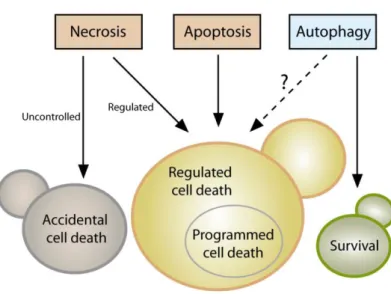
Even though it is now clear that yeast can indeed
un-dergo cellular suicide, the corresponding terminology to
describe this multifaceted process remains heterogenous
and potentially misleading. Thus, we believe that there is
timely need for a more precise and consistent
nomencla-ture that clearly defines the concept of “yeast cell death”,
considering morphological, enzymological, and functional
aspects. Such standardization seems of importance, given
that the field of yeast cell death is continuously expanding
with significant progress being made at the phenotypical
and mechanistic levels, including the finding that, akin to
higher eukaryotes, yeast can also engage in distinct cell
death modalities (
Figure 1
). In this paper we thus attempt
for the first time to formulate a series of recommendations
and caveats with respect to cell death-related results
ob-tained in yeast. To this aim, we have followed the
direc-tions of the Nomenclature Committee on Cell Death
(NCCD)
[92
–
95]
and adapted them to the particularities of
Saccharomyces cerevisiae
, which we think can be extended
to other yeast species and to multicellular filamentous
fungi. Our goal is to frame a uniform set of guidelines that
facilitate the communication among yeast cell death
re-searchers, ultimately supporting and accelerating scientific
advance (
Box 1
). In that respect, the nomenclature
pre-sented herein will likely need to be revised and updated as
the field of yeast cell death moves forward and even more
precise definitions are required.
YEAST CELL DEATH AND SURVIVAL
A crucial issue that demands a clear definition is the
ques-tion of cell death itself. When is a cell dead? According to
the NCCD guidelines this is only the case upon irreversible
plasma membrane breakdown or complete cellular
frag-mentation, because only then the cell is factually
disinte-grated, irrespectively of which upstream pathway or
rou-tine has been engaged
[93]
. In fact, no earlier marker can
be defined that reliably determines death in all settings.
Thereby, this lethal irreversibility might start with the
col-lapse of the electrochemical membrane potential across
the plasma membrane through formation of a leak. In
yeast, the most common method to monitor cell
mem-brane integrity
in vivo
is to use propidium iodide (PI). PI is a
fluorescent nucleic acid intercalator that can only enter
cells with a ruptured cell membrane, and can be routinely
employed in both low and high throughput formats
[96
–
98]
. Along similar lines, colorimetric dyes like trypan blue
may be used, but are less common
[99
–
101]
. Further
po-tential alternatives exist (e.g., 7-aminoactinomycin D), but
will need to be thoroughly tested with respect to their
suitability for yeast cell death applications in future studies.
As mentioned, assessing cell membrane disintegrity is the
only technique to quantify actual cell death and must be
performed irrespectively of the lethal setting being
ana-lyzed. This is imperative, since lethal signaling does not
imply that the final stage (cell death) is reached or even
that it will be reached at a later stage (see below). In fact,
specific subpopulations engaged in lethal pathways that
maintain plasma membrane integrity (e.g., early apoptotic
cells, see below) are by definition not (yet) dead. In that
respect, timecourse experiments are important to monitor
both the lethal subroutine-specific phenotypes and the
actual occurrence of cell death over time. Of note,
indica-tions exist that upon specific stress insults, a small
subpop-ulation of yeast retains the ability to repair cell membrane
damage even after having stained positive for PI
[102]
.
Given the lack of other comparably well established dyes in
this context and the large body of data supporting PI
stain-ing as a valid method to quantify loss of survival, we
con-clude that determining PI positivity is
–
at this point - the
best technique to quantitatively approach yeast cell death.
Still, for the sake of accuracy and waiting for further
evi-dence supporting the above-mentioned indications, we
suggest expressing a corresponding quan
tification as “% PI
-positivity” or “%
cell death ( PI positive
)”
instead of
“%
d
eath” or “% survival” upon using this method.
In the
long term, the development and establishment of
alterna-tive dyes should be explored in order to validate data
ob-tained with PI. A number of approaches allow to
experi-mentally assess (i) cell viability, which reflects the ability of
a cell to divide, and (ii) cell vitality, defined as the
physio-logical capabilities of a cell
[100]
. Nonetheless, an
im-paired/compromised (i) proliferation or (ii) metabolic
ca-pacity does not necessarily result in cellular demise. Thus,
these techniques alone cannot be used to demonstrate cell
death. Still, they are very useful to complement and
cor-roborate data obtained with PI or alternative dyes.
Assessing clonogenicity with plating assays is the most
commonly used method to quantify cell viability
[62, 103]
.
Here, a defined number of cells from a given culture are
plated on rich medium agar plates that are further
incu-bated to allow colony formation. The ratio between the
resulting colony-forming units (CFUs) and the originally
plated number of cells reflects the viability state in the
culture. Theoretically, however, it is possible that under
specific conditions (of genetic nature, for instance), colony
formation may be blocked in cells that
per se
are still alive
(a condition usually refered to as senescence). Additional
caveats include the possibility that live cells at the point of
plating might die before forming a colony and/or that the
plating procedure itself might drive (a fraction of) cells into
death, which would be indistinguishable from cell
senes-cence. Nonetheless, the literature suggests clonogenic
capacity as a very good correlate to cell death in a plethora
of different settings
[69, 96, 104, 105]
and thus represents
a valid approximation to quantify survival in yeast
popula-tions. Of note, clonogenicity can also be measured by
mon-itoring CFU formation at the microcolony level (time-lapse
photomicroscopy)
[106, 107]
. Even though cell and colony
counting can be automated, clonogenicity assays are
ra-ther time-consuming and used for low- to
medium-throughput analyses.
A further technique to assess yeast viability follows the
growth rate of a given culture, which may decrease as a
consequence of increased cell death. For this purpose, an
aliquot is inoculated into fresh liquid medium and the
growth is monitored, for instance, via photometric
meas-urement of optical densities over a specific period of time
[108, 109]
. Optionally, spot dilution assays can be
per-formed, where cultures are spotted in serial dilutions on
agar plates
[110]
. Here, the growth ability is compared
between cultures at the various dilution steps in a
sequantitative manner, although automated readout of
mi-crocolonies can be used to yield a quantitative result
[111]
.
Monitoring growth can be scaled up and performed either
manually or using robotics support, which makes it an
at-tractive technique, especially for screen-based analyses. As
with other viability assays, an important disadvantage is
that a decreased growth rate can also result from a
non-lethal event such as modulation of cell cycle progression or
a reduced metabolism due to an alteration in the use of
media components.
One possibility to evaluate yeast cell vitality is to
direct-ly assess the activity of specific enzymes directdirect-ly. Although
this is not widely employed in yeast cell death research, it
represents an avenue to assay the physiological state of a
metabolic pathway within the cell
[100, 112, 113]
. As
pointed out below, a caveat of this approach is the possible
distortion of results by residual activity in dead cells. A
further option is to use vital dyes, like the two-color
fluo-rescent probe FUN-1, which diffuses into cells,
irrespective-ly of their viability status, and results in green fluorescence
of the cytoplasm. Dead cells fluoresce green while (live)
cells that have both plasma membrane integrity and
meta-bolic capability, can further process the probe, resulting in
red vacuolar fluorescence
[114, 115]
. Similarly, several
tetrazolium salts are reduced into colored formazan
crys-tals
[116]
. Methylene blue is converted to the colorless
leucomethylene blue only in metabolically active cells
[117]
,
while the red dye phloxine B is only retained in
metaboli-cally inactive cells that are unable to actively export it
[100,
118]
. Other methods aim at assessing further aspects of
BOX 1: DEFINITIONS OF KEY CONCEPTS IN YEAST CELL DEATH
Accidental cell death describes cellular death following exposure to very harsh microenvironmental conditions.
Apoptosis represents a regulated cell death subroutine charac-terized by specific morphologic and biochemical features and executed via different pro-apoptotic factors; eventually, it culminates in secondary necrosis.
Autophagy defines a predominantly cytoprotective process that orchestrates the digestion of intracellular material (e.g. proteins, organelles) in the vacuole.
Autophagy-dependent cell death describes a lethal subrou-tine, in which the molecular machinery of autophagy (or parts thereof) causally contributes to cellular demise.
Cell death defines a status of irreversible plasma membrane breakdown (only then, the cell is factually disintegrated, irre-spectively of which upstream pathway or routine has been engaged).
Cell viability reflects the ability of a cell to divide and thus to proliferate.
Cell vitality reflects the physiological capabilities of a cell and thus its metabolic activity.
Necrosis is a cell death instance mainly characterized by plas-ma membrane permeabilization; priplas-mary necrosis (cellular necrosis occurring ab initio) may take place in an accidental or regulated manner; secondary necrosis (combined necrotic and apoptotic features) is the final stage of the apoptotic process.
Programmed cell death designates a specific type of regulated cell death, which occurs in strictly physiological scenarios (e.g., development, aging).
Regulated cell death describescellular death occurring in the context of a failing response to internal or external mild stress.
cellular physiology, including the cellular ATP content (e.g.,
based on the luciferin-luciferase reaction)
[119]
or
mito-chondrial transmembrane potential (e.g., upon staining
with rhodamine 123, JC-1, TMRM/E, DiOC
6(3))
[120, 121]
. It
should be noted that the readout of metabolic signatures
has considerably improved with new generation
extracellu-lar flux analyzers, offering the possibility to simultaneously
measure mitochondrial respiration and glycolysis (and thus
mitochondrial function). A drawback of metabolic assays
resides in the fact that cells may be able to maintain some
metabolic activities until cell membrane rupture occurs,
and that some rely on specific metabolic processes such as
oxidative phosphorylation that are not mandatory for cell
survival. Thus, such techniques may fail to detect
subpopu-lations of dead (or alive) cells, reflecting the notion that a
decrease in growth or metabolic activity (i.e
.
, viability or
vitality) cannot be placed on a par with an increase in cell
death. In conclusion, as mentioned above, the term cell
death should be used only upon observing breakdown of
the plasma membrane and thus loss of cell integrity (e.g.,
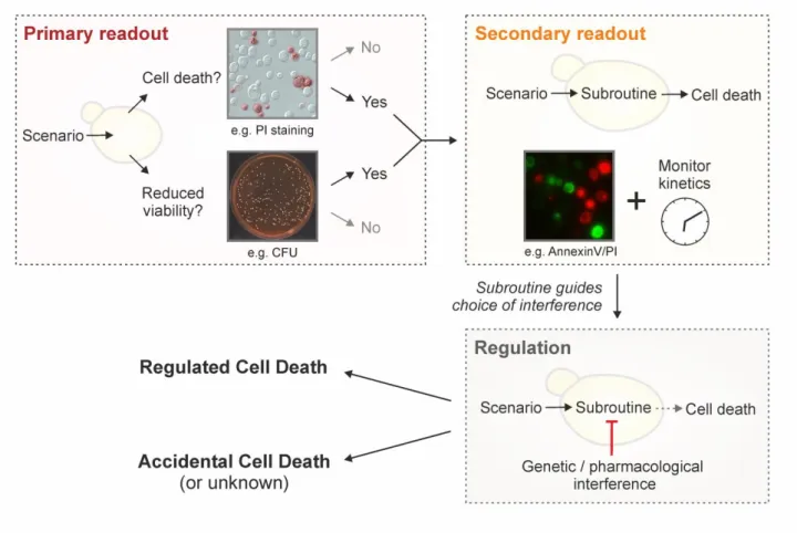
upon PI staining). In addition, we suggest to strengthen this
observation by simultaneously assessing clonogenic
capaci-ty (
Figure 2
), since (i) it represents the best-established
output to accurately monitor overall cellular viability and
(ii) it empirically correlates very strongly with actual cell
death markers. Importantly, both methods are easy, quick
and relatively inexpensive. The use of additional
dyes/stainings/assays provides valuable complementary
information, but cannot be used alone to unequivocally
define a cell as dead.
Yeast cell death is often accompanied by oxidative
damage and thus, a widely employed method in the field is
the detection of reactive oxygen species (ROS)
[122]
.
In-deed, a number of different ROS, like the superoxide anion,
hydroxyl radical, and hydrogen peroxide, can accumulate
upon mitochondrial disturbances, ER stress or other
cellu-lar derangements
[96, 122
–
125]
. ROS can generally be
de-tected using membrane-permeable dyes that are oxidized
to fluorescent products in a ROS-dependent manner.
Im-portantly, these stains do not measure ROS as a group, but
rather react with specific species. For instance,
dihydroeth-idium (DHE) preferentially reacts with superoxides, while
dihydrorhodamine
123
(DHR123)
and
2,7-dichlorodihydrofluorescein diacetate (H
2-DCF-DA) are
con-verted by a broad range of other ROS, but only poorly by
superoxides
[126]
. Such specificities should be taken into
account when measuring ROS with a particular stain, since
distinct lethal triggers might result in the production of a
differential ROS subset
[123]
. Thus, we recommend to
spe-cifically indicate the ROS subtype that is being monitored
instead of generally referring to ROS production. Of note,
to a certain degree, DHE may also be oxidized
unspecifical-ly (independentunspecifical-ly of superoxide). In order to exploit the full
potential of DHE as a superoxide-specific dye, a range of
methodological possibilities (e.g., the use of optimized
spectra) exist
[127, 128]
. The standardization of such
re-finements for DHE assays, which are a preferred tool in
yeast cell death research, should be addressed in the
fu-ture. While ROS measurements allow for high-throughput
approaches due to their simplicity and relatively low cost,
it is imperative to realize that this method does not
dis-criminate between living and dead cells, although ROS
usually precede and are often causative for cell death in
yeast
[125]
. In fact, ROS play a crucial role in intracellular
signaling
[129
–
132]
, functioning, for instance, as direct and
indirect regulators of diverse physiologically relevant
tar-gets
[133
–
135]
. In addition, limited ROS generation might
be beneficial under certain conditions, since the resulting
adaptive responses can promote stress resistance as a
form of preconditioning (hormesis)
[131, 136
–
139]
. Thus,
an increase in ROS should be regarded as a cell
death-correlated phenotype only in connection with assays that
directly determine increased plasma membrane
disintegra-tion and loss of clonogenicity (see above). Similarly, a
de-crease in ROS production by incubation with anti-oxidants
might support the mechanistic involvement of ROS in the
lethal process, but only when cell death is adequately
mon-itored.
ACCIDENTAL VERSUS REGULATED CELL DEATH
Cellular demise in yeast may occur in two mutually
exclu-sive variants: either as an accidental event or through a
regulated pathway. Accidental cell death (ACD) occurs
up-on exposure to severe cup-onditiup-ons, resulting in a rapid,
un-controllable and unavoidable form of death. ACD may
fol-low a series of extreme stimuli, including physical
condi-tions, such as very high temperatures or pressures, severe
chemical insults like strong detergents and high
concentra-tions of acids or bases as well as mechanical challenges, for
instance, vigorous shearing or ultrasonic treatment. The
immediate nature of ACD, which is characterized by a
vir-tually immediate structural breakdown of cells, allows no
form of pharmacologic or genetic inhibition. Thus, this
form of cell death does not constitute a direct target for
modulation or prevention. However, it remains unclear
whether yeast cells undergoing ACD may release
endoge-nous, bioactive molecules to the extracellular space
[75,
79]
. If so, such molecules could interact with local cells that
have survived the primary insult and ignite a response
within the whole yeast population. Such a consequence of
ACD may resemble the release of damage-associated
mo-lecular patterns (DAMPs) by dying human cells. DAMPs can
stimulate a direct or indirect (via innate immune effectors)
cytotoxic response in surrounding bystander cells that have
survived ACD
[140
–
144]
. In such a case, interfering with
the effects of ACD on the rest of the population remains
possible.
ACD is often equated with necrosis, which in yeast is
usually identified as a cellular condition of early plasma
membrane permeabilization in the absence of typical
apoptotic markers and of complete disintegration of
sub-cellular structures
[103]
. Indeed, ACD usually exhibits
mor-phological features of necrosis, but mounting evidence
suggests that
–
as it is the case in human cells
–
a
physio-logically relevant, regulated type of necrosis does also exist
in yeast. Thus, we recommend to avoid using the term
“necrosis” to define an accidental and uncontroll
able type
of death, and to favor the term “ACD”. We believe that this
will avoid any potential misunderstandings regarding the
two fundamentally dinstinct (accidental
versus
regulated)
modalities of yeast cell death manifesting with a necrotic
morphology (see below).
That said, many lethal stimuli result in a form of yeast
cell death that
–
at odds with ACD - is executed by a
genet-ically encoded, dedicated molecular machinery. In higher
eukaryotes, a distinction is made between such a
con-trolled form of cell death when it occurs (i) in the
frame-work of a purely physiological program, e.g., during (post-)
embryonic development or tissue homeostasis, or (ii) as a
response to either a perturbation of intracellular or
extra-cellular homeostasis, e.g
.
, upon exposure to mild stress or
as a consequence of mutations. Cell death occurring in the
former scenario is termed “programmed cell death” (PCD),
while the expression “regulated cell death” (RCD) enco
m-passes both PCD as well as all other instances of cell death
that depend on a molecular machinery
[145
–
148]
.
For yeast cell death, many authors have used the term
PCD to interchangeably refer to all types of cellular demise
that are not accidental (i.e., to all instances of RCD).
How-ever, emerging evidence is confirming that a yeast
popula-tion, be it a liquid culture or a solid colony, bears a degree
of complexity reminiscent of multicellular organisms that
demands a revision of this terminology. For instance,
dur-ing yeast gametogenesis (or sporulation), immature
meiot-ic products as well as the mother cell itself succumb via
activation of vacuolar rupture
[149, 150]
. Interestingly, the
mother cell’s demise is delayed until spores have reached a
program,
de facto
representing an instance of PCD. During
yeast chronological aging, the cellular community
main-tains homeostasis thanks to the programmed death of
dysfunctional or old cells, which spares and provides
nutri-ents to the fitter individuals
[75, 76]
. In yeast colonies,
stationary-phase or slow-growing cells differentiate into
specific subpopulations with unique metabolic properties
and particular functions within the colony
[151, 152]
.
These examples show that, indeed, yeast populations can
harness cell death to control coordinated development,
homeostasis and differentiation. Hence, we propose to
define PCD in yeast as a specific instance of RCD that is
executed in the frame of such physiologic programs. All
other forms of regulated demise (e.g., cell death induction
upon stress, or as a consequence of specific genetic
altera-tions) should be referred to with the superordinate term of
RCD.
Importantly, since RCD depends on a defined molecular
machinery, it can be modulated with pharmacologic or
genetic means. The extent of such modulation depends on
the progression of the process across a hitherto poorly
defined point-of-no-return. According to the NCCD, the
processes preceding such point are part of cellular stress
responses, while those following it belong to actual cell
death signaling
[93]
. Adopting this rationale, RCD can be
accelerated or delayed (but not avoided) if the
point-of-no-return has been trespassed. Instead, prior to that point,
modulating stress responses or avoiding stress can prevent
RCD. However, the definition of this point-of-no-return has
not been established yet, implying that the exact boundary
between the reversibility of a stress stimulus and the
irrev-ocable engagement in a lethal cascade remains to be
speci-fied.
Yeast RCD may follow different subroutines (see below)
that can be differentiated from each other by a series of
morphological and biochemical features. To precisely
char-acterize the lethal phenotype, we recommend (i) to first
determine if cell death actually occurs (as opposed to only
reduced viability/vitality), (ii) if it does, to then examine the
subroutine(s) involved via morphological and biochemical
observations,
using at least two different detection
meth-ods
[155]
, and (iii) finally, to corroborate the implicated
mechanism(s) via genetic and pharmacological
interven-tions
(Figure 2).
Finally, it should be noted that in cell
death research, it is generally advisable to determine the
kinetics of the parameters under scrutiny
[156]
. In order to
detect the differential appearance of apoptotic or necrotic
characteristics, we recommend assessing such features at
different time points to yield a better resolution of cell
death events. Importantly, subroutine-specific markers
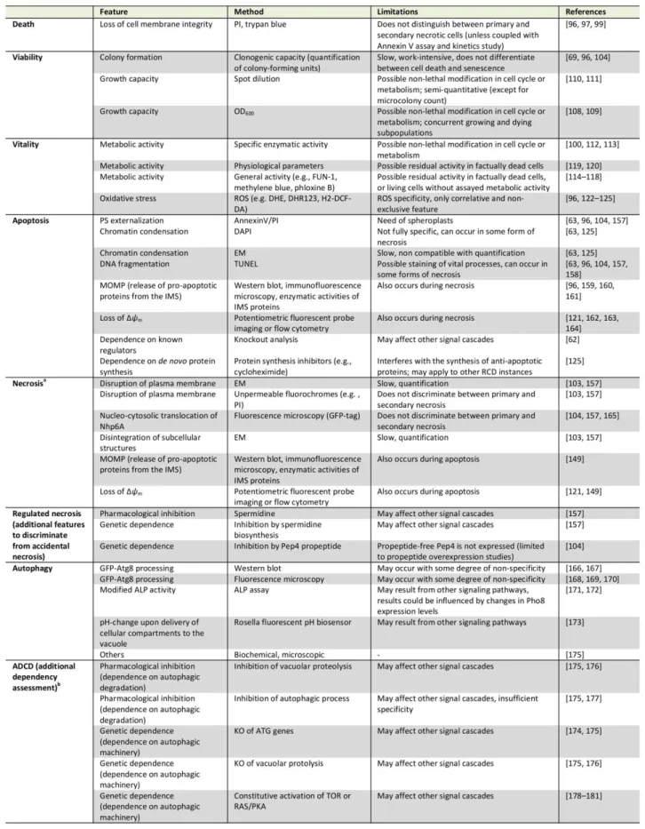
should precede cell death. In the following sections, we will
describe yeast RCD subroutines and the techniques to
pre-cisely discriminate amongst them (
Table 1
). Beyond the
specificities outlined below, a number of general issues
and notes of caution also need to be considered (
Box 2
).
APOPTOSIS
Most studies on RCD in yeast have been conducted in the
budding yeast
Saccharomyces cerevisiae
. This includes the
first observation of an apoptotic phenotype in yeast,
spe-cifically in a strain with a point mutation in the gene coding
for the cell cycle protein Cdc48
[63]
. One of the early
indi-cations for an active cellular participation in the yeast
apoptotic process was that RCD in this setting can be
pre-vented by inhibiting
de novo
protein synthesis, e.g. by
cy-cloheximide
[125]
. Ever since these discoveries, a set of
methods has been established, validated and refined that
allows to specifically determine whether a yeast cell has
engaged in an apoptotic pathway
[62]
. These techniques
are mainly based on the key morphologic and biochemical
features of an apoptotic cell. We suggest employing at
least two of these apoptosis-specific methods (one of them
should be Annexin V staining, see below) and include at
least one viability assay (preferably clonogenic capacity) to
describe a corresponding phenotype.
One of the events most commonly associated with
apoptosis is the exposure of phosphatidylserine (PS) on the
outer leaflet of the plasma membrane
[182]
. However, PS
externalization might be context-dependent to a certain
degree, at least within the complexity of the human
cellu-lar network
[93, 183, 184]
. It remains unclear whether this
is also the case in yeast, although the current evidence
suggests that PS externalization is a universal feature of
yeast cells undergoing apoptosis. PS externalization can be
assessed via monitoring PS-binding to Annexin V, which is
usually fluorescently labeled for quantitative (e.g.,
fluores-cence reader-based or flow cytrometric analyses) and
qual-itative (microscopic) evaluation. To this aim, the cell wall
needs to be (partially) digested in order to make the
exter-nalized PS accessible to Annexin V and permit binding.
Usually, the Annexin V assay is performed as a co-staining
with a marker for plasma membrane rupture like PI
[63, 96,
104, 157]
. This allows for the discrimination between
sev-eral subpopulations as they occur in yeast: (i) Annexin V/PI
double negative, (ii) Annexin V positive, (iii) PI positive, and
(iv) Annexin V/PI double positive cells.
We believe that the second (Annexin V positive) and
third (PI positive) subpopulations can be readily
interpret-ed as apoptotic and primary necrotic, respectively,
provid-ed that at least one more assay is performprovid-ed to validate
this assumption. For the fourth subpopulation (Annexin
V/PI double positive cells), we favor the following
intertation: unlike multicellular animals, a yeast population
pre-sumably does not eliminate apoptotic cells via the
phago-cytic activity of other yeast cells. In the absence of such
clearance by scavengers, an apoptotic cell eventually
un-dergoes a metabolic collapse that results in breakdown of
the plasma membrane integrity and thus a necrotic
pheno-type.
This phenomenon is termed “secondary necrosis” to
Still, these cells might also have undergone secondary
necrosis following other cell death subroutines, but at this
point there is no evidence for this possibility, which should
be evaluated earlier in the cascade of events leading to
cellular demise. Importantly, the phenotypical shift from
apoptosis to secondary necrosis might reflect defined
mo-ecular events and thus be experimentally distinguishable
from ACD with necrotic features and primary necrosis also
at the functional level
[187, 188]
. It could be argued that,
in turn, primary necrotic cells might eventually stain for
apoptotic markers like Annexin V, thus also yielding
An-nexinV/PI double-stained cells. However, necrotic markers
do appear without apoptotic characteristics and such
pri-mary necrotic populations are stably maintained during
long-term physiological conditions like chronological aging.
This strongly suggests that primary necrosis can be
distin-guished from secondary necrosis by the absence or
pres-ence of apoptotic markers. Still, no study has yet
systemat-ically evaluated this distinction at the cellular level, for
instance, via cell sorting analysis. Until such further analysis,
this interpretation remains a valid approximation. In any
case, we suggest determining the kinetics of the cell death
process (see above) to accurately resolve the appearance
of these subpopulations. In general, any approaches that
facilitate monitoring death scenarios time-dependently
represent a helpful improvement, for instance replicative
age-associated changes using microfluidic platforms
[189
–
193]
.
In multicellular animals, clearance of apoptotic cells is a
central physiological feature for maintenance of
organis-mal homeostasis. Still, secondary necrosis does occur
un-der certain circumstances
[186]
.
In vitro
, cultured
metazo-an cells that are left to finalize the apoptotic process
with-out interruption (e.g., withwith-out interference of phagocytic
scavenging) eventually succumb with features of secondary
necrosis
[186, 194, 195]
.
In vivo
, secondary necrosis may
occur in multicellular animals, for example, when apoptotic
cells are shed into the lumina of hollow organs with low
probability to encounter scavengers or when apoptotic cell
death occurs at a pace that surpasses the local scavenging
capacity
[186, 196, 197]
. These observations suggest that
secondary necrosis following apoptosis is a conserved
out-come upon exposure to pro-apoptogenic stimuli if
clear-ance mechanisms are absent or insufficient.
Besides PS externalization, apoptotic cells exhibit
chromatin condensation, which can be readily assessed by
nuclear staining with dies such as
4',6-diamidino-2-phenylindole (DAPI) followed by microscopic inspection
[63,
125]
. Another characteristic that accompanies yeast
apop-tosis
–
especially at late steps of the process - is DNA
frag-mentation. It is often assessed via the “
terminal
deoxynu-cleotidyl transferase-
mediated dUTP nick end labeling”
(TUNEL) test, which allows for the fluorescent labelling of
fr
ee 3′
-hydroxyl ends that can be easily monitored via
mi-croscopy analysis and quantified using a fluorescent plate
reader or a flow cytometer
[63, 96, 104, 157]
. In many
yeast cell death scenarios, TUNEL positivity matches
apop-totic markers determined by other assays
[96, 104, 157,
198]. However, TUNEL staining detects free 3′
-hydroxyl
ends regardless of the molecular mechanism involved in
generating them. In fact, in some conditions, necrosis, DNA
repair, or active gene transcription have all been shown to
yield TUNEL positivity, at least in human cells
[199-204]
. In
yeast, the nature and the kinetics of DNA fragmentation
detected by the TUNEL test need further investigation,
even though previous studies have partly addressed these
issues
[79, 205]
. In summary, we recommend using the
TUNEL test as a method to determine the occurrence of
DNA fragmentation associated with yeast apoptosis rather
than a technique for quantifying apoptosis on its own. In
addition, the TUNEL test may provide an assay to screen
for cellular demise in high-throughput assays. In this
set-ting, hits must be confirmed by testing cellular membrane
integrity and clonogenic capacity. Furthermore, apoptotic
DNA damage may be tested using the so-
called “comet
assay”, or single cell gel electro
phoresis, whereby
physio-logic DNA strand breaks are distinguished from apoptotic
DNA dissolution in individual cells (the latter forms a
dis-tinct cluster of fragmented DNA at the ‘tail’ of the comet)
[206]
. In addition, the flow cytometric detection of a
sub-population with hypoploid DNA content (sub-G
0/G
1) has
been previously employed as an alternative to assess
apop-totic DNA degradation
[207]
. However, such results should
be interpreted carefully, since apparent hypoploidy may
also reflect an artefact from the debris associated with
necrotic cells, unless discarded by cell sorting analyses
[208]
.
Apoptotic cell death often follows mitochondrial outer
membrane permeabilization (MOMP), which culminates
with the release of pro-apoptotic proteins from the
inter-membrane space and irreversible loss of mitochondrial
transmembrane potential (Δ
ψm
)
[96, 159, 160, 162-164,
209,210]
. A detailed analysis of these mitochondrial
sube-vents requires precise kinetic determinations. For instance,
in acetic-acid induced RCD, pro-apoptotic cytochrome
c
release, which depends on the ADP/ATP carrier
[211]
,
oc-curs before mitochondrial integrity is lost
[212]
. All of these
biochemical features might be evaluated to determine an
apoptotic phenotype, though it should be kept in mind that
mitochondria have also been associated with at least one
other RCD subroutine (regulated necrosis)
[149]
. Thus, we
recommend the involvement of mitochondria in apoptosis
to be validated by at least two specific methods (one of
them should be assessing PS externalization) and at least
one viability assay (preferentially clonogenic capacity).
69, 96, 159, 160, 213-216]
. Importantly, the molecular
network underlying apoptosis regulation in yeast is starting
to be uncovered and additional regulators and subroutines
that are yet unknown are expected to emerge. Thus, if a
given cell death phenotype is not dependent on any of the
known apoptotic regulators this does not exclude
apopto-sis as a possible cell death modality.
For exploring a putative apoptotic mechanism in a
giv-en cell death scgiv-enario, the deletion strains of known
totic regulators should be harnessed, since distinct
apop-totic subroutines exist that rely on different factors that
may act independently from each other to orchestrate
cellular demise. For instance, the yeast metacaspase Yca1
is involved in many apoptotic RCD and PCD settings
[62,
69, 75]
. Thus, cell death inhibition in
yca1
knockout
strains may point towards an apoptotic mechanism.
How-ever, under certain conditions, apoptosis is not executed
via Yca1, but instead relies on other factors, including Aif1,
Nuc1, the human cyclophilin D ortholog Cpr3, the BH3-only
protein Ybh3 or
ceramides [96, 160, 217-223].
Im-portantly
, while yeast harbors a single
metacaspase-encoding gene (
YCA1
), it is possible that other proteases
might functionally substitute for metacaspases
[224-226 ].
Thus, in cases where Yca1 is not involved in cell death
regu-lation, we favor the expression “Yca1
-
independent” i
n-stead of “metacaspase
-
” or “caspase
-
independent” cell
death. For cell death stimuli that are dependent on Yca1,
we consider that the terms “Yca1
-
“, “metacaspase
-
“, and
“caspase
-
dependent” are all a
ppropriate. In fact, though
much controversy has accompanied the denomination of
metacaspases as true homologs of caspases, recent
ad-vances strongly indicate that this is the case [71]. Indeed,
caspases and metacaspases seem to be evolutionary
dis-tinct variants with a functional commonality that do fulfill
the criteria of homology, since they both share (i) a
com-mon cellular program (RCD) and (ii) comcom-mon or at least
overlapping substrates [70, 2 27, 228].
In human cells, extrinsic apoptosis defines a
caspase-dependent cell death subroutine that is induced by
extra-cellular lethal ligands. These ligands are sensed and
trans-mitted either via specific transmembrane death receptors
or through so-
called ‘dependence receptors'. Dependence
receptors can trigger two opposite signaling pathways: in
the presence of ligand, they elicit signals involved in cell
survival, migration and differentiation, but in the absence
of ligand, they promote apoptotic RCD. Thus, dependence
receptors only exert lethal functions when the
concentra-tion of their specific ligands falls below a critical threshold
level
[229]
. While in yeast no such dedicated receptors are
known, cases of metacaspase-dependent apoptosis
induc-tion by molecules that may operate from the extracellular
microenvironment have been described. For instance,
tox-ins secreted by virus-infected killer stratox-ins and a number of
drugs have been shown to trigger apoptosis executed by
Yca1
[82, 230-233]
. Yet, it remains unknown whether these
factors act on intracellular targets, or whether they may
also bind to plasma membrane-localized receptors. Given
the complexity and interactivity of a yeast population, it is
BOX 2: GENERAL NOTES OF CAUTION
Besides the specific points to be addressed for appropriately classifying an observed cell death phenotype, various general issues need to be considered, as well.
(i) In general, we recommend to sequentially test the following: first, whether cell death occurs (defined as loss of plasma membrane integrity, which may be accompanied by decreased proliferation and/or diminished metabolic activity), second, the hallmarks of cell death subroutines as implied at the descriptive level (morphology, biochemistry), and third, the mechanisms of cell death as determined at the interventional level (genetic, pharmacological). Thus, it is imperative to combine multiple and complementary approaches, also with respect to kinetics (markers should preceed cell death), to characterize a specific cell death type. We recommend performing at least two independent and subroutine-specific assays, preferably not just restricted to the assessment of morphological features.
(ii) While mainly qualitative or arduously quantifiable methods (e.g., electron microscopy) offer the possibility to accurately define spe-cific cell death phenotypes, they may be poorly representative of the general sample conditions. Thus, we encourage using these me-thods for exploratory purposes, but strongly suggest accompanying them with quantitative assays.
(iii) In many cell death settings, the dependence on specific factors may be tested by inhibiting their function. Where possible, we recommend employing genetic tools (i.e., knockout, temperature-sensitive mutants) instead of pharmacological inhibitors. Indeed, the specificity of such compounds might not be sufficient to precisely block the activity of a single pathway/factor that characterizes a cell death subroutine [153].
(iv) If knockout strains are used to inquire the involvement of the corresponding gene/protein in a given cell death scenario, we recom-mend employing self-generated deletion strains and control results by complementation analysis (i.e., ectopic re-expression of detleted genes etc.). Those available at public strain collections constitute useful starting tools for experimentation, but may have accumulated secondary mutations that might lead to misinterpretations [154].
(v) For the quantification of fluorescence-based detection methods, we recommend using flow cytometry rather than a fluorescent plate reader. Data obtained with a plate reader may indeed be influenced by the fluorescence of the entire culture, which may vary with several parameters including strain-specific cell size. Few highly fluorescent cells may yield the same signals compared to a substantial fraction of moderately or low fluorescent but still positively stained cells. Thus, even upon normalization to the OD600, bulk results are