Research Article
The Chemical Profile of
Senna velutina
Leaves and
Their Antioxidant and Cytotoxic Effects
Jaqueline Ferreira Campos,
1David Tsuyoshi Hiramatsu de Castro,
1Marcio José Damião,
1Heron F. Vieira Torquato,
2Edgar J. Paredes-Gamero,
2,3Carlos Alexandre Carollo,
4Leticia M. Estevinho,
5Kely de Picoli Souza,
1and Edson Lucas dos Santos
11School of Environmental and Biological Science, Federal University of Grande Dourados, Rodovia Dourados Itahum, Km 12,
79.804-970 Dourados, MS, Brazil
2Department of Biochemistry, Federal University of S˜ao Paulo, Rua Pedro de Toledo 669, 04039-032 S˜ao Paulo, SP, Brazil
3Interdisciplinary Center of Biochemistry Investigation, University of Mogi das Cruzes, Av. Dr. Cˆandido Xavier de Almeida Souza,
No. 200, Mogi das Cruzes, SP, Brazil
4Center of Biological and Health Sciences, Federal University of Mato Grosso do Sul, Campo Grande, MS, Brazil
5Department of Biology and Biotechnology, Agricultural College of Braganc¸a, Polytechnic Institute of Braganc¸a,
Campus Santa Apol´onia, 5301-855 Braganc¸a, Portugal
Correspondence should be addressed to Edson Lucas dos Santos; [email protected]
Received 4 June 2016; Accepted 14 September 2016
Academic Editor: Jerzy Kruk
Copyright © 2016 Jaqueline Ferreira Campos et al. This is an open access article distributed under the Creative Commons Attribution License, which permits unrestricted use, distribution, and reproduction in any medium, provided the original work is properly cited.
Natural products can be a source of biomolecules with antioxidant activity which are able to prevent oxidative stress-induced diseases and show antitumor activity, making them important sources of new anticancer drug prototypes. In this context, this study aimed to analyze the chemical composition of an ethanol extract ofSenna velutinaleaves and to assess its antioxidant and cytotoxic activities in leukemic cells. The antioxidant properties were evaluated using a DPPH free radical scavenging assay and by examining the extract’s inhibition of AAPH-induced lipid peroxidation in human erythrocytes. Its cytotoxicity and possible mechanisms of action were assessed in Jurkat and K562 leukemic cell lines. The ethanol extract contained flavonoids, such as epigallocatechin, epicatechin, kaempferol heteroside, rutin, and dimeric and trimeric proanthocyanidin derivatives. The extract exhibited antioxidant activity by scavenging free radicals and antihemolytic action, and it decreased malondialdehyde content in human erythrocytes. Furthermore, the extract also induced leukemic cell death by activating intracellular calcium and caspase-3, decreasing mitochondrial membrane potential, and arresting the cell cycle in S and G2 phases. Hence,S. velutinaleaf extract contains antioxidant and antileukemic biomolecules with potential applications in diseases associated with oxidative stress and in the inhibition of tumor cell proliferation.
1. Introduction
Several diseases, including cancer, diabetes, atherosclerosis, inflammatory diseases, and premature aging, are related to oxidative stress [1]. Oxidative stress stems from excess of free radicals in the body and low antioxidant activity, resulting in damage to essential biomolecules such as nucleic acids, proteins, and lipids [2].
Cancer is an oxidative stress-related disease that causes high rates of morbidity and mortality in the global population [3]. Leukemias are cancers that affect the cells of the hema-topoietic system; depending on their cellular origin and maturity stage, leukemias can be classified as either myeloid or lymphoid and as acute or chronic [4]. Surgery, radio-therapy, and chemotherapy [5] are among the main types of treatment for these cancers.
Biomolecules with anticancer activity at low therapeutic doses and with reduced side effects have been increasingly sought in recent decades [6]. Between 1940 and 2014, 49% of the 174 anticancer drugs that were made available on the mar-ket were either natural products or their derivatives [7]. Thus, there is a trend among the general population and the medical community to regard medicinal plants as alternative sources of antitumor drugs, provided that the therapeutic properties of such plants have been scientifically researched and proven [8]. The discoveries of paclitaxel [9], an anticancer drug, and of homoharringtonine [10], which is used in the treatment of acute and chronic myeloid leukemia, are examples of suc-cessful cases in the development of medicinal plant-derived drugs.
The search for new molecules with therapeutic properties, including antioxidant and anticarcinogenic activities, is facil-itated by the vast biodiversity and bioprospecting potential in
Brazil.Sennagenus has been used in Brazilian folk medicine
for its antioxidant, antimicrobial [11], anti-inflammatory [12], antidiabetic [13], and antitumor [14] activities, among other uses.
Taxonomically, some species have been transferred from
Cassiagenus toSennagenus [15]. This taxon currently
com-prises 500–600 species [14, 16], of which many have not been yet characterized with respect to their chemical compositions
and biological properties as the arboreal species Senna
velutina (Vogel) H. S. Irwin & Barneby (Fabaceae,
Cae-salpinioideae). In this context, the aim of the present study was to determine the chemical composition of an ethanol
extract ofS. velutinaleaves and to evaluate its antioxidant and
antileukemic activities.
2. Materials and Methods
2.1. Plant Material and Extract Preparation. S. velutinaleaves
were collected following the identification of the plant and authorization of the SISBIO (Sistema de Autorizac¸˜ao e Informac¸˜ao em Biodiversidade, permit number 54470-1)
in Dourados, Mato Grosso do Sul (S 22∘05545 and W
055∘20746), Brazil, oven-dried with the air circulation at a
temperature of45±5∘C, and then ground in a Willy-type knife
mill. An exsiccated sample was deposited in the Herbarium of the Federal University of Grande Dourados, Mato Grosso do Sul, Brazil, with registration number 4665.
The extract was then prepared by macerating the plant material in an ethanol 95% mixture at room temperature in the dark for 7 days. Then, the extract was filtered, and the residue was further extracted twice using the same process. After 21 days, the filtrate was concentrated in a rotary vacuum evaporator (Gehaka, S˜ao Paulo, SP, Brazil) to obtain the
ethanol extract ofS. velutinaleaves (ESV). The dry extract
yield was 29%, calculated using the following formula:
extrac-tion yield (%) = (weight of the freeze-dried extract×100)/
(weight of the original sample). The ESV was stored at−20∘C
protected from light.
2.2. Chemical Analysis. The extract was analyzed by Ultra
Fast Liquid Chromatography (UFLC) (Shimadzu) coupled to Diode Array Detector (DAD) (240–800 nm, Shimadzu)
and electrospray ionization time-of-flight (ESI-QTOF-micrOTOF QII) (operating in positive and negative mode, 120–1200 Da, Bruker Daltonics). A C-18 column was used
(Kinetex, 2.6𝜇m, 150 × 2.2 mm, Phenomenex) protected
by a guard column of the same material. The mobile phase was as follows: water (solvent A) and acetonitrile (solvent B) both with 0.1% of formic acid in a gradient of 0–2 min 3% B, 2–25 min 3–25% B, and 25–40 min 25–80% B followed by washing and reconditioning of the column (8 minutes).
The flow rate was 0.3 mL/min and 1𝜇L (1 mg/mL) of extract
was injected. The other micrOTOF-QII parameters were as
follows: temperature, 200∘C; N2drying gas flow rate, 9 L/min;
Nebulizer, 4.0 bar; capillary voltage,−3500 V (negative) and
+4500 V (positive); and internal calibration with TFA-NA injected at the end of the chromatographic analysis. The rutin and epicatechin standards were obtained from Sigma-Aldrich
with a purity of≥95%.
2.3. Antioxidant Activity
2.3.1. DPPH Free Radical Scavenging Activity. The
2,2-diphenyl-1-picrylhydrazyl (DPPH) radical scavenging activ-ity of ESV was evaluated as described in D. Gupta and R. K. Gupta [17], with modifications. In this assay, 0.2 mL of ESV at
different concentrations (1–1000𝜇g/mL) was added to 1.8 mL
of DPPH solution (0.11 mM) in 80% ethanol. The mixture was incubated for 30 minutes at room temperature in the dark. Absorbance at 517 nm was then measured spectrophotomet-rically. Ascorbic acid and butylhydroxytoluene (BHT) were used as reference antioxidants. As a control, 0.2 mL of solvent used to dilute the extract was added to 1.8 mL of DPPH solution (0.11 mM) in 80% ethanol. Two independent experi-ments were performed in triplicate. The percentage inhibition was calculated relative to the control using the following equation:
inhibition of DPPH radical(%)
= (1 − Abssample
Abscontrol
) × 100. (1)
2.3.2. Inhibition of Lipid Peroxidation in Human Erythrocytes
(1) Preparation of Erythrocyte Suspension. After approval
by the Research Ethics Committee (Comitˆe de ´Etica em Pesquisa, CEP) of the University Center of Grande Dourados (UNIGRAN, Brazil (CEP process number: 123/12)), periph-eral blood from healthy donors was collected into tubes
con-taining sodium citrate which were then centrifuged at 400×g
for 10 min. The plasma and leukocyte layer were discarded, and the erythrocytes were washed 3 times with 0.9% sodium chloride solution (NaCl) and centrifuged. Finally, 10% ery-throcyte suspension was prepared in 0.9% NaCl solution to attain a 2.5% final concentration for further analysis.
Oxidative Medicine and Cellular Longevity 3
evaluated using an antihemolytic assay in human
erythro-cytes that were incubated with the oxidant 2,2
-azobis-(2-amidinopropane) dihydrochloride (AAPH), a thermoin-ducible initiator of lipid peroxidation, as described by Cam-pos et al. [18]. To evaluate hemolytic activity and inhibition of oxidative hemolysis, erythrocytes were preincubated with
either ESV or ascorbic acid (25–125𝜇g/mL) at 37∘C for 30 min
before the addition of 0.5 mL of either 0.9% NaCl or 50 mM AAPH (dissolved in 0.9% NaCl). In the assay, 1% ethanol was used as a solvent control, while 0.9% NaCl was used as the baseline hemolysis control. Total hemolysis was induced by incubating erythrocytes with distilled water. Samples were
incubated at 37∘C with regular stirring. The extent of
hemol-ysis was determined at 60-minute intervals for an incubation
period of 5 h. Tubes were centrifuged at 700×g for 5 minutes;
0.2 mL of the supernatant was collected and added to 1.8 mL of 0.9% NaCl for spectrophotometric reading at 540 nm. The hemolysis percentage was calculated using the formula
𝐴/𝐵 × 100, where𝐴is the sample absorbance and𝐵is the absorbance of the total hemolysis sample. Three independent experiments were performed in triplicate.
(3) Malondialdehyde (MDA) Measurements.The inhibition of
malondialdehyde (MDA) production, which is a byproduct of lipid peroxidation, was evaluated according to the method described by Campos et al. [18]. Erythrocytes were
preincu-bated at 37∘C for 30 min with either ESV or ascorbic acid (25–
125𝜇g/mL) before the addition of 0.5 mL of 50 mM AAPH
solution. The mixtures were incubated at 37∘C with regular
stirring; 1% ethanol was used as a negative control. The MDA concentration was determined at 60-minute intervals for a total of 5 h. To determine the MDA concentration, samples
were centrifuged at 700×g for 5 min; 0.5 mL of each
super-natant was collected and transferred to a tube containing 1 mL of 10 nM thiobarbituric acid (TBA), dissolved in 75 mM monobasic potassium phosphate buffer at pH 2.5. The
stan-dard controls used were 500𝜇L of a 20 mM MDA solution
and 1 mL of TBA. Samples were incubated at 96∘C for 45 min
and allowed to cool before adding 4 mL of n-butyl alcohol and
centrifuging at 1600×g for 5 min. The resulting supernatant
was read at 532 nm in a spectrophotometer. Two independent experiments were performed in triplicate. Sample MDA levels were expressed in nM/mL, according to the following formula:
MDA=Abssample× (20 × 220.32Abs standard ) .
(2)
2.4. Cytotoxic Activity
2.4.1. Cell Culture. Leukemia human cell lines Jurkat and
K562 were cultivated in RPMI 1640 (Sigma-Aldrich, Ger-many) culture medium, supplemented with 10% fetal bovine serum, 100 U/mL penicillin (Sigma-Aldrich, Germany), and
100𝜇g/mL streptomycin (Sigma-Aldrich, Germany). Cells
were cultured in a humidified incubator containing 5% CO2
at 37∘C.
2.4.2. Cytotoxicity and Cell Death Profile. Cytotoxic activity
and the cell death profile were evaluated according to the
method described by Paredes-Gamero et al. [19]. Leukemic
cells were seeded at 105cells/mL in 96-well microplates and
treated with ESV (0–100𝜇g/mL) for 24 h. Then, the cells were
centrifuged 600×g for 6 min and resuspended in binding
buffer (0.14 M NaCl, 2.5 mM CaCl2, 0.01 M HEPES, and pH
7.4) and incubated at room temperature with 1𝜇L of Annexin
V-FITC (BD Biosciences, San Diego, CA, USA) and 5𝜇g/mL
propidium iodide (PI) Becton Dickinson, USA for 20 min. Sample analysis was performed using Accuri C6 flow cytome-ter (Becton Dickinson, San Diego, CA, USA), with acquisi-tion of 3,000 events.
2.4.3. Measurement of Mitochondrial Membrane Potential. To evaluate the possible effects of ESV on mitochondrial membrane potential, leukemic cells were incubated with
the fluorescent marker JC-1 (5,5,6,6-tetrachloro-1,1,3,3
-tetraethylbenzimidazolylcarbocyanine iodide; Molecular
Probes, Eugene, OR, USA) according to the method described by Moraes et al. [20]. JC-1 probe accumulates in mitochondria in a potential-dependent manner. Viable cells with high mitochondrial membrane potential are stained red. Upon reduction of the mitochondrial membrane potential, cells appear green. In this assay, cells were seeded into
24-well plates (105cells/mL) containing supplemented media
and were then incubated with 27.6𝜇g/mL (Jurkat cells) or
67.5𝜇g/mL (K562 cells) ESV for 24 h. The cells were then
centrifuged and incubated with JC-1 (1𝜇g/mL) for 15 min at
room temperature. Fluorescence readings were performed in a FACSCalibur flow cytometer using CellQuest software (Becton Dickinson, San Diego, CA, USA). A total of 10,000 events were collected per sample.
2.4.4. Caspase-3 Activity. Caspase-3 activity was assessed
according to the method described by Moraes et al. [20], with minor modifications. Caspase activity was measured by flow cytometer. Leukemic lineages were treated with ESV
(27.6𝜇g/mL) in 24-well microplates (105cells/mL) for 24 h.
Then the cells were fixed with 2% paraformaldehyde in PBS for 30 min and permeabilized with 0.01% saponin for 15 min at room temperature. Next, the cells were incubated for 1 h
at 37∘C with anti-cleaved-caspase 3-FITC antibody (Becton
Dickinson, USA). After incubation for 40 min, the fluores-cence was analyzed by Accuri C6 flow cytometer (Becton Dickinson, USA). A total of 10,000 events were acquired.
2.4.5. Intracellular Calcium and Pan-Caspase Inhibitors. The
roles of intracellular calcium and caspases in ESV-promoted cytotoxicity were evaluated according to Bechara et al. [21], with minor modifications. Jurkat cells were pretreated for
1 h at 37∘C under a 5% CO2 atmosphere with either the
intracelullar calcium chelator BAPTA-AM or the pan-caspase
inhibitor Z-VAD-FMK; ESV (27.6𝜇g/mL) was then added to
the cells and allowed to incubate for 24 h. Control and treated cells were resuspended in culture medium containing 0.05% trypan blue and were counted in a hemocytometer chamber to determine cell viability (trypan blue exclusion assay).
2.4.6. Cell Cycle Phases. Distribution of cell cycle was
lineages (105cells/mL) were treated with ESV (27.6𝜇g/mL) for 24 h and then were fixed and permeabilized as previously described and treated with 4 mg/mL RNase (Sigma-Aldrich,
Germany) for 45 min at 37∘C. For DNA labeling, cells were
incubated with 5𝜇g/mL of PI (Sigma Aldrich, Germany).
Per-centages of cells within cell cycle compartments (G1, S, and G2/M) were determined by Accuri C6 flow cytometer (Bec-ton Dickinson, USA). A total of 10,000 events were acquired.
2.5. Statistical Analyses. The data are shown as the mean±
standard error of the mean (SEM) and were analyzed for statistical significant differences between the groups using
Student’st-test for comparison between two groups and
one-way analysis of variance (ANOVA) followed by Dunnett’s test for comparison of more than two groups using Prism 5 GraphPad Software. The results were considered significant when𝑃 < 0.05.
3. Results
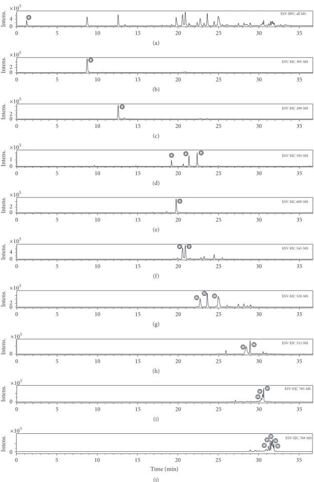
3.1. Chemical Composition of ESV. The metabolites in ESV
were identified by interpreting the UV absorption and mass spectra and comparing them with data in the literature. When available, compounds were compared with authentic standards for confirmation.
The identification of compound 2, withm/z305.0660
[M-H]−, was based on the fragmentation pattern of
epigallo-catechin proposed by Dou et al. [22], namely,m/z261
[M-CO2]−,m/z219 [M-C4H6O2]−,m/z179 [M-C6H6O3]−,m/z
167 [M-C7H6O3]−, andm/z165 [M-C7H8O3]−, and also on
the detection of UV absorption at 270 nm.
Compounds 4, 8, and 9 showed UV absorption patterns that were characteristic of flavonols (270 and 340 nm). Their fragmentation patterns were consistent with those of hetero-side derivatives of kaempferol, whose main fragment consists
ofm/z285 [C15H9O6]−. Several compounds of this class have
been described inSennagenus [23, 24].
Dimeric (compounds 6, 7, and 10–14) and trimeric (com-pounds 15–22) proanthocyanidins, comprising cassiaflavan, afzelechin, epicatechin, epigallocatechin, and naringenin subunits, were also observed. These derivatives, although rare
in nature, are often reported inSennagenus; some authors
consider them to be chemical markers for the genus [25–27]. The UV absorption maxima at 280 nm and the increase in reverse-phase retention time concomitant with decreased hydroxyl group content or increased degree of polymeriza-tion are in agreement with the study by Callemien and Collin [28]. Fragmentation patterns obtained by retro-Diels-Alder (RDA) fission, heterocyclic ring fission (HRF), and quinone methide (QM) can be used to characterize the subunits that make up proanthocyanidins [29]. A complete discussion on the elucidation of this class of compounds may be found in the literature [29, 30]. Thus, based on retention times, fragmentation profiles, and comparisons with previously published data, 22 compounds in ESV were characterized (Figure 1 and Table 1).
3.2. DPPH Free Radical Scavenging Activity. ESV was able to
scavenge the DPPH free radical, with a 2.5-fold higher IC50
and maximum activity values relative to ascorbic acid; how-ever, these values were lower than those obtained with BHT (Table 2).
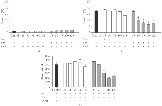
3.3. Hemolytic Activity and Inhibition of Oxidative Hemolysis. Over the range of tested concentrations, ESV showed no hemolytic activity in human erythrocytes, as no hemolysis was observed after up to 5 h of incubation (Figure 2(a)).
The control antioxidant, ascorbic acid, was able to protect erythrocytes from hemolysis for up to 4 h of incubation when they were exposed to the oxidant AAPH (data not shown). ESV was able to protect erythrocytes for 5 h over the tested
50–125𝜇g/mL concentration range (Figure 2(b)),
demon-strating its powerful antihemolytic activity.
3.4. MDA Measurements. The degree of protection conferred
by ESV against AAPH-induced lipid peroxidation in human erythrocytes was evaluated by measuring MDA levels. At
ESV concentrations of 100 and 125𝜇g/mL, the MDA levels
were decreased throughout the course of the assay (data not shown) and after 5 h of incubation (Figure 2(c)).
3.5. Cytotoxic Activity and Cell Death Profile. ESV promoted
cell death in both tested cell lines. IC50 values indicated
that ESV was more effective in Jurkat cells than in the
ery-throleukemic cell line K562 (IC50= 27.6𝜇g/mL and 67.5𝜇g/
mL, resp.) (Figures 3(a) and 3(b)). ESV treatment promoted double staining in both cell lines. This type of death was
evident in 71.9 ± 5.7% of Jurkat cells and 30.1 ± 2.8% of
K562 cells after treatment with 40 and 80𝜇g/mL of extract,
respectively (Figures 4(a) and 4(b)).
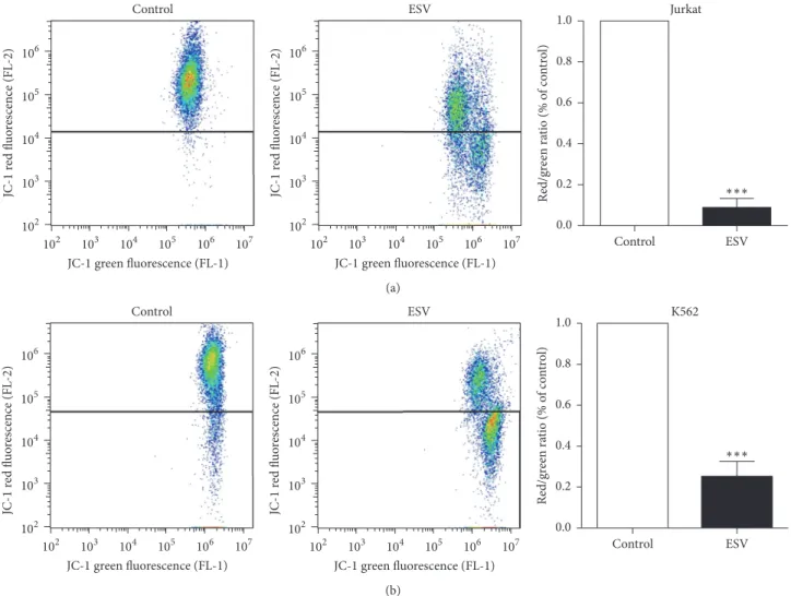
3.6. Mitochondrial Membrane Potential. The mitochondrial
membrane potentials of Jurkat and K562 leukemic cells decreased after 24 h of incubation with ESV, as evidenced by a decrease in red fluorescence and an increase in green fluorescence compared to untreated cells. The mitochondrial
membrane potential was reduced by91.0±4.3% in Jurkat cells
and by74.7 ± 7.3% in K562 cells after treatment with 27.6 and
67.5𝜇g/mL of extract, respectively (Figures 5(a) and 5(b)).
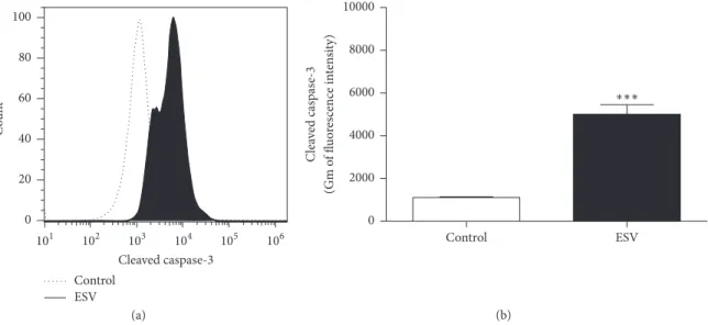
3.7. Caspase-3 Activity. The Jurkat cell line, which was more
sensitive to ESV activity, was used to investigate ESV-pro-moted cell death mechanisms. The fluorescence histogram (Figure 6(a)) showed a rightward shift (greater fluorescence values), indicating the activation of the apoptosis-inducing enzyme caspase-3. Cleaved caspase-3 levels increased 4.5-fold in ESV-treated cells relative to untreated cells (Figure 6(b)).
3.8. Pan-Caspase Inhibition and Intracellular Calcium
Chela-tion. Jurkat cells were preincubated with the pan-caspase
inhibitor Z-VAD-FMK to assess whether ESV cytotoxicity was mainly mediated by caspase activation. Z-VAD-FMK pretreatment did not decrease ESV-induced cell death
(Fig-ure 7). However, the intracellular Ca2+chelator BAPTA-AM
O
xida
tiv
e
M
edicine
and
C
ell
u
la
r
L
o
n
ge
vi
ty
5
Table 1: Compounds identified in ESV by UFLC-DAD-ESI-QTOF-micrOTOF QII.
ID Time (min) UV [M-H]
−
(𝑚/𝑧) Molecular formula Error (ppm) MS/MS Compound
1 1.1 — 341.1086 C12H20O11 0.6 341: 179 Sugar derivative
2 8.6 270 305.0660 C15H14O7 2.3 305: 261, 219, 179, 167, 165 Epigallocatechin
3 12.5 280 289.0714 C15H14O6 1.3 289: 245, 205, 203 Epicatechin
4 19.1 270/346 593.1524 C27H30O15 2.1 593: 447, 285 Kaempferol-O-hexoside-deoxyhexoside
5 19.7 270/346 609.1450 C27H30O16 1.9 609: 463, 301 Rutin
6 20.5 280 545.1440 C30H26O10 2.5 545: 305, 239, 219, 167, 165 Cassiaflavan-epigallocatechin
7 20.8 280 545.1440 C30H26O10 2.5 545: 305, 239, 219, 167, 165 Cassiaflavan-epigallocatechin
8 21.2 268/338 593.1522 C27H30O15 1.6 593: 447, 285 Kaempferol-O-hexoside-deoxyhexoside
9 22.3 270/342 593.1521 C27H30O15 1.5 593: 447, 285 Kaempferol-O-hexoside-deoxyhexoside
10 22.6 280 529.1489 C30H26O9 2.9 529: 289, 245, 239, 203 Cassiaflavan-epicatechin
11 23.5 280 529.1484 C30H26O9 3.7 529: 289, 245, 239, 203 Cassiaflavan-epicatechin
12 24.9 280 529.1489 C30H26O9 2.9 529: 267, 257, 239, 151 Naringenin-afzelechin
13 28.3 280 513.1551 C30H26O8 0.8 513: 267, 255, 239 Cassiaflavan-afzelechin
14 28.8 280 513.1541 C30H26O8 2.8 513: 267, 255, 239 Cassiaflavan-afzelechin
15 30.2 280 785.2266 C45H38O13 3.4 785: 435, 305, 239 Cassiaflavan-cassiaflavan-epigallocatechin
16 30.4 280 785.2285 C45H38O13 1.7 785: 435, 305, 239 Cassiaflavan-cassiaflavan-epigallocatechin
17 30.5 280 785.2255 C45H38O13 2.0 785: 435, 305, 239 Cassiaflavan-cassiaflavan-epigallocatechin
18 31.1 280 769.2310 C45H38O12 2.6 769: 529, 419, 289 Cassiaflavan-cassiaflavan-epicatechin
19 31.3 280 769.2303 C45H38O12 1.6 769: 529, 419, 289 Cassiaflavan-cassiaflavan-epicatechin
20 31.5 280 769.2295 C45H38O12 0.6 769: 377, 267, 239 Cassiaflavan-naringenin-afzelechin
21 31.7 280 769.2310 C45H38O12 2.6 769: 377, 267, 239 Cassiaflavan-naringenin-afzelechin
ESV-BPC-all MS 1
5 10 15 20 25 30 35
0
×105
0 4
Int
en
s.
(a)
2 ESV-EIC 305-MS
5 10 15 20 25 30 35
0
×105
0 2
Int
en
s.
(b)
3 ESV-EIC 289-MS
5 10 15 20 25 30 35
0
×105
0 2
Int
en
s.
(c)
4 8 9 ESV-EIC 593-MS
5 10 15 20 25 30 35
0
×105
0 1
Int
en
s.
(d)
5 ESV-EIC 609-MS
5 10 15 20 25 30 35
0
×105
0 2
Int
en
s.
(e)
6 7 ESV-EIC 545-MS
5 10 15 20 25 30 35
0
×105
0 4
Int
en
s.
(f)
10 12
11
ESV-EIC 529-MS
5 10 15 20 25 30 35
0
×105
0 2
Int
en
s.
(g)
13 14 ESV-EIC 513-MS
5 10 15 20 25 30 35
0
×105
0
Int
en
s.
(h)
15
16 17 ESV-EIC 785-MS
5 10 15 20 25 30 35
0
×105
0
Int
en
s.
(i)
18 19
22 21
20 ESV-EIC 769-MS
5 10 15 20 25 30 35
0
Time (min)
×105
0
Int
en
s.
(j)
Oxidative Medicine and Cellular Longevity 7
H
emo
lysis (%)
0 10 20 30 40 50
75 100
75 100 125 25 50
25 50 125
Control
+ + + + + − − − − − −
AA
− − − − − + + + + + −
ESV
− − − − − − − −
− − −
AAPH
(a)
H
emo
lysis (%) ∗
∗ ∗ ∗
0 10 20 30 40 50
25 50 75 100 125 25 50 75 100 125
Control
+ + + + +
− − − − − −
AA
− − − − − + + + + + −
ESV
+ + + + + + + + + + +
AAPH
(b)
∗ ∗
MD
A (nM/mL)
0 500 1000 1500 2000 2500 3000 3500
25 50 75 100 125 25 50 75 100 125
Control
+ + + + + − − − − − −
AA
− − − − − + + + + + −
ESV
+ + + + + + + + + + +
AAPH
(c)
Figure 2: Hemolysis and MDA content in human erythrocytes incubated for 5 hours with ascorbic acid (AA) and ESV (50–125𝜇g/mL). (a) Hemolytic activity of ESV in the absence of AAPH. (b) Antihemolytic activity after addition of AAPH. (c) Malondialdehyde (MDA) concentration (nM/mL) after addition of the oxidizing agent.∗𝑃 < 0.05compared to the AAPH-only control (erythrocytes incubated with oxidant only).
Jurkat
∗
∗
∗
∗ ∗
C
ell via
b
ili
ty (%)
0 20 40 60 80 100
20 40 60 80 100
0
Concentration (𝜇g/mL)
(a)
C
ell via
b
ili
ty (%)
K562
∗
∗
∗ ∗
20 40 60 80 100
0
Concentration (𝜇g/mL)
0 20 40 60 80 100
(b)
Figure 3: Viability of leukemic Jurkat (a) and K562 (b) cells after treatment with different concentrations of ESV.∗𝑃 < 0.05compared to the untreated control group.
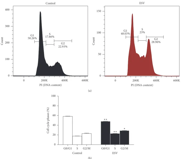
3.9. Cell Cycle Phases. Histograms were used to show the
distributions of cell cycle phases in control and ESV-treated Jurkat cells after 24 h of incubation (Figure 8(a)). The results
show a decreased portion of cells in G0/G1 phase (17.4±0.6%)
and increased portions of cells in S and G2/M phases (30.7 ±
1.8% and 26.5 ± 1.9%, resp.) (Figure 8(b)). These results
Jurkat
Control
ESV (40𝜇g/mL) ESV (20𝜇g/mL) 0 20 40 60 80 100 C ell p o p u la tio n (%) ∗ ∗ ∗
Anx+/PI+ Anx−/PI− Anx+/PI− Anx−/PI+
(a)
K562
Control
ESV (80𝜇g/mL) ESV (40𝜇g/mL) 0 20 40 60 80 100 C ell p o p u la tio n (%) ∗ ∗ ∗
Anx+/PI+ Anx−/PI− Anx+/PI− Anx−/PI+
(b)
Figure 4: Cell death profiles of ESV-treated Jurkat (a) and K562 (b) cells. Anx−/PI−, viable cells; Anx+/PI−, apoptotic cells; Anx−/PI+, necrotic cells; and Anx+/PI+, late apoptotic cells.∗𝑃 < 0.05compared to the respective control groups.
Jurkat
Red/gr
een ra
ti
o (% o
f co n tr o l) ESV Control 0.0 0.2 0.4 0.6 0.8 1.0 ∗∗∗ Control ESV 102 103 104 105 106 JC-1 re d fl u o res cence (FL -2 ) 107
104 105 106 103
102
JC-1green fluorescence (FL-1)
102 103 104 105 106 JC-1 re d fl u o res cence (FL -2 ) 107
104 105 106 103
102
JC-1green fluorescence (FL-1)
(a)
K562
Red/gr
een ra
ti
o (% o
f co n tr o l) ESV Control 0.0 0.2 0.4 0.6 0.8 1.0 102 103 104 105 106 JC-1 re d fl u o res cence (FL -2 ) 107
104 105 106 103
102
JC-1green fluorescence (FL-1)
102 103 104 105 106 JC-1 re d fl u o res cence (FL -2 ) 107
104 105 106 103
102
JC-1green fluorescence (FL-1)
Control ESV
∗∗∗
(b)
Figure 5: Mitochondrial membrane potential of leukemic Jurkat (a) and K562 (b) cells treated with different ESV concentrations.∗∗∗𝑃 <
Oxidative Medicine and Cellular Longevity 9
ESV Control
0 20 40 60 80 100
C
o
un
t
103 104 105 106 101 102
Cleaved caspase-3
(a)
Cle
av
ed caspas
e-3
(G
m o
f fl
u
o
res
cence in
te
n
si
ty)
ESV Control
0 2000 4000 6000 8000 10000
∗∗∗
(b)
Figure 6: Histogram (a) and representative graph (b) of caspase-3 activation in ESV-treated Jurkat cells.∗∗∗𝑃 < 0.0001compared to the untreated control group.
Table 2: IC50and maximal DPPH radical scavenging activity of standard antioxidants and of ESV.
Sample IC50(𝜇g/mL) Maximal inhibition
% 𝜇g/mL
Ascorbic acid 2.6 ± 0.8 90.9 ± 1.6 10
BHT 21.3 ± 1.2 92.4 ± 1.2 250
ESV 6.3 ± 1.3 92.4 ± 0.4 25
Values are means±SEM.
#
∗
0 20 40 60 80 100
C
ell via
b
ili
ty (%)
− − + + +
−
ESV
+ − − + −
−
ZVAD-FMK
− + − − +
−
BAPTA-AM
Figure 7: Involvement of caspases (via the pan-caspase inhibitor Z-VAD-FMK) and intracellular calcium (using the chelator BAPTA-AM) in ESV-induced Jurkat cell cytotoxicity.∗𝑃 < 0.05compared to the untreated control group.#
𝑃 < 0.05compared to the ESV group.
4. Discussion
Brazilian biodiversity is rich in active compounds with high potential for development of new therapeutic drugs, particu-larly antioxidants and anticancer agents. Several plant species found in Brazil have been characterized for their antioxidant and cytotoxic activities in several tumor cell lines [31–33].
In the present study, an ethanol extract of S. velutina
leaves exhibited antioxidant activity and showed cytotoxic effects against two leukemic cell lines, Jurkat and K562. The antioxidant activity of ESV was demonstrated in AAPH-incubated human erythrocytes; DPPH free radical scaveng-ing and inhibition of lipid peroxidation led to decreases in oxidative hemolysis and malondialdehyde production. This activity of ESV is likely related to the presence of flavone derivatives with antioxidant activity, such as epigallocatechin, epicatechin, rutin, kaempferol glycosides, and dimeric and trimeric proanthocyanidins, in the leaves [34–37].
Flavonoids can donate hydrogen atoms to radicals, pro-tecting against lipid peroxidation, and this ability is associated with the presence of a dihydroxylated B-ring [38]. Similar to other phenolic compounds, the antioxidant activity of flavonoids is ascribed to the presence of free hydroxyl groups in the molecule, and the level of antioxidant activity increases concomitantly with the number of hydroxyl groups [39].
Excess free radicals in the body can promote not only lipid peroxidation but also oxidative DNA damage, leading to the development of early stages of mutagenesis and carcinogen-esis [40, 41]. Thus, compounds with antioxidant properties have fundamental roles in preventing diseases such as cancer. The cytotoxic activity of ESV against leukemic lines was evaluated with an Annexin/PI-stain cell death assay. Further-more, activation of caspase-3 was observed but was not con-firmed to be a main mechanism of cell death when the pan-caspase inhibitor Z-VAD-FMK was used. Different extracts can promote several different death mechanisms simultane-ously because of their different compositions. This fact com-plicates the analysis and identification of specific cell death pathways.
G1 S
G2
G1 S
G2 58.26% 17.66%
22.93%
48.03% 23%
28.96%
Control ESV
0 100 200 300
C
o
un
t
400
0 50
C
o
un
t
100 150
0 200K 400K 600K PI (DNA content)
0 200K 400K 600K PI (DNA content)
(a)
∗ ∗∗ ∗∗
S G2/M
G0/G1 Control
S G2/M
G0/G1 ESV 0
20 40 60 80 100
C
ell c
yc
le p
has
es (%)
(b)
Figure 8: Histogram (a) and representative graph (b) of cell cycle distribution after 24 h of treatment with ESV.∗𝑃 < 0.05and∗∗𝑃 < 0.001 compared to the untreated control group.
arising from an increase in intracellular calcium levels, a characteristic of necrotic cell death [42, 43].
The present study demonstrated the involvement of calcium in cell death, as the reduced cell viability of extract-treated cells was reversed by incubation with the calcium chelator BAPTA-AM. High calcium levels promote the open-ing of mitochondrial permeability transition pores; these pores are nonselective and thus release the contents of the intermembrane space of the mitochondrion [44].
Another mechanism of the ESV-induced cytotoxicity against Jurkat cells consists of cell cycle arrest. ESV promoted a decrease in the number of cells in G0/G1 phase and an increase in the number of cells in S and G2 phases. Established anticancer drugs, such as cisplatin and doxorubicin, exert a similar profile change in the tumor cell cycle [14, 45]. Mueller et al. [46] observed that cisplatin is likely to be active in G2/M phases because cells in these phases are more sensitive to
DNA damage, as DNA repair mechanisms are less active than in G1/S phases. Flavonoids are phytochemicals that are known to induce cell cycle arrest by decreasing cellular levels of cyclin B and cyclin-dependent kinase 1, which are respon-sible for controlling cell cycle progression between S and M stages [47]. Furthermore, one of the major anticancer mech-anisms ascribed to flavonoids is their ability to induce cell cycle changes in tumor cell lines [48]. Thus, cyclin-dependent kinase inhibitors (CDKIs) have generated great interest for their ability to arrest the tumor cell cycle and prevent tumor cell proliferation; such cell cycle arrest may be the main mech-anism through which ESV operates [49]. However, plant extracts are natural products with complex chemical compo-sition, and biologically active compounds in extracts may act alone or synergistically through different pathways.
In conclusion, an ethanol extract of S. velutina leaves
Oxidative Medicine and Cellular Longevity 11
on leukemic cells by activating intracellular calcium and caspase-3, decreasing mitochondrial membrane potential, and arresting the cell cycle in S and G2 phases.
Abbreviations
AA: Ascorbic acid
AAPH: 2,2
-Azobis-(2-amidinopropane) dihydrochloride
Abs: Absorbance
Anx: Annexin V-FITC
BHT: Butylhydroxytoluene
Ca2+: Calcium
CaCl2: Calcium chloride
DAD: Diode Array Detector
DPPH: 2,2-Diphenyl-1-picrylhydrazyl
ESI-QTOF-micrOTOF QII: Electrospray ionization time-of-flight
ESV: Ethanol extract ofS. velutina
leaves
JC-1: 5,5,6,6-Tetrachloro-1,1,3,3
- tetraethylbenzimidazolylcar-bocyanine
iodide
MDA: Malondialdehyde
NaCl: Sodium chloride
PI: Propidium iodide
SEM: Standard error of the mean
TBA: Thiobarbituric acid
UFLC: Ultra Fast Liquid
Chromatography.
Competing Interests
The authors declare that they have no competing interests.
Acknowledgments
This work was supported by grants of the Fundac¸˜ao de Apoio ao Desenvolvimento do Ensino, Ciˆencia e Tecnologia do Mato Grosso do Sul (FUNDECT), Conselho Nacional de Desenvolvimento Cient´ıfico e Tecnol´ogico (CNPq), and Coordenac¸˜ao de Aperfeic¸oamento de Pessoal de N´ıvel Supe-rior (CAPES). Edson Lucas dos Santos, Carlos Alexandre Carollo, and Edgar J. Paredes-Gamero are recipients of fel-lowships from Brazilian National Research Council (CNPq), Brazil.
References
[1] S. Sultan, “Reviewing the protective role of antioxidants in oxidative stress caused by free radicals,”Asian Pacific Journal of Health Science, vol. 1, pp. 401–406, 2014.
[2] V. Lobo, A. Patil, A. Phatak, and N. Chandra, “Free radicals, antioxidants and functional foods: impact on human health,” Pharmacognosy Reviews, vol. 4, no. 8, pp. 118–126, 2010. [3] R. Siegel, D. Naishadham, and A. Jemal, “Cancer statistics, 2013,”
CA: A Cancer Journal for Clinicians, vol. 63, no. 1, pp. 11–30, 2013.
[4] American Cancer Society,Leukemia-Chronic Myeloid (Myeloge-nous) Overview, 2013.
[5] M. J. S. Asmaa, H. A. N. Al-Jamal, C. Y. Ang, J. M. Asan, A. Seeni, and M. F. Johan, “Apoptosis induction in MV4-11 and K562 human leukemic cells byPereskia sacharosa(Cactaceae) leaf crude extract,”Asian Pacific Journal of Cancer Prevention, vol. 15, no. 1, pp. 475–481, 2014.
[6] T. Srdic-Rajic, N. Tisma-Miletic, M. Cavic et al., “Sensitization of K562 leukemia cells to doxorubicin by theViscum album extract,”Phytotherapy Research, vol. 30, no. 3, pp. 485–495, 2016. [7] D. J. Newman and G. M. Cragg, “Natural products as sources of new drugs from 1981 to 2014,”Journal of Natural Products, vol. 79, no. 3, pp. 629–661, 2016.
[8] M. J. Balunas and A. D. Kinghorn, “Drug discovery from medic-inal plants,”Life Sciences, vol. 78, no. 5, pp. 431–441, 2005. [9] A. Nagle, W. Hur, and N. S. Gray, “Antimitotic agents of natural
origin,”Current Drug Targets, vol. 7, no. 3, pp. 305–326, 2006. [10] O. Prakash, A. Kumar, P. Kumar, and Ajeet, “Anticancer
poten-tial of plants and natural products: a review,”American Journal of Pharmacological Sciences, vol. 1, no. 6, pp. 104–115, 2013. [11] Y. W. Mak, L. O. Chuah, R. Ahmad, and R. Bhat, “Antioxidant
and antibacterial activities of hibiscus (Hibiscus rosa-sinensisL.) and Cassia (Senna bicapsularisL.) flower extracts,”Journal of King Saud University-Science, vol. 25, pp. 275–282, 2013. [12] A. C. Susunaga-Notario, S. P´erez-Guti´errez, M. ´A.
Zavala-S´anchez et al., “Bioassay-guided chemical study of the anti-inflammatory effect of Senna villosa (Miller) H.S. Irwin & Barneby (Leguminosae) in TPA-induced ear edema,”Molecules, vol. 19, no. 7, pp. 10261–10278, 2014.
[13] E. Thilagam, B. Parimaladevi, C. Kumarappan, and S. C. Mandal, “𝛼-Glucosidase and𝛼-amylase inhibitory activity of
Senna surattensis,”Journal of Acupuncture and Meridian Studies,
vol. 6, no. 1, pp. 24–30, 2013.
[14] R. M. Pereira, G. ´A. Ferreira-Silva, M. Pivatto et al., “Alkaloids derived from flowers ofSenna spectabilis, cassine and (–)-spectaline, have antiproliferative activity on HepG2 cells for inducing cell cycle arrest in G1/S transition through ERK inacti-vation and downregulation of cyclin D1 expression,”Toxicology in Vitro, vol. 31, pp. 86–92, 2016.
[15] C. Viegas Junior, A. de Rezende, D. H. S. Silva et al., “Aspectos qu´ımicos, biol´ogicos e etnofarmacol´ogicos do gˆeneroCassia,” Qu´ımica Nova, vol. 29, no. 6, pp. 1279–1286, 2006.
[16] L. Delumlle and K. Demeyer,Anthraquinones in Plants—Source, Safety and Applications in Gastrointestinal Health, Nottingham University Press, 2008.
[17] D. Gupta and R. K. Gupta, “Bioprotective properties of Dragon’s blood resin: in vitro evaluation of antioxidant activity and antimicrobial activity,”BMC Complementary and Alternative Medicine, vol. 11, article 13, pp. 1–9, 2011.
[18] J. F. Campos, U. P. Das Santos, P. D. S. Da Rocha et al., “Antimi-crobial, antioxidant, anti-inflammatory, and cytotoxic activities of propolis from the stingless beeTetragonisca fiebrigi(Jata´ı),” Evidence-Based Complementary and Alternative Medicine, vol. 2015, Article ID 296186, 11 pages, 2015.
[19] E. J. Paredes-Gamero, M. N. C. Martins, F. A. M. Cappabianco, J. S. Ide, and A. Miranda, “Characterization of dual effects induced by antimicrobial peptides: regulated cell death or membrane disruption,”Biochimica et Biophysica Acta—General Subjects, vol. 1820, no. 7, pp. 1062–1072, 2012.
myeloid leukemia cells,”Cell Death & Disease, vol. 4, article e658, 8 pages, 2013.
[21] A. Bechara, C. M. V. Barbosa, E. J. Paredes-Gamero et al., “Palladacycle (BPC) antitumour activity against resistant and metastatic cell lines: the relationship with cytosolic calcium mobilisation and cathepsin B activity,” European Journal of Medicinal Chemistry, vol. 79, pp. 24–33, 2014.
[22] J. Dou, V. S. Y. Lee, J. T. C. Tzen, and M.-R. Lee, “Identification and comparison of phenolic compounds in the preparation of oolong tea manufactured by semifermentation and drying pro-cesses,”Journal of Agricultural and Food Chemistry, vol. 55, no. 18, pp. 7462–7468, 2007.
[23] K. V. Rao, A. G. Damu, B. Jayaprakasam, and D. Gunasekar, “Flavonol glycosides fromCassia hirsuta,”Journal of Natural Products, vol. 62, no. 2, pp. 305–306, 1999.
[24] N. H. El-Sayed, A. M. A. Dooh, S. A. M. El-Khrisy, and T. J. Mabry, “Flavonoids ofCassia italica,”Phytochemistry, vol. 31, no. 6, p. 2187, 1992.
[25] S. Morimoto, G. Nonaka, R. Chen, and I. Nishioka, “Tannins and related compounds. LXI. Isolation and structures of novel bi- and triflavanoids from the leaves of Cassia fistula L.,” Chemical and Pharmaceutical Bulletin, vol. 36, no. 1, pp. 39–47, 1988.
[26] T. Hatano, A. Yamashita, T. Hashimoto et al., “Flavan dimers with lipase inhibitory activity fromCassia nomame,” Phyto-chemistry, vol. 46, no. 5, pp. 893–900, 1997.
[27] J. Coetzee, L. Mciteka, E. Malan, and D. Ferreira, “Structure and synthesis of butiniflavan-epicatechin and -epigallocatechin probutinidins,”Phytochemistry, vol. 52, no. 4, pp. 737–743, 1999. [28] D. Callemien and S. Collin, “Use of RP-HPLC-ESI (-)-MS/MS to differentiate various proanthocyanidin isomers in lager beer extracts,”The Journal of the American Society of Brewing Chemists, vol. 66, pp. 109–115, 2008.
[29] L. Gu, M. A. Kelm, J. F. Hammerstone et al., “Liquid chromato-graphic/electrospray ionization mass spectrometric studies of proanthocyanidins in foods,”Journal of Mass Spectrometry, vol. 38, no. 12, pp. 1272–1280, 2003.
[30] S. A. Lazarus, G. E. Adamson, J. F. Hammerstone, and H. H. Schmitz, “High-performance liquid chromatography/mass spectrometry analysis of proanthocyanidins in foods and bever-ages,”Journal of Agricultural and Food Chemistry, vol. 47, no. 9, pp. 3693–3701, 1999.
[31] J. C. Casagrande, L. F. B. Macorini, K. A. Antunes et al., “Antiox-idant and cytotoxic activity of hydroethanolic extract from
Jacaranda decurrensleaves,”PLoS ONE, vol. 9, no. 11, Article ID
e112748, pp. 1–8, 2014.
[32] J. M. Serpeloni, A. F. Leal Specian, D. L. Ribeiro et al., “Antimu-tagenicity and induction of antioxidant defense by flavonoid rich extract ofMyrcia bellaCambess. in normal and tumor gastric cells,”Journal of Ethnopharmacology, vol. 176, pp. 345– 355, 2015.
[33] A. S. N. Formagio, D. D. Ramos, M. C. Vieira et al., “Phenolic compounds of Hibiscus sabdariffaand influence of organic residues on its antioxidant and antitumoral properties,” Brazil-ian Journal of Biology, vol. 75, no. 1, pp. 69–76, 2015.
[34] J. Yang, J. Guo, and J. Yuan, “In vitroantioxidant properties of rutin,”LWT—Food Science and Technology, vol. 41, no. 6, pp. 1060–1066, 2008.
[35] L. S. Chua, “A review on plant-based rutin extraction methods and its pharmacological activities,”Journal of Ethnopharmacol-ogy, vol. 150, no. 3, pp. 805–817, 2013.
[36] E. D. Pauli, G. B. Malta, P. M. Sanchez, I. C. Moreira, and I. S. Scarminio, “Mixture design analysis of solvent extractor effects on epicatechin, epigallocatechin gallate, epigallocatechin and antioxidant activities of theCamellia sinensisL. leaves,” Analytical Chemistry Research, vol. 2, pp. 23–29, 2014.
[37] G. W. Plumb, S. De Pascual-Teresa, C. Santos-Buelga, V. Cheynier, and G. Williamson, “Antioxidant properties of cat-echins and proanthocyanidins: effect of polymerisation, galloy-lation and glycosygalloy-lation,”Free Radical Research, vol. 29, no. 4, pp. 351–358, 1998.
[38] M. Valko, C. J. Rhodes, J. Moncol, M. Izakovic, and M. Mazur, “Free radicals, metals and antioxidants in oxidative stress-induced cancer,”Chemico-Biological Interactions, vol. 160, no. 1, pp. 1–40, 2006.
[39] A. Kurek-G´orecka, A. Rzepecka-Stojko, M. G´orecki, J. Stojko, M. Sosada, and G. Swierczek-Zieba, “Structure and antioxidant activity of polyphenols derived from propolis,”Molecules, vol. 19, no. 1, pp. 78–101, 2014.
[40] B. Frei, “Reactive oxygen species and antioxidant vitamins: mechanisms of action,”The American Journal of Medicine, vol. 97, no. 3, pp. 5–13, 1994.
[41] M. Valko, D. Leibfritz, J. Moncol, M. T. D. Cronin, M. Mazur, and J. Telser, “Free radicals and antioxidants in normal physio-logical functions and human disease,”The International Journal of Biochemistry & Cell Biology, vol. 39, no. 1, pp. 44–84, 2007. [42] J. J. Lemasters, T. Qian, C. A. Bradham et al., “Mitochondrial
dysfunction in the pathogenesis of necrotic and apoptotic cell death,”Journal of Bioenergetics and Biomembranes, vol. 31, no. 4, pp. 305–319, 1999.
[43] N. Festjens, T. Vanden Berghe, and P. Vandenabeele, “Necrosis, a well-orchestrated form of cell demise: signalling cascades, important mediators and concomitant immune response,” Biochimica et Biophysica Acta (BBA)—Bioenergetics, vol. 1757, no. 9-10, pp. 1371–1387, 2006.
[44] K. W. Kinnally, P. M. Peixoto, S.-Y. Ryu, and L. M. Dejean, “Is mPTP the gatekeeper for necrosis, apoptosis, or both?” Biochimica et Biophysica Acta—Molecular Cell Research, vol. 1813, no. 4, pp. 616–622, 2011.
[45] W. Wang, K. Chen, Q. Liu et al., “Suppression of tumor growth
byPleurotus ferulaeethanol extract through induction of cell
apoptosis, and inhibition of cell proliferation and migration,” PLoS ONE, vol. 9, no. 7, Article ID e102673, pp. 1–14, 2014. [46] S. Mueller, M. Schittenhelm, F. Honecker et al., “Cell-cycle
progression and response of germ cell tumors to cisplatinin
vitro,”International Journal of Oncology, vol. 29, no. 2, pp. 471–
479, 2006.
[47] M. Carocho and I. C. F. R. Ferreira, “The role of phenolic compounds in the fight against cancer—a review,”Anti-Cancer Agents in Medicinal Chemistry, vol. 13, no. 8, pp. 1236–1258, 2013. [48] R. P. Singh and R. Agarwal, “Natural flavonoids targeting deregulated cell cycle progression in cancer cells,”Current Drug Targets, vol. 7, no. 3, pp. 345–354, 2006.
Submit your manuscripts at
http://www.hindawi.com
Stem Cells
International
Hindawi Publishing Corporationhttp://www.hindawi.com Volume 2014
Hindawi Publishing Corporation
http://www.hindawi.com Volume 2014
MEDIATORS
INFLAMMATIONof
Hindawi Publishing Corporation
http://www.hindawi.com Volume 2014
Behavioural
Neurology
Endocrinology
International Journal of Hindawi Publishing Corporationhttp://www.hindawi.com Volume 2014
Hindawi Publishing Corporation
http://www.hindawi.com Volume 2014
Disease Markers
Hindawi Publishing Corporation
http://www.hindawi.com Volume 2014 BioMed
Research International
Oncology
Journal of Hindawi Publishing Corporationhttp://www.hindawi.com Volume 2014
Hindawi Publishing Corporation
http://www.hindawi.com Volume 2014
Oxidative Medicine and Cellular Longevity Hindawi Publishing Corporation
http://www.hindawi.com Volume 2014
PPAR Research
The Scientific
World Journal
Hindawi Publishing Corporation
http://www.hindawi.com Volume 2014
Immunology Research Hindawi Publishing Corporation
http://www.hindawi.com Volume 2014
Journal of
Obesity
Journal ofHindawi Publishing Corporation
http://www.hindawi.com Volume 2014
Hindawi Publishing Corporation
http://www.hindawi.com Volume 2014
Computational and Mathematical Methods in Medicine
Ophthalmology
Journal ofHindawi Publishing Corporation
http://www.hindawi.com Volume 2014
Diabetes Research
Journal ofHindawi Publishing Corporation
http://www.hindawi.com Volume 2014
Hindawi Publishing Corporation
http://www.hindawi.com Volume 2014
Research and Treatment
AIDS
Hindawi Publishing Corporation
http://www.hindawi.com Volume 2014
Gastroenterology Research and Practice
Hindawi Publishing Corporation
http://www.hindawi.com Volume 2014
Parkinson’s
Disease
Evidence-Based Complementary and Alternative Medicine
Volume 2014 Hindawi Publishing Corporation