Histopathological and ultrastructural aspects of mice lungs
experimentally infected with dengue virus serotype 2
Débora Ferreira Barreto/
+, Christina Maeda Takiya
*, Hermann Gonçalves Schatzmayr,
Rita Maria Ribeiro Nogueira, José da Costa Farias-Filho, Ortrud Monika Barth
Departamento de Virologia, Instituto Oswaldo Cruz -Fiocruz, Av. Brasil 4365, 21040-000 Rio de Janeiro, Brasil *Departamento de Histologia e Embriologia, Instituto de Ciências Biomédicas, Universidade Federal do Rio de Janeiro, Rio de Janeiro, RJ, Brasil
Histological and ultrastructural alterations in lung tissue of BALB/c mice infected with dengue virus sero-type 2 (non-neuroadapted), by intraperitoneal and intravenous routes were analyzed. Lung tissues were pro-cessed following the standard techniques for photonic and electron transmission microscopies. Histopatho-logical and ultrastructural studies showed interstitial pneumonia, characterized by the presence of mono-nuclear cells. In the mouse model, the dengue virus serotype 2 seems to led to a transient inflammatory process without extensive damage to the interalveolar septa, but caused focal alterations of the blood-exchange bar-rier. Endothelial cells of blood capillaries exhibited phyllopodia suggesting activation by presence of dengue virus. Morphometrical analysis of mast cells showed an expressive increase of the number of these cells in peribronchiolar spaces and adjacent areas to the interalveolar septa. Alveolar macrophages showed particles dengue virus-like inside rough endoplasmic reticulum and Golgi complex, suggesting viral replication. The tissue alterations observed in our experimental model were similar to the observed in human cases of dengue fever and dengue hemorrhagic fever. Our results show that BALB/c mice are permissive host for dengue virus serotype 2 replication and therefore provides an useful model to study of morphological aspects of dengue virus infection.
Key words: dengue-2 virus - BALB/c mice - C6/36 cell line - macrophage - mast cell - ultrastructure
Dengue fever (DF) is an acute infectious disease caused by dengue virus (DENV) (Halstead 1988, Henchal & Putnak 1990) that belong to the Flavivirus genus of the Flaviviridae family of single-stranded, positive-po-larity, enveloped RNAviruses (Chambers et al. 1990). Dengue disease (DEN) has a spectrum of clinical signs and symptoms, ranging from asymptomatic infection to severe and lethal manifestations. The four DENV sero-types (DENV-1, -2, -3, -4) may cause dengue hemor-rhagic fever/dengue shock syndrome (DHF/DSS) in hu-mans, with an estimated 100 million new cases of DF and over 250,000 cases of DHF/DSS per year world-wide. The major difficulty in studying DENV infection in humans and for developing a vaccine, is the absence of a suitable animal model which develops a disease with similar aspects of the DHF and DSS (Bhamarapravati 1993). Several studies suggest that mice are a permis-sive host for DENV (Meiklejhon et al. 1952, Lin et al.1998, Johnson & Roehrig 1999, An et al. 1999), but in the majority of these models the animals are
im-Financial support: CNPq, Capes, PDTSP +Corresponding author: [email protected] Received 9 October 2006
Accepted 31 January 2007
munocompromised and inoculated by invasive routes with neuroadapted DENV in mice (Table). In the present study we characterized the injuries caused by DENV-2 infection in lung tissue of BALB/c mice using photonic and electron transmission microscopies. Differing from the majority of the mice models, the DENV-2 (non-neuroadapted) were obtained from a patient serum, propagated once in the C6/36 cell line and inoculated by intravenous (i.v.) and intraperitoneal (i.p.) routes.
MATERIALS AND METHODS
Virus - The virus strain used in our experiments was isolated from a patient serum during an epidemic of DENV-2 in the state of Rio de Janeiro in 1995 and propa-gated in the Aedes albopictus mosquito cell line (C6/ 36). The serum was tested by the indirect immunofluo-rescence technique using a type specific DENV-2 mono-clonal (3H5, DENV-2) antibody. The virus had not un-dergone passage in mouse brain. The titer of the virus (106.3 TCID50/0.1 ml) was calculated by the method of Reed and Muench (1938).
Cells - The C6/36 monolayers were grown in L-15 medium supplemented with 1% non-essencial ami-noacids, 10% tryptose phosphate broth, and 10% fetal bovine serum. The tubes were kept at 28oC.
as controls and sacrificed at the same time. The experi-ments were previously approved by the Animal Experi-mentation Ethical Committee of the Instituto Oswaldo Cruz-Fiocruz.
Processing of tissues for photonic microscopy analysis - The animals were peritoneally anaesthetized and lung tissue fragments were collected from infected and non-infected mice. Samples were fixed in Millonig’s fixative, dehydrated in ethanol, and paraffin-embedded. Sections (5 µm thick) were stained with haematoxilyn and eosin, giemsa, and toluidin Perez blue.
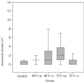
Morphometrical analysis - Numerical density of mast cells was obtained from histological sections of lung fragments from mice 48 h p.i. stained with toluidin Perez blue. Thirty four images of non-infected animals and 34 images of pulmonar parenchyma of infected ani-mals were obtained and mast cells on each image were quantified. Images were obtained with a 20x objective of a Nikon Eclipse 104 light microscope and digitallized using a Nikon Coolpix 990 camera. Data were submit-ted to statistical analysis using t-test or Mann Whitney rank sum test considering p < 0.05.
Processing of tissues for transmission electron mi-croscope analysis -Lung fragments were collected af-ter 48 and 72 h, 7, 14, and 49 days p.i. The infected ani-mals were peritoneally anaesthetized and fixed by per-fusion with 4% paraformaldeyde in sodium phosphate buffer (0.2 M, pH 7.2) by 30 min. In sequence the lungs were carefully collected, the fragments post-fixed by im-mersion in 2% glutaraldehyde in sodium cacodylate buffer (0.2 M, pH 7.2), followed by 1% buffered os-mium tetroxide, dehydrated in crescent concentrations of acetone, embedded in epoxy resin, and polymerized at 60oC during three days. Semithin section of 0.5 µm were obtained using a diamond knife (Diatome) adapted to a Reichert-Jung Ultracut E microtome. The sections were stained with methylene blue and azure II solution (Humprey & Pittman 1974) and observed at a Zeiss Axiophot light microscopy. Ultrathin sections of 50-70 nm thickness were picked up onto copper grids and stained with uranyl acetate and lead citrate (Reynolds 1963) and observed at a Zeiss EM-900 TEM.
Isolation of DENV-2 in the C6/36 cell line inocu-lated with the supernatant of lung tissue macerates from infected BALB/c mice - Lung tissue fragments were washed in phosphate saline buffer (PBS, pH 7.2) and macerated in Leibovitz medium (L-15), supplemented with antibiotics. The suspension was incubated for 1 h for antibiotic action, centrifuged at 1400 g for 5 min in a refrigerate centrifuge and the supernatant was collected. Cell monolayers were inoculated with 100 µl of the cell supernatant and incubated for 1 h at 28oC for virus ad-sorption. Subsequently monolayers were grown in L-15 medium supplemented with 1% non-essencial ami-noacids, 10% tryptose phosphate broth, and 10% fetal bovine serum. The tubes were kept at 28oC and observed daily for viral cytopathic effects for 15 days. C6/36 nor-mal cell monolayers were used as negative control, while the positive control consisted of cell monolayers
inocu-lated with DENV-2. After the periods of observation, the monolayers were divided into two groups: the first was tested using the indirect immunofluorescence technique with a type-specific monoclonal antibody for dengue serotype 2 (3H5, DENV-2) and the second was fixed in 1% buffered glutaraldehyde, dehydrated, and embedded in epoxy resin as described above.
Broncho-alveolar aspirate - Infected mice were peritoneally anaesthetized and 1 ml of PBS (pH 7.2) was injected by trachea and immediately collected carefully from the mice lungs. The cells resultant of these collect were fixed in 1% glutaraldehyde in sodiumcacodylate buffer (0.2 M, pH 7.2), centrifuged at 1500 rpm for three minutes, and processed for ETM observations.
RESULTS
Clinical signs - Both DENV-2 infected mice groups did not show deaths.
Morphology - Histological (paraffin and semithin sections) and ultrastructural (ultrathin sections) obser-vations of the infected lung tissues demonstrated the presence of an interstitial pneumonia, which was simi-lar for both routes of inoculation.
Intravenous route - At 48 h p.i. swelling of interal-veolar septa, vascular congestion, presence of alinteral-veolar macrophages and erythrocytes inside alveolar spaces, and peribronchiolar infiltrate were detected; recruitment of platelets, mononuclear, and polymorphonuclear cells inside blood vessels (Fig. 5) could be also observed. Presence of phyllopodia in endothelial cells (Fig. 6), but without signs of injury or necrosis.
In 72 h p.i., swollen interalveolar septa (more severe than at 48 h p.i.), vascular congestion, presence of al-veolar macrophages and erythrocytes inside alal-veolar spaces, small focus of hemorrhagy, oedema and infil-trate in the peribronchiolar space and hyperplasia of the bronchiolar epithelium were observed. The presence of platelets, mononuclear, and polymorphonuclear cells in-side blood vessels. Endothelial cells showed a similar profile as observed at 48 h p.i.
At 7 days p.i., swollen interalveolar septa (more mild than 72 h p.i.), vascular congestion and presence of al-veolar macrophages and erythrocytes inside alal-veolar spaces were observed as well as the presence of plate-lets, mononuclear, and polymorphonuclear cells inside blood vessels.
Endothelial cells showed a similar profile as ob-served at the initial stage of infection (Fig. 7).
In 49 days p.i., few foci of peribronchiolar hemor-rhagy and swollen interalveolar septa (Fig. 1), vascular congestion, presence of alveolar macrophages and eryth-rocytes inside the alveolar spaces and also platelets, mononuclear, and polymorphonuclear cells persisting inside and outside the blood vessels.
Endothelial cells showed a similar profile as ob-served at the initial stage of infection.
TABLE
Experimental mouse models of dengue virus infection
Animals Viruses RI Histopathology Clinical Viruses Detection References manifestations isolation of antigen
Mice DENV-2 IP Increase of - - - Chaturvedi et al.
vascular 1991
permeability
Suckling DENV-1 IC - Encephalitis Brain Neuron Cole &
weanling neuro- Wisseman
and adult adapted 1969
mice
Nude mice DENV-1 IC - Encephalitis Brain Neuron Hotta et al.
IP muscle muscle 1981a
lymph nodes kupffer cells Hotta et al. several organs 1981b
Adult mice DENV-2 IP - - - Macrophages Boonpucknavig
neuro- IV of some et al. 1981
adapted tissues
"scid-hu" DENV-1 IP - - Blood, some Some tissues Marchette et al.
mice a tissues (one animals) 1973
Several DENV-2 IC - Obit, aneroxia, In C6/36 cells - Hotta et al.1996 species of neuro- petechial inoculated of
rodents adapted gastrointestinal macerate of haemorrhage, tissues of paralysis, infected atrophy of animals spleen
AG129 b DENV-2 IP - Obit, Serum - Johnson
mice neuro- neurological spleen & Roehrig 1999
adapted abnormality
SCID c DENV-2 IP - Anorexia, Liver serum Hep G2 cells An et al. 1999
mice asthenia, brain of the liver,
paralysis neuron
Several
species of DENV-2 IV - Paraplegia, Blood - Huang et al.
rodents thrombocytopenia 2003
BALB/c DENV-2 IP Necrosy Shock Blood - Atrasheuskaya
mice neuro- haemorrhage of paralysis et al. 2003
adapted the spleen thrombocytopenia
ICR mice DENV-2/-4 IC - Anaemia Glial cells - Lucia & Kangwanpong 1994
Newborn DENV-2 IC Neuron apoptose Fatal Neurons - Deprès et al.
SNC mice neuro- encephalitis 1998
adapted
Immunodefi- DENV-2 IC - - Brain Brain Na et al. 2003
cient mice spinal cord spinal cord
BALB/c DENV-2 IP Focal alterations - C6/36 cells C6/36 cells Barreto et al.
mice d in lung, liver, inoculate inoculate 2004
cerebellum, and of macerate of macerate kidney of tissues of tissues
BALB/c DENV-2 IP Focal alterations - Serum Liver Paes et al.
mice d in liver 2005
RI: route of infection; IC: intracranial; IP: intraperitoneal; IV: intravenous; a: immunocompromised mice; b: mice without receptores for IFN α, β, and
Fig. 1:lung of BALB/c mice 49 days p.i. by i.v. route. Swollen alveolar septa and presence of a hemorrhage focus (h) in peribronchiolar space. Semi-thin section stained with methylene blue and azure II solution. Bar = 0.025 µm. Fig. 2:lung of BALB/c mice 72 h p.i. by i.p. route. Swollen alveolar septa by presence of inflammatory cells and alveolar space diminished can be observed. Histological section stained with haematoxilyn and eosin. Bar = 0.02
µm. Fig. 3:lung of BALB/c mice 72 h p.i. by i.p. route. Presence of inflammatory cells in alveolar septa and a hemorrhage focus (h) were observed. Histological section stained with giemsa. Bar = 0.001 µm. Fig. 4:lung of BALB/c mice 72 h p.i. by i.p. route. Presence of mast cells (Mt) in peribronchiolar space. Histological section stained with toluidin Perez blue. Bar = 0.09 µm. A: alveolar space; S: alveolar septa, Ep: bronchiolar epithelium; B: bronchiole.
also depicted in a peribronchiolar localization (bronchi-olitis), admixed with platelets. Recruitment of this type was still evident later, at 14 and 49 days of infection, but decreases progressively, with platelets persisting inside blood vessels (Fig. 8). Polymorphonuclear cells were recruited at a later stage, up to 14 days p.i. Up to 72 h p.i. swollen of interalveolar septa (Fig. 2), hemorrhage fo-cus (Fig. 3), and hyperplasia of the bronchiolar epithe-lium were observed. Foci of subepithelial oedema were depicted at 7 days p.i., but rare pneumocytes showed evidence of injury at this stage. They exhibited a dense cytoplasm with swollen mitochondria, whereas in the alveolar septa interstitial fibroblasts containing lipid droplets became evident.
The endothelial cells maintained their structure with-out signs of reversible injury or necrosis.
Morphometrical analysis - The infected lung tissue showed significant increase in the density of mast cells in 48 (p < 0.05) and 72 h p.i. (p < 0.05) by both, the i.p. and the i.v. routes of infection, when compared to con-trols (Fig. 17). These mast cells were observed in the peribronchiolar space (Fig. 4) and adjacent to the inter-alveolar septa. The presence of an interstitial oedema was also detected in these area.
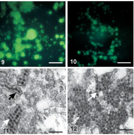
Isolation of DENV-2 in the C6/36 cell line inocu-lated with the supernatant of lung tissue macerates from infected BALB/c mice - In cell cultures of the
posi-tive controls (Figs 9, 11) and in monolayer cultures in-oculated with the supernatant of the lung tissues (Figs 10, 12), the DENV-2 antigen and virus particles inside cysterns of the rough endoplasmic reticulum were ob-served.
Monolayer cells of the negative control showed no morphological alterations, exhibiting neither DENV-2 antigen nor virus particles.
Broncho-alveolar aspirate -All alveolar macroph-ages, collected 48 and 72 h p.i. showed pseudopodia (Figs 13, 14). These macrophages were supposely acti-vated by presence of DENV-2.
In 48 h p.i. (i.p. route) DENV-like particles, were observed inside citoplasmatic vesicles of alveolar mac-rophages.
In alveolar macrophages of animals 72 h p.i., particles DENV-like, inside the rough endoplasmic reticulum (i.p. route) (Fig. 15) and Golgi complex (i.v. route) (Fig. 16) were observed.
DISCUSSION
In this work BALB/c mice infected with DENV-2 (non-neuroadapted) by the intraperitoneal and the intra-venous routes, did not show neither clinical signs nor mortality. In experimental studies with defficient mice strains, severe clinical signs and mortality were observed (Raut et al. 1996). In these studies the defficient
Figs 13-16:alveolar macrophages collected of the BALB/c mice by broncho-alveolar aspirate. Fig. 13: macrophages collected 72 h p.i. by i.p. route. Note presence of pseudopodia (arrow) and particles DENV-like (V) inside cytoplasmatic vesicles. Bar = 0.7 µm. Fig. 14:macrophages collected 72 h p.i. by i.v. route. Note presence of pseudopodia (arrow). Bar = 0.73 µm. Fig. 15:macrophages collected 72 h p.i. by i.p. route. Note presence of particles DENV-like (V) inside rough endoplasmic reticulum. Bar = 2.4 µm. Fig. 16:macrophages collected 72 h p.i. by i.p. route. Note presence of particles DENV-like (V) inside Golgi complex. Ultra-thin section. Bar = 2 µm. M: mitochondria, N: nucleus, rER: rough endoplasmic reticulum.
mals were infected by the intracerebral route with neuroadapted DENV strain. The severity of clinical signs in this work must be correlated with the immunodefi-cience of these animals and the adaptation of DENV in mice that increase the virulence of this dengue virus strain.
In our studies the histological analysis of lung tis-sues revealed interstitial pneumonia associated with vas-cular congestion, rare focal zones of parenquimal haemorrhage, increase of alveolar macrophages number, recruiting of platelets, mononuclear, and polymor-phonuclear cells. Histological alterations were still ob-served in 49 days p.i, being less severe in time. The course of the infection was similar in both routes. The tissular alterations were similar the observed in others animal models (Bhamarapravati 1989, Hotta et al. 1981a, Atra-sheukaya et al. 2003). Similar injuries have been observed in necropsies of human pulmonar tissues of DEN fatal cases (Burke et al. 1988, Miagostovich et al.1997).
In the ultrastructural analysis of alveolar macroph-ages of infected mice by the intravenous and intraperi-toneal routes, the presence of DENV-like particles in-side vesicles the rough endoplasmic reticulum and Golgi complex were observed suggesting viral replication. All macrophages presented pseudopodia, probably due to activation by the presence of DENV. DENV antigen and virus particles were detected in monolayers of C6/36 cells inoculated with pulmonar tissue macerate
super-natants of infected animals. In immunocompromised mice, the replication of DENV was verified in pulmonar tissue by titulation of cell cultures inoculated with lung tissue macerate (Hotta et al. 1981a). Studies reported that monocytes are more permissive cells for infection by DENV in vitro (Hastead & O’ Rourke 1977). In stud-ies with monocytes gotten of patients with DEN, the DENV were often detected (Scott et al. 1980). Based on these data, several authors suggested that monocytes and macrophages resident in tissues can be target cells predominantly in the infection in vivo by DENV (Rothman & Ennis 1999). Histochemical studies of human fatal cases of DEN demonstrated that cells of the phagocytic system can be infected by DENV (Miagostovich et al.
1997).
sug-Fig. 17: quantitative analysis of mast cells of BALB/c mice lungs infected with DENV-2. Note increase of the numerical density of mast cells in lungs of infected animals 48 and 72 h post-infection. h: hours, i.p.: intra-peritoneal route; i.v: intravenous route.
gests that the histamine can be the mediator of the vas-cular permeability in DHF and in DSS. In studies with cases of DHF and DSS, increase of levels of histamine in urine was observed (Tuchinda et al. 1977). Studies with human mast cell culture infected with dengue virus sug-gest a role for these cells in the initiation of chemokine-dependent host responses to dengue virus infection (King et al. 2002). Studies with mice inoculated with a cyto-toxic factor obtained of spleen of mice infected with DENV showed alterations of the blood-brain barrier, leading to oedema, which was mediated via liberation of histamine (Chaturvedi 1991).
In our mouse model focal alterations of the blood-exchange barrier was verified. The endothelial cells of blood capillaries exhibited phyllopodia but necrosis of this cellular type not was observed. According to Feroze (1997), the presence a of great number of endocytic vacuoles and phyllopodia in endothelial cells can be an indication of activation. It was suggested that endothe-lial cells can support DENV replication and liberation of several inflammatory mediators including a interleukin 8 (IL-8) and RANTES (Avirutnan et al. 1998, Juffrie et al. 2000). These substances are capable to enlist neu-trophiles and to promote vascular permeability increase. In the present work the recruitment of polymorpho-nuclear cells that occurred in a higher number in a later period of infection was verified. The recruitment of these cells, probablly is due to the release of IL-8 and RANTES by activated endothelial cells.
Several studies utilizing mice as a experimental model for DENV infection have been carried out. The suscep-tibility of mice for DENV was demonstrated in several species (Meiklejhon et al. 1952, Lin et al. 1998, Johnson & Roehrig 1999, An et al. 1999, Atrasheukaya et al. 2003). The susceptibility of BALB/c mice inoculated
with neuroadapted DENV by intraperitoneal and intrave-nous route was demonstrated (Atrasheukaya et al. 2003, Huang et al. 2000). In our studies, DENV were ultra-structurally indentified and immunolocalized in C6/36 cell cultures inoculated with the supernatant of lung tis-sue macerates of BALB/c mice. Ultrastructural studies showed the presence of DENV-like particles inside vesicles of the rough endoplasmic reticulum and Golgi complex.
This findings are a proof of DENV-2 infection, and con-firms that BALB/c mice are susceptible for DENV-2.
ACKNOWLEDGMENTS
To the staff of the Flavivirus Laboratory of the Departament of Virology for virus isolation and identification, to the Departa-ment of Pathology, and to the Laboratory of Image Processing of the Instituto Oswaldo Cruz; to Ms Vanessa Elen de França Valle for technical assistance.
REFERENCES
An J, Kimura-Kuroda J, Hirabayashi Y, Yasui K 1999. Develop-ment of novel mouse model for dengue virus infection. Virol-ogy263: 70-77.
Atrasheuskaya A, Petzelbauer P, Fredeking TM, Ignatyev G 2003. Anti-TNF antibody treatment reduces mortality in experimen-tal dengue virus infection. FEMS Immunol Med Microbiol 35: 33-42.
Avirutnan P, Malasit P, Seliger B, Bhakdi S, Husman M 1998. Dengue virus infection in human endothelial cells leads to chemokine production, complement activation and apoptosis.
J Immunol161: 6338-6346.
Barreto DF, Takiya CM, Paes MV, Farias-Filho J, Pinhão AT, Alves AM, Costa SM, Barth OM 2004. Histopathological aspects of dengue-2 virus infected mice tissues and comple-mentary virus isolation. J Submicrosc Cytol Pathol36: 121-30.
Bhamarapravati N 1989. Homostatic defects in dengue hemor-rhagic fever. Rev Infect Dis 11(Suppl. 4): S826-829.
Bhamarapravati N 1993. Pathology of dengue haemorrhagic fe-ver. In P Thong-charoen, Monograph on Dengue/Dengue
Haemorrhagic Fever, WHO-SEARO, New Delhi 22, p.
72-79.
Boonpucknavig S, Vuttiviroj O, Boonpucknavig V 1981. Infec-tion of young adult mice with dengue virus type 2. Trans R Soc Trop Med Hyg75: 647-653.
Burke DS, Nisalak A, Johnson DE, Scott RM 1988. A prospec-tive study of dengue infections in Bangkok. Am J Trop Med Hyg 38: 172-180.
Chambers TJ, Weir RC, Grakoui A, McCourt DW, Bazan JF, Fletterick RJ, Rice CM 1990. Evidence that the N-terminal domain of nonstructural protein NS3 from yellow fever virus is a serine protease responsible for site-specific cleaveages in the viral polyprotein. Proc Natl Acad Sci USA 87: 8898-8902.
Chaturvedi UC, Dhawan R, Khanna M, Mathur A 1991. Break-down of the blood-brain barrier during dengue virus infection of mice. J Gen Virol72: 859-866.
Cole GA, Wisseman Jr CL 1969. Pathogenesis of type 1 dengue virus infection in suckling weanling and adult mice. The rela-tion of virus replicarela-tion interferon and antibody formarela-tion.
Desprès P, Frenkiel M-P, Ceccaldi P-E, Dos Santos CD, Deubel V 1998. Apoptosis in the mouse central nervous system in response to infection with mouse-neurovirulent dengue vi-ruses. J Virol72: 823-829.
Feroze NG 1997. Ultrastructural Pathology on the Cell and Matrix, 4th ed., p. 619-1414.
Fox B, Bull TB, Guz A 1981. Mast cells in the human alveolar wall: an electronmicroscopic study. J Clin Pathol 34:
1333-1342.
Halstead SB 1988. Pathogenesis of dengue: challenges to mo-lecular biology. Science239(4839): 476-481.
Halstead SB, O’Rourke EJ 1977. Dengue viruses and mono-nuclear phagocytes. Infection enhacement by non-neutraliz-ing antibody. J Exp Med146: 201-217.
Henchal FA, Putnak JR 1990.The dengue viruses.Clin Microbiol Rev 3: 376-396.
Hotta H, Murakami I, Miyasaki K, Takeda Y, Shirane H, Hotta S 1981a. Inoculation of dengue virus into nude mice. J Gen Virol 52: 71-76.
Hotta H, Murakami I, Miyasaki K, Takeda Y, Shirane H, Hotta S 1981b. Localization of dengue virus in nude mice. Microbiol Immunol25: 89-93.
Huang CY, Butrapet S, Tsuchya KR, Bhamarapravati N, Gubler DJ, Kinney RM 2003. Dengue 2 PDK-53 virus as a chimeric carrier for tetravalent dengue vaccien development. J Virol 77: 11436-47.
Huang K-J, Li S-Y L, Chen S-C, Liu H-S, Lin Y-S, Yeh T-M, Liu C-C, Lei H-Y 2000. Manifestation of thrombocytopenia in dengue-2 virus-infected mice. J Gen Virol81: 2177-2182.
Humprey CD, Pittman EE 1974. A simple methylene blue-azure-II basic fuchsin for epoxy-embedded tissue sections. Stain Technol49: 9.
Innis B 1995. Dengue and dengue hemorrhagic fever. In JS Porterfield, Kass Handbook of Infectious Disease; Ex-otic Viral Infections, Chapman Hall, London, p. 103-146.
Johnson AJ, Roehrig JT 1999. New mouse model for degue virus vaccine testing. J Virol73: 783-786.
Juffrie M, Meer DM, Hack CE, Haasnoot K, Sutaryo, Veerman AJ, Thijs LG 2000. Inflammatory mediators in dengue virus infection in children: interleukin-8 and its relationship to neu-trophil degranulation. Infect Immun68: 702-707.
Khanna M, Chaturvedi UC, Sharma MC, Pandey VC, Mathur A 1990. Increased capillary permeability mediated by dengue virus-induced lymphokine. Immunology 69: 449-453.
King CA, Anderson R, Marshalli JS 2002. Dengue virus
selec-tively induces human mast cell chemokine production. J Virol
8408-8419.
Lin Y, Liao CL, Chen LK, Yeh CT, Liu CI, Ma SH, Huang YY, Huang YL, Kao CL, King CC 1998. Study of dengue virus infection in SCID mice engrafted with human K562 cells.
J Virol72: 9729-9737.
Lucia HL, Kangwanpong D 1994. Identification of dengue virus-infected cells in paraffin-embedded tissue using in situ poly-merase chain reaction and DNA hybridization. J Virol Meth-ods48: 1-8.
Marchette NJ, Halstead SB, Falkler WA Jr, Stenhouse A, Nash D 1973. Studies on the pathogenesis of dengue infection in monkeys. 3. Sequencial distribuition of the virus in primary and heterologous infection. J Infect Dis 128: 23-30. Meiklejohn G, England B, Lennette EH 1952. Adaptation of
den-gue virus strains in unweaned mice. Am J Trop Med Hyg1: 51-58.
Miagostovich MP, Ramos RG, Nicol AF, Nogueira RMR, Cuzzi-Maya T, Oliveira AV, Marchevsky RS, Mesquita RP, Schatzmayr HG 1997. Retrospective study on dengue fatal cases. Clin Neuropathol 16: 204-208.
Na J, Zhou DS, Kawasaki K, Yasui K 2003. The pathogenesis of spinal cord involvement in dengue virus infection. Virchows Arch442: 472-481.
Paes MV, Pinhão AT, Barreto DF, Costa SM, Oliveira MP, Nogueira AC, Takiya CM, Farias-Filho JC, Schatzmayr HG, Alves AMB, Barth OM 2005. Liver injury and viremia in mice infected with dengue-2 virus. Virology 338: 236-246.
Raut CG, Deolankar RP, Kolhapure RM, Goverdhan MK 1996. Susceptibility of laboratory-bred rodents to the experimental infection with dengue virus type 2. Acta Virol 40: 143-146.
Reed LJ, Muench H 1938. A simple method of stimating fifty percent endpoints. Am J Trop Med Hyg 27: 493-497. Reynolds 1963. The use of lead citrate at high pH as an electron
opaque stain in electron microscopy. J Cell Biol17: 208-212.
Rothman AL, Ennis FA 1999. Immunopathogenesis of dengue hemorrhagic fever. Virology 257: 1-6.
Russel PK 1971. Immunopathological mechanism in the dengue shock syndrome. Prog Immunol1: 831.
Scott RM, Nisalak A, Cheamudon U, Seridhoranakul S, Nimmannitya S 1980. Isolation of dengue viruses from pe-ripheral blood leukocytes of patients with hemorrhagic fever. J Infect Dis141: 1-6.