Characterization and expression analysis of chymotrypsin after bacterial
challenge in the mud crab,
Scylla paramamosain
Jie Gong
1, Yinjie Xie
1, Kun Yu
1, Ya’nan Yang
1, Huiyang Huang
1and Haihui Ye
1,21
College of Ocean and Earth Sciences, Xiamen University, Xiamen, Fujian, China.
2Center for Marine Biotechnology, Xiamen University, Xiamen, Fujian, China.
Abstract
Chymotrypsin is one of the serine proteases families that have various biological functions. A chymotrypsin gene was isolated from hepatopancreas of the mud crab,Scylla paramamosain (designated SpCHY) in this study. The full-length cDNA ofSpCHY contained 942 nucleotides with a polyadenylation sequence and encoded a peptide of 270 amino acids with a signal peptide of 17 amino acids. TheSpCHY gene contains seven exons, six introns, a TATA box and several transcription factor binding sites that were found in 5’-promoter region which is 1221 bp in length. Real-time quantitative PCR analysis indicated that the expression level ofSpCHY mRNA in hepatopancreas was significantly higher than that in other tissues. Immunocytochemistry andin situ hybridization exhibited the CHY-like reactivity presented in resorptive cells of the hepatopancreas. After bacterial challenge withVibrio alginolyticus, the expression level ofSpCHY mRNA was extremely up-regulated at 3 h in hepatopancreas. Our results suggest that SpCHY might play an important role in the mud crab’s immune response.
Keywords: chymotrypsin,Scylla paramamosain, immune response, immunocytochemistry,in situhybridization.
Received: August 21, 2013; Accepted: November 13, 2013.
Introduction
Belonging to one of the largest gene family in the ani-mal kingdom, serine proteases (SP) have a tryp-spc do-main, which is conserved with the catalytic triad (His, Asp and Ser), part of an extensive hydrogen bonding network (Szabo and Bugge, 2008; Zhouet al., 2012). In the human genome, approximate 500 protease-encoding genes have been identified, of which about 30% are SP or SP homo-logues (SPH) (Southan, 2001). In Drosophila melanogaster, around 200 SP- and SPH-encoding genes have been identified (Rosset al., 2003). SPs participate in various biological processes, including protein digestion (Mazumdar and Broadway, 2001; Broehanet al., 2008), immune response (Jiang et al., 2003a, b), and molting (Samuel and Reynolds, 1993; Heet al., 2009).
As one of the SP, the chymotrypsin family includes chymotrypsin A and chymotrypsin B, two structurally re-lated, but phylogenetically distinct subfamilies (Rawlings et al., 2008). Chymotrypsin B plays an important role in intracellular protein turnover, while chymotrypsin A is prevalent in the extracellular space and performs different functions (Broehanet al., 2010). The chymotrypsin A sub-family contains a variety of enzymes, such as chymo-trypsin, chymo-trypsin, elastase, granzyme and different matrix
peptidases, with different cleavage specificities. The sub-strate-binding pocket near the catalytic site determines these types of specificity (Perona and Craik, 1995). These proteins are all synthesized as inactive zymogens, which can be activated by specific proteolytic cleavage. The ca-nonical catalytic triad residues (Ser, His and Asp) form the active site (Hedstrom, 2002).
In invertebrates, studies on chymotrypsin are mostly focused on the digestive system of some pest insects. In the lepidopteran,Spodoptera exigua, chymotrypsin was found likely to mediate the proteolytic remodeling in the gut dur-ing larval-pupal transition (Herreroet al., 2005). The injec-tion of dsRNA for chymotrypsin 5C/6C in the red flour beetle,Tribolium castaneum, resulted in severe molting de-fects, which indicate that chymotrypsin plays an important role in molting process (Broehanet al., 2010). In addition, chymotrypsin was associated with immune defense reac-tions against bacteria inD. melanogaster(de Moraiset al., 2005). In crustaceans, only few studies report on chymo-trypsin (Sellos and Wormhoudt, 1992; Shi et al., 2008; Serrano, 2013), and only few chymotrypsin cDNA and genomic DNA sequences have been cloned and character-ized. The polymorphism and evolution of this gene have been analyzed in the pacific white shrimp, Litopenaeus vannamei(Sellos and Wormhoudt, 1992, 1999). Chymo-trypsin in Chinese shrimp,Fenneropenaeus chinensis, was observed to be involved in innate immune reactions after bacterial and viral challenges (Shiet al., 2008).
Send correspondence to Haihui Ye. College of Ocean and Earth Sciences, Xiamen University, Siming Nanlu No. 422, Xiamen, 361005 Fujian, China. E-mail: [email protected].
The mud crabs of the genusScyllaare important cul-tured crustaceans that live in intertidal and subtidal shel-tered soft-sediment habitats (Keenan, 1999). In Southeast Asia, mud crabs are a valuable source of income for coastal communities (Le Vay, 2001; Yeet al., 2011). The bacte-rium,Vibrio alginolyticus, can cause many diseases (such as exoskeleton ulcer disease, black gill disease) that seri-ously affect crustacean aquaculture and thus receive in-creasing attention in recent years (Zhuet al., 2008).
In this study, we first cloned the cDNA, 5-promoter re-gion and genomic DNA of a chymotrypsin gene from the mud crab,Scylla paramamosain(designatedSpCHY), and investi-gated its expression in various tissues by real-time quantitative PCR. The localization of chymotrypsin protein and mRNA in hepatopancreas was detected by immunocytochemistry andin situhybridization. The temporal responses ofSpCHYto the bacteriumV. alginolyticuswere investigated to study the role ofSpCHYin the immune response.
Materials and Methods
Sample collection
Vigorous female crabs (~250 g), with both claws in-tact and antennae in movement, were purchased from a lo-cal fish market in Xiamen city, China. Brain, thoracic ganglion, heart, gill, hepatopancreas, stomach, muscle, and ovary tissues were dissected and immediately preserved in liquid nitrogen. Total RNA was extracted using Trizol re-agent (Invitrogen, USA) according to the manufacturer’s protocol and potential genomic contamination was
re-moved by DNase I treatment. RNA quality was determined by agarose gel electrophoresis and quantification was done with an ND-1000 NanoDrop UV spectrophotometer (NanoDrop Technologies, USA). RNA aliquots of 1 mg were reversely transcribed using a reversed first strand cDNA synthesis kit (Fermentas, USA) and stored at -20 °C.
Cloning of full-lengthSpCHYcDNA
The degenerate primersCHYf1 andCHYr1 (Table 1), directed to highly conserved sequences of various trypsin orthologs, were used to amplify a partial chymo-trypsin-like sequence of S. paramamosain. The SpCHY sequence was completed by 3’ and 5’ rapid amplification of cDNA ends (RACE) by means of a 3’, 5’ full race kit (Takara, Dalian, China). The specific primersCHY3’ and CHY5’ are listed in Table 1.
Polymerase chain reactions (PCR) were carried out in a total volume of 25mL that contained 1mL of cDNA tem-plate, 2.5mL of 10xPCR buffer (containing Mg2+), 1mL of each primer (10mM), 2.5mL of dNTP (2.5 mM), 0.2mL (2.5 U) of LATaqpolymerase (Takara, Dalian, China) and 16.8mL of PCR-grade water. PCR conditions were as fol-lows: 94 °C for 3 min; 32 cycles of 94 °C for 30 s, 58 °C for 30 s and 72 °C for 1 min; followed by a final extension at 72 °C for 10 min. After agarose gel electrophoresis, the DNA fragment of expected size was ligated into pMD19-T vectors (Takara, Dalian, China) and then used to transform competent cells ofEscherichia coli.Positive recombinant clones were sequenced using the specific primers RV-M
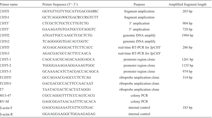
Table 1- Summary of primers used in this study.
Primer name Primer Sequence (5’- 3’) Purpose Amplified fragment length
CHYf1 GGYGTYGTYTGCATYGACGGHRC fragment amplication 203 bp
CHYr1 GCTCAGGGWKTGACRCCRGTCTT fragment amplication
CHY3’ CTCGCTCTGCTCCTTGTCTG 3’ amplication 904 bp
CHY5’ GAAAGATGTGATGCCGTAGGTC 5’ amplication 728 bp
CHYf2 ATGATTGCCAAGCTCGCTCTG genomic DNA amplify 1994 bp
CHYr2 TCAGGGGGTGACACCGGTC genomic DNA amplify
CHYf3 ACGAGCAGGGACTTCTTCACC real-time RT-PCR forSpCHY 286 bp
CHYr3 AGACGACGCCACTTCCAACA real-time RT-PCR forSpCHY
CHY5-1 CAGCAACGCAGACAAGGAGCA promoter region clone 1261 bp
CHY5-2 TGGGGAAAGAAGGAAAGTGGC promoter region clone 1155 bp
CHY5-3 GCAAAACATCTACGACCACAGCA promoter region clone 974 bp TCHYf1 GCCAGAACGAGCCCTCTCAG riboprobe amplication clone 314 bp TCHYr1 GACGACGCCACTTCCAACAAT riboprobe amplication clone
T7 TAATACGACTCACTATAGGG riboprobe amplication clone M13-47 CGCCAGGGTTTTCCCAGTCACG colony PCR RV-M GAGCGGATAACAATTTCACACA colony PCR
b-actin F GAGCGAGAAATCGTTCGTGAC internal control 183 bp
and M13-47 (Table 1) at Sangon Biotech Co, Ltd (China). Finally, the full-length of SpCHY cDNA was assembled from 3’ end and 5’ end sequences.
Genomic DNA and promoter cloning ofSpCHY
Genomic DNA was extracted from muscle tissue of the mud crab by means of a DNA extraction kit (Takara, Dalian, China) PCR amplified by two specific primers CHYf2 andCHYr2 (Table 1) PCR and cloned as described above. The promoter region was cloned by genome walk-ing uswalk-ing the Universal Genome Walker kit (Takara, Da-lian, China). Nested PCR was performed with primers CHY5-1,CHY5-2,CHY5-3 (Table 1) according to the man-ufacturer’s protocol. The PCR product was purified and se-quenced as before.
Phylogenetic and sequence analysis ofSpCHY
A homology analysis ofSpCHYwithCHYgenes of other species was performed using the Blastp algorithm. Characteristics of the protein were predicted using algo-rithms.of the Expasy site. The putative signal peptide was identified with SignalP software (Nielsenet al., 1997), and the ClustalW program was used to perform multiple se-quence alignments. The neighbor-joining method imple-mented in MEGA3.1 software was used to construct the phylogenetic tree based on protein sequences (Kumaret al., 2004), with a bootstrapping replication of 1000. SSRHunter software was used to search for microsatellite sequences.
Tissue expression ofSpCHY
mRNA transcripts ofSpCHYin different tissues were examined by real-time quantitative PCR (Applied Bio-systems 2770 Thermal Cycle, New York, USA). The reac-tions were performed in a 20mL reaction volume contain-ing 10mL of SYBR premix, 2mL of cDNA template (1/10x dilution of cDNA), 0.8mL of each primer (10mMCHYf3 andCHYr3; Table 1) which amplify a product of 286 bp, and 6.4mL of PCR-grade water. PCR conditions were as follows: 94 °C for 10 min; 40 cycles of 94 °C for 20 s, 56 °C for 30 s and 72 °C for 40 s; final extension at 72 °C for 10 min. A 183 bp b-actin (GU99242) fragment of S. paramamosainwas amplified as the internal control. Stan-dard curves were run for each primer and the cDNA tem-plates were tested in a graded dilution series (1, 1/10, 1/100, 1/1000). Based on these analyses, PCR efficiency was cal-culated to be > 96% (according to the PCR amplification formula E = 10(-1/slope)-1; where E is the PCR efficiency). The negative control was performed with PCR-grade water replacing the cDNA template. All samples were run in trip-licate and relative expression was calculated as 2-DDCt (Livak and Schmittgen, 2001).
Immunocytochemistry
Hepatopancreas tissue removed from adult female crabs was fixed in Bouin’s fixative overnight, dehydrated,
embedded in paraffin, and then sectioned at 7mm thickness. The sections were immunocytochemically stained by the streptavidin-peroxidase method with a primary antiserum generated in mouse against CHY (1:100 dilution, Abcam, UK) following an immunocytochemical protocol of the supplier (Transgen, China). The presence of CHY-like immunoreactivity in the tissues was visualized by a DAB enhanced liquid substrate system (Sigma-Aldrich, USA). Thereafter, the sections were dehydrated and observed on an Olympus multifunction microscope BX51 (Olympus, Japan). Control sections were prepared simultaneously by substituting PBS buffer solution in place of the primary an-tibody.
In situhybridization
Digoxigenin-labeled cRNA riboprobes were synthe-sized with a DIG-RNA labeling Kit (Roche, Switzerland) using a 314 bp template ofSpCHYthat was ligated into the pGEM-T easy vector (Promega, USA). Hepatopancreas tis-sue was dissected and immediately fixed overnight in 4% paraformaldehyde (PFA) in phosphate-buffered saline (PBS) made in diethypyrocarbonate (DEPC) water. Tissue sections of 7mm thickness were hybridized with the digo-xigenin-labeled riboprobes at 57 °C overnight followed by incubation in an anti-DIG alkaline phosphatase-conjugated antibody (Roche, Switzerland). Hybridization signals were visualized with the colorimetric substrates nitroblue tetra-zolium/4-bromo-4-chloro-30-indolylphosphate
(NBT/BCIP). The riboprobe templates for SpCHY were generated by RT-PCR from hepatopancreas cDNA using the specific primers TCHYf1, TCHYr1 containingT7 adap-ters. Photographs were taken on an Olympus multifunction microscope BX51 (Olympus, Japan).
Temporal expression ofSpCHYin hepatopancreas after immune challenge
cDNA synthesis and real-time quantitative assays were per-formed according to the procedures described above.
Statistical analysis
One-way analysis of variance (ANOVA) and Stu-dent’s t-test done with SPSS 11.5 software were used to de-termine the statistical significance ofSpCHYexpression in different tissues and challenge experiment respectively (SPSS, Chicago, IL, USA). Before the comparisons, Kol-mogorov-Smirnov and Cochran tests were run to test for normality and homogeneity of variances. P values of < 0.05 were considered statistically significant.
Results
Cloning of theSpCHYgene
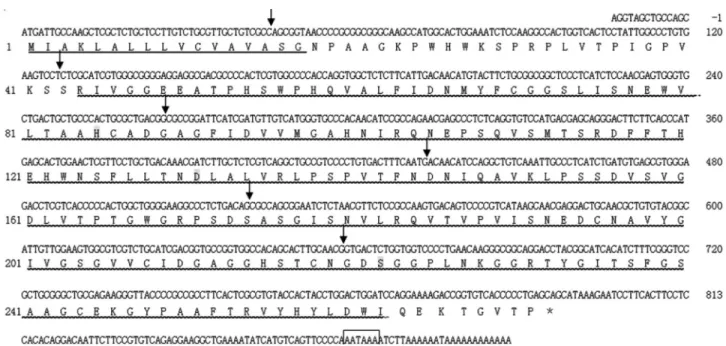
A 942 bp cDNA sequence ofSpCHY(GenBank ac-cession number: JF831535.1) was obtained in this study. It comprises an 813 bp open reading frame (ORF) encoding 270 amino acids with a signal peptide of 17 amino acids, an 115 bp 3’-untranslated region (UTR) with a polyA tail, and a 14 bp 5’UTR (Figure 1). The deduced molecular weight of mature SpCHY protein was 28.5 kDa and its isoelectric point 6.11. Conserved domain analysis done online in NCBI showed that SpCHY contained a trypsin-like SP do-main including one cleavage site I-45, three active site (H-85, D-131, S-222), three substrate binding sites (S-216, S-237, G-239), and six cysteine residues, which were simi-lar to other chymotrypsin members.
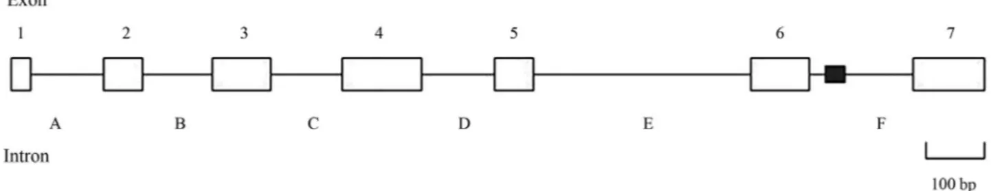
Similar to other chymotrypsin genes,SpCHYis com-posed of seven exons interrupted by six introns. In addition,
all the intron-exon boundaries conformed to the GT-AG rule, which belonged to a 0-type intron/exon junction. Moreover, a 36 CA repeat microsatellite sequence was found by screening with SSRHunter software (Figure 2).
In order to study the regulation ofSpCHYexpression in the mud crab, we used a cloned 1221 bp fragment of the 5’ flanking region of theSpCHYgene. Using the program Promoter 2.0, we found a putative TATA box that was lo-cated at 45 bp upstream of the translation start site. In addi-tion, several putative transcriptional factor binding sites or cis-regulatory elements including HSF, Hb, Dfd, SP1, Bcd, CF1 and Ubx were also identified.
Phylogenetic analysis of SpCHY
Blastp data showed that the deduced amino acid se-quence shared high similarity with chymotrypsins of L. vannamei CHYA (GenBank accession no. CAA71672, 82%), F. chinensis (ACC68669.1, 80%), M. japonicus (BAI49929.1, 79%),L. vannamei CHYB (CAA71673.1, 79%). The phylogenetic analysis suggested that three dif-ferent groups were formed, representing CHYs from inver-tebrates, vertebrates and urochordates respectively. The vertebrate CHY group could be further separated into three distinct and well-supported clades: CHYA, CHYB, and CHYC (caldecrin). The invertebrate group contained 2 sub-groups. As showed in Figure 3, crustacean CHY was well separated from insect CHY and formed a separate cluster.
Tissue distribution ofSpCHYmRNA
Real-time quantitative PCR showed that SpCHY mRNA is expressed in a wide variety of tissues, including
brain, thoracic ganglion, heart, gill, hepatopancreas, stom-ach, muscle, and ovary. The mRNA expression level in hepatopancreas was considerably higher than that of other tissues, with the expression level in muscle being the lowest (Figure 4).
Immunocytochemistry andin situhybridization
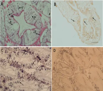
The histological results showed that hepatopancreas ofS. paramamosainconsists of many blind ending tubules (hepatopancreatic tubules). The hepatopancreas cells could be classified into four types: embryonic cells, fibrillar cells, resorptive cells, and blister cells (Figure 5A).
Using immunocytochemistry, SpCHY protein was detected in resorptive cells of the hepatopancreas, and the positive signals were mottled (Figure 5B).SpCHYgene ex-pression was determined byin situhybridization. Positive hybridization signals with the antisenseSpCHYriboprobe were also mainly localized in resorptive cells (Figure 5C). However, specific signals were also detected in some small cells around the blind ending tubules. No positive signal was detected with the sense SpCHY riboprobe in hep-atopancreas (Figure 5D).
Expression ofSpCHYin hepatopancreas following bacterial challenge
In order to determine whether SpCHY may be in-volved in innate immune reactions, the expression profiles ofSpCHYafter bacterial challenge were evaluated. Total
Figure 2- Organization of theSpCHYgene. The positions of the exons (open boxes 1-7), introns (A-F), and CA repeat sequence (filled box) are denoted.
Figure 3- Phylogenetic analysis of SpCHY with other chymotrypsins. A NJ tree was produced with Mega3.1 software. One thousand bootstraps were carried out to check the repeatability of the result.L. vannamei -CHYA (CAA71672),L. vannamei-CHYB (CAA71673),F. chinensis -CHY (ACC68669), P. humanus corporis-CHY (AAV68346), P. cochleariae-CHY (CAA76928), T. molitor-CHY (DQ356031.1), M. sexta-CHY (2120321A),A. grandis-CHY (AAT09847.1),A. gambiae -CHY1 (CAA79325),A. gambiae-CHY2 (CAA79326),B. Taurus- CHYB (P00767),G. morhua-CHYB (P80646),S. aurata-CHYB (AAT45258),
M. musculus-CHY (AAL11034), H. sapiens-CHY (CAA74031.1),B. taurus-CHYC(AAI51507.1), X. laevis-CHYC(NP_001085458), B. taurus-CHYB (NP_001098800.1),D. rerio-CHYB1 (NP_997783.1), and
O. dioica-CHYB (AAT47850).
Figure 4- The results of quantitative real-time PCR analysis ofSpCHY
hepatopancreas RNA was extracted from control and bacte-rial challenged crab at 0, 3, 6, 12, 24, 48 and 72 h. Com-pared to the control group, in crabs injected with the bacteriumV. alginolyticusthe SpCHYmRNA expression level increased distinctly about 20-fold at 3 h (p < 0.01) and then decreased to normal level (Figure 6). During this 72 h time intervalSpCHYexpression levels in the control group fluctuated slightly but not significantly.
Discussion
In present study, a new chymotrypsin gene was iden-tified from the mud crab,S. paramamosain, and was desig-nated as SpCHY. The full-length cDNA contained an 813 bp open reading frame which encoded a putative chy-motrypsin of 270 amino acids. The putative amino acids se-quence has high identity with the other known crustacean chymotrypsins such as L. vannamei and F. chinensis. ClustalX alignment of the CHY sequence revealed that the tryp-spc domain was conserved among arthropod chymo-trypsins. In addition, the catalytic triad (H, D, S) character-istic of chymotrypsins was observed in the deduced amino sequence. Furthermore, three disulfide bonds formed by six cysteines were found at the same location as in other chy-motrypsins. This indicates the importance of secondary structure conservation for the enzymatic activity of this family. Another free cysteine residue found in the signal peptide was also identical to chymotrypsins from other in-vertebrates. The high similarity, together with the
conser-vation of tryp-spc domain and catalytic triad, indicated that SpCHYis a true member of the chymotrypsin family.
The genomic sequence ofSpCHY, here first reported in crabs, is composed of seven exons and six introns, with the first intron inserted near the end of the putative signal peptide. The locations of introns were almost the same as in the white shrimp,L. vannamei, chymotrypsin gene (Sellos and Wormhoudt, 1992). The active site residues (His85, Asp131 and Ser222) involved in catalysis, as well as the residues (Ser216, Ser237 and Gly239) forming the binding pocket to interact with the hydrophobic side chains of the substrate, were encoded by separate exons. These function-ally important amino acids and binding regions in separate exons are typical for theSPgenes that have been described (Swiftet al., 1984; Craiket al., 1984). Hence, the joining of different exons, encoding intrinsically catalytically inac-tive protein segments, resulted in the substrate specificity and catalytic activity of the enzyme. Moreover, the similar-ity betweenSpCHYand otherSPgenes in the number and location of intron/exon junctions revealed an evolutionary conservation of chymotrypsin gene.
In our study,SpCHYexpression was detected in vari-ous tissues and strongly so in hepatopancreas. The high ex-pression level ofSpCHYin hepatopancreas was consistent with the role of the hepatopancreas as the main site for syn-thesizing digestive enzymes in crustaceans (Shi et al., 2008). Furthermore, crustacean hepatopancreas plays im-portant roles in initiating humoral immunity and mediating cellular immune responses performed by certain special-ized cells and phagocytes (Grosset al., 2001), which is sup-ported by the discovery of several immunity-related genes in crustacean hepatopancreas post bacterial infection (Pan et al., 2005; Zhaoet al., 2007).
The results obtained by immunocytochemistry andin situhybridization indicated that the hepatopancreas is the
Figure 5- Location ofSpCHYby immunocytochemistry andin situ hy-bridization in hepatopancreas ofS. paramamosain. (A) histological obser-vation; R resorptive cells, B blister cells, E embryonic cells, F fibrillar cells, Nu nucleus. (B) immunocytochemistry results; the arrows point to immunocytochemical positive signals. (C)in situhybridization results; ar-rows indicate the specificSpCHYmRNA hybridization signal with the antisense riboprobe. (D) The negative control with the sense riboprobe showed no specific signal. Scale bars: 50mm.
Figure 6 - Transcript profiles of SpCHY in hepatopancreas of S. paramamosainfollowing challenge withV. alginolyticus,. The relative
SpCHYtranscript levels in crabs challenged withV. alginolyticuswere compared to those of saline injected animals. The expression of ab-actin gene was used as endogenous control. Significant differences ofSpCHY
site of expression and translation of SpCHY. CHY-immunoreactivity was found in resorptive cells, supplying morphological evidence for the secretory function of resor-ptive cells. The localization ofSpCHYmRNA in resorptive cells byin situhybridization further strengthens this con-clusion. All these findings indicated thatSpCHYis synthe-sized in resorptive cells and might be secreted to implement the digestive and immune roles.
Lacking an acquired specific immune system, the in-nate immune system in crustaceans is considered as the ma-jor microbial infection defense mechanism (Chaikeeratisak et al., 2012; Kiruthigaet al., 2012). In recent years, non-specific immune system has been found to be of equal im-portance as a specific immune system, especially for the production of anti-bacterial and anti-viral proteins (Liuet al., 2010). Pathogen molecules can trigger these immune responses by pattern recognition proteins (PRPs) (Medz-hitov and Janeway, 1997). These PRPs bind to microbes and then activate the prophenoloxidase system (proPO-system), stimulate the release of antimicrobial peptides (AMPs), or initiate other biological defense processes. Re-cently, the clip domain SP was demonstrated to be cofactor for the activation of the proPO cascade in invertebrates (Cerenius and Söderhäll, 2004; Gaiet al., 2009). For exam-ple, in Sydney rock oysters,Saccostrea glomerata, the in-crease in chymotrypsin could activate ProPO to PO (Aladailehet al., 2007).
The immune function of chymotrypsin has been re-ported inF. chinensis(Shiet al., 2008). However, little re-search has focused on the function of innate immunity in crabs. In this study,SpCHYwas strongly up-regulated inS. paramamosainat 3 h after infection with the bacteriumV. alginolyticus. In appropriate hosts, this kind of bacteria could proliferate unceasingly. The infection caused by un-ceasing reproduction of bacteria could induce the formation of reactive oxygen species (ROS) and severely destroy the functionality of crab cells (Liet al., 2011). Similar results showing thatSpCHYexpression is significantly shortly af-ter bacaf-terial infection were also obtained in other crusta-ceans (Amparyupet al., 2007; Qinet al., 2009; Cuiet al., 2010). Hence we hypothesize that increasing the expres-sion ofSpCHYcould activate PO production triggering an immune response and killing the bacteria.
In conclusion, our data suggest clearly for the first time thatSpCHYis involved in the immune reaction against invading bacteria in the mud crab,S. paramamosain.The result should be helpful to understand the antibacterial de-fense mechanisms of crabs and provide biological informa-tion for mitigating crab diseases. Notwithstanding, the exact role ofSpCHY in the activation of the immune re-sponse cascade needs further investigation.
Acknowledgments
This work was funded by grants from the National Natural Science Foundation of China (No. 41076081,
31272632) and the Innovative Research Funds in Xiamen University (No. 201112G009).
References
Amparyup P, Jitvaropas R, Pulsook N and Tassanakajon A (2007) Molecular cloning, characterization and expression of a masquerade-like serine proteinase homologue from black ti-ger shrimp Penaeus monodon. Fish Shellfish Immunol 22:535-546.
Aladaileh S, Rodney P, Nair SV and Raftos DA (2007) Character-ization of phenoloxidase activity in Sydney rock oysters
(Saccostrea glomerata). Comp Biochem Physiol B
148:470-480.
Broehan G, Kemper M, Driemeier D, Vogelpohl I and Merzen-dorfer H (2008) Cloning and expression analysis of midgut chymotrypsin-like proteinases in the tobacco hornworm. J Insect Physiol 54:1243-1252.
Broehan G, Arakane Y, Beeman RW, Kramer KJ, Muthukrishnan S and Merzendorfer H (2010) Chymotrypsin-like peptidases fromTribolium castaneum: A role in molting revealed by RNA interference. Insect Biochem Mol Biol 40:274-283. Cerenius L and Söderhäll K (2004) The
prophenoloxidase-activating system in invertebrates. Immunol Rev 1981:16-26. Chaikeeratisak V, Somboonwiwat K, Wang HC, Lo CF and
Tas-sanakajon A (2012) Proteomic analysis of differentially ex-pressed proteins in the lymphoid organ of Vibrio harveyi-infectedPenaeus monodon. Mol Biol Rep 39:6367-6377. Cheng W, Liu CH, Ye ST and Chen JC (2004) The immune
stimulatory effect of sodium alginate on the white shrimp Litopenaeus vannamei and its resistance against Vibrio alginolyticus. Fish Shellfish Immunol 17:41-51.
Craik CS, Choo QL, Swift GH, Quinto C, MacDonald RJ and Rutter WJ (1984) Structure of two related rat pancreatic trypsin genes. J Biol Chem 259:14255-14264.
Cui ZX, Liu Y, Wu DH, Luan WS, Wang SY, Li QQ and Song CW (2010) Molecular cloning and characterization of a serine proteinase homolog prophenoloxidase-activating fac-tor in the swimming crab Portunus trituberculatus. Fish Shellfish Immunol 29:679-686.
Gai YC, Qiu LM, Wang LL, Song LS, Mu CK, Zhao JM, Zhang Y and Li L (2009) A clip domain serine protease (cSP) from the Chinese mitten crabEriocheir sinensis: cDNA charac-terization and mRNA expression. Fish Shellfish Immunol 27:670-677.
Gross PS, Bartlett CL, Browdy CL, Chapman RW and Warr GW (2001) Immune gene discovery by expressed sequence tag analysis of hemocytes and hepatopancreas in the pacific white shrimp,Litopenaeus vannamei, and the atlantic white shrimp,L. setiferus. Dev Comp Immunol 25:565-577. Hedstrom L (2002) Serine protease mechanism and specificity.
Chem Rev 102:4501-4524.
Herrero S, Combes E, Van OMM, Vlak JM, deMaagd RA and Beekwilder J (2005) Identification and recombinant expres-sion of a novel chymotrypsin fromSpodoptera exigua. In-sect Biochem Mol Biol 35:1073-1082.
Jiang H, Wang Y, Yu XQ, Zhu Y and Kanost MR (2003a) Prophenoloxidase-activating proteinase-2 (PAP-2) from hemolymph ofManduca sexta: A bacteria-inducible serine proteinase containing two clip domains. J Biol Chem 278:3552-3561.
Jiang H, Wang Y, Yu XQ, Zhu Y and Kanost MR (2003b) Prophenoloxidase-activating proteinase-3 (PAP-3) from Manduca sextahemolymph: A clip-domain serine protei-nase regulated by serpin-1J and serine proteiprotei-nase homologs. Insect Biochem Mol Biol 33:1049-1060.
Keenan C (1999) Aquaculture of mud crab, genusScylla- past, present and future. In: Keenan C and Blackshaw (eds) A Mud Crab Aquaculture and Biology. ACIAR Proceedings, No. 78. Watson Ferguson and Company, Canberra, pp 9-13. Kiruthiga C, Rajesh S, Rashika V, Priya R and Narayanan RB
(2012) Molecular cloning, expression analysis and charac-terization of peroxiredoxin during WSSV infection in shrimpFenneropenaeus indicus. J Invert Pathol 109:52-58. Kumar S, Tamura K and Nei M (2004) MEGA3: Integrated soft-ware for molecular evolutionary genetics analysis and se-quence alignment. Brief Bioinform 5:150-163.
Le Vay L (2001) Ecology and management of mud-crabScylla spp. Asian Fish Sci 14:101-112.
Li JT, Chen P, Liu P, Gao BQ, Wang QY and Li J (2011) Molecu-lar characterization and expression analysis of extracelluMolecu-lar copper-zinc superoxide dismutase gene from swimming crabPortunus trituberculatus. Mol Biol Rep 38:2107-2115. Liu HP, Chen RY, Zhang M and Wang KJ (2010) Isolation, gene
cloning and expression profile of a pathogen recognition protein: A serine proteinase homolog (Sp-SPH) involved in the antibacterial response in the crabScylla paramamosain. Dev Comp Immunol 34:741-748.
Livak KJ and Schmittgen TD(2001) Analysis of relative gene ex-pression data using real time quantitative PCR and the 2 (-Delta Delta C (T)) method. Methods 25:402-408. Mazumdar LS and Broadway RM (2001) Identification of six
chymotrypsin cDNAs from larval midguts ofHelicoverpa zeaandAgrotis ipsilonfeeding on the soybean Kunitz tryp-sin inhibitor. Insect Biochem Mol Biol 31:633-644. Medzhitov R and Janeway CA (1997) Innate immunity: The
vir-tues of a nonclonal system of recognition. Cell 91:295-298. de Morais GS, Vitorino R, Domingues R, Tomer K, Correia AJF, Amado F and Domingues P (2005) Proteomics of immune-challenged Drosophila melanogaster larvae hemolymph. Biochem Biophys Res Commun 328:10-15.
Nielsen H, Engelbrecht J, Brunak S and vonHeijne G (1997) Identi-fication of prokaryotic and eukaryotic signal peptides and prediction of their cleavage sites. Protein Eng Des Sel 10:1-6. Pan D, He N, Yang Z, Liu H and Xu X (2005) Differential gene
expression profile in hepatopancreas of WSSV-resistant shrimp (Penaeus japonicus) by suppression subtractive hy-bridization. Dev Comp Immunol 29:103-112.
Perona JJ and Craik CS (1995) Structural basis of substrate speci-ficity in the serine proteases. Protein Sci 4:337-360. Qin C, Chen L, Qin JG, Zhao D, Zhang H and Wu P (2009)
Char-acterization of a serine proteinase homologous (SPH) in Chinese mitten crabEriocheir sinensis. Dev Comp Immunol 34:14-18.
Rawlings ND, Morton FR, Kok CY, Kong J and Barrett AJ (2008)
MEROPS: The peptidase database. Nucleic Acids Res
36:320-325.
Ross J, Jiang H, Kanost MR and Wang Y (2003) Serine proteases and their homologs in the Drosophila melanogaster ge-nome: An initial analysis of sequence conservation and phylogenetic relationships. Gene 304:117-131.
Samuel RI and Reynolds SE (1993) Molting fluid enzymes of the tobacco hornworm Manduca sexta: Timing of proteolytic and chitinolytic activity in relation to preecdysial develop-ment. Arch Insect Biochem 24:33-44.
Sellos D and Wormhoudt A (1992) Molecular cloning of a cDNA that encodes a serine protease with chymotryptic and colla-genolytic activities in the hepatopancreas of the shrimp
Penaeus vanameii (Crustacea, Decapoda). FEBS Lett
309:219-224.
Sellos D and Wormhoudt A (1999) Polymorphism and evolution of collagenolytic serine protease genes in crustaceans. Bio-chim Biophys Acta 1432:419-424.
Serrano AE (2013) Ontogenetic changes in the activity of chymo-trypsin and carboxypeptidases A and B in mud crab,Scylla serrata. Isr J Aquacult-Bamid 65:1-6.
Shi XZ, Zhao XF and Wang JX (2008) Molecular cloning and ex-pression analysis of chymotrypsin-like serine protease from the Chinese shrimp,Fenneropenaeus chinensis. Fish Shell-fish Immunol 25:589-597.
Southan C (2001) A genomic perspective on human proteases as drug targets. Drug Discov Today 6:681-688.
Swift GH, Craik CS, Stary SJ, Quinto C, Lahaie RG, Rutter WJ and MacDonald RJ (1984) Structure of the two related elastase genes expressed in the rat pancreas. J Biol Chem 259:14271-14278.
Szabo R and Bugge TH (2008) Type II transmembrane serine pro-teases in development and disease. Int J Biochem Cell B 40:1297-1316.
Ye HH, Tao Y, Wang GZ, Lin QW, Chen XL and Li SJ (2011) Ex-perimental nursery culture of the mud crab Scylla para-mamosain(Estampador) in China. Aquacult Int 19:313-321. Zhao ZY, Yin ZX, Weng SP, Guan HJ, Li SD, Xing K, Chan SM
and He JG (2007) Profiling of differentially expressed genes in hepatopancreas of white spot syndrome virus-resistant shrimp (Litopenaeus vannamei) by suppression subtractive hybridization. Fish Shellfish Immunol 22:520-534. Zhou LM, Wu SG, Liu DC, Xu B, Zhang XF and Zhao BS (2012)
Characterization and expression analysis of a trypsin-like serine protease from planarianDugesia japonica. Mol Biol Rep 39:7041-7047.
Zhu L, Song LS, Mao YZ, Zhao JM, Li CH and Xu W (2008) A novel serine protease with clip domain from scallop Chlamys farreri. Mol Biol Rep 35:257-264.
Internet Resources
ORF Finder, http://www.ncbi.nlm.nih.gov/gorf (July 3, 2013). NCBI, http://www.ncbi.nlm.nih.gov (July 3, 2013).
Expasy, http://www.expasy.ch/ (July 3, 2013).
SignalP 4.0 software, http://www.cbs.dtu.dk/services/SignalP (July 3, 2013).
ClustalW, http://www.ebi.ac.uk/Tools/msa/clustalw2/.
Associate Editor: Juan Lucas Argueso Almeida