Preliminary
in vitro
evaluation of the anti-proliferative activity
of guanylhydrazone derivatives
Guanylhydrazones have shown promising antitumor activity in preclinical tumor models in several studies. In this study, we aimed at evaluating the cytotoxic eff ect of a series of synthetic guanylhydrazones. Diff erent hu-man tumor cell lines, by including HCT-8 (colon carci-noma), MDA-MB-435 (melanoma) and SF-295 (glioblas-toma) were continuous exposed to guanylhydrazone derivatives for 72 hours and growth inhibition of tumor cell lines and macrophages J774 was measured using tet-razolium salt (MTT) assay. Compounds 7, 11, 16 and 17
showed strong cytotoxic activity with IC50values lower
than 10 μmol L–1 against four tumor cell lines. Among
them, 7 was less toxic to non-tumor cells. Finally,
ob-tained data suggest that guanylhydrazones may be re-garded as potential lead compounds for the design of novel anticancer agents.
Keywords: guanylhydrazone, cytotoxic eff ect, synthesis, MTT assay
PAULO H. B. FRANÇA2 EDEILDO F. DA SILVA-JÚNIOR2 PEDRO G. V. AQUINO1 ANTÔNIO E. G. SANTANA1 JAMYLLE N. S. FERRO3
EMILIANO DE OLIVEIRA BARRETO3 CLÁUDIA DO Ó PESSOA4 ASSUERO SILVA MEIRA4 THIAGO M. DE AQUINO2 MAGNA S. ALEXANDRE-MOREIRA5 MARTINE SCHMITT6
JOÃO X. DE ARAÚJO-JÚNIOR2*
1 Natural Resources Research Laboratory
Institute of Chemistry and Biotechnology Federal University of Alagoas, Maceio, Brazil
2 Laboratory of Medicinal Chemistry
School of Nursing and Pharmacy
Federal University of Alagoas, Maceio, Brazil
3 Cell Biology Laboratory, Biological and
Health Sciences, Federal University of Alagoas, Maceio, Brazil
4 Experimental Oncology Laboratory
Department of Physiology and Pharmacology Federal University of Ceara, Fortaleza, Brazil
5 Institute of Biological and Health Sciences
Laboratory of Pharmacology and Immunity Federal University of Alagoas, Maceio, Brazil
6 Laboratoire d’innovation Thérapeutique
Faculté de Pharmacie, Université de Strasbourg Illkirck, France
Accepted November 25, 2015 Online published February 11, 2016
Guanylhydrazones represent a class of compounds showing interesting pharmaco-logical activities on several levels; they derive from the same aminoguanidine chemotype with mixed hydrogen bond acceptor and donor properties as well as being able to establish electrostatic interactions (1, 2). Moreover, they already led to various active derivatives, as illustrated in Fig. 1 (3). Guanidine derivatives have been reported to be e cient anti-cancer agents. Previous reports showed that m-iodobenzylguanidines (MIBG),
pyridylcyanogua-nidine (CHS 828) and mitoguazone (MGBG) have shown promising antitumor activity in preclinical tumor models, while mitoguazone showed useful clinical activity for the treat-ment of malignant lymphoma, carcinoma of the head, neck and esophageal and non-small cell lung cancer (4). More recently, a series of imidazo[2,1-b]thiazoleguanylhydrazones was
reported for their antitumor activity and was able to induce apoptosis in HT29 and HL-60 cell lines (5). However, no structure-activity relationship of imidazothiazole compounds was available. Sixteen guanylhydrazone coactivator binding inhibitors for estrogen recep-tor showed good to moderate activity, but no cytotoxicity data were shown for these com-pounds (6).
Fig. 1. Cytotoxic guanylhydrazones previously reported in literature (4).
At er identifi cation of the guanidine moiety, as the key pharmacophoric group for
activ-ity, we decided to replace the imidazothiazole ring by a phenyl ring, which off ered the ad-vantage of scaff old modifi cation. In particular, our objective was to perform a systematic
exploration of the aryl group allowing introduction of various substituents at the phenyl ring. The phenyl ring was replaced by heteroaromatics in another series of compounds.
EXPERIMENTAL
General
chamber. NMR spectra (1H: 400 MHz; 13C: 100 MHz) were obtained from a Bruker® DPX 500 (Germany) device. All chemical shit values were recorded as d (ppm), the coupling
constant value J was measured in hertz; the peaks are presented as s (singlet), d (doublet),
t (triplet), dd (double doublet), and m (multiplet).
Synthesis of target compounds
Guanylhydrazones 1-20 were prepared by the reaction of an appropriate aryl alde-hyde with aminoguanidine hydrochloride (1:1.25 equiv). The reactants were dissolved in a minimal amount of methanol, heated to refl ux, and stirred overnight. Upon cooling to
room temperature, products crystallized out from the methanol solution. Products were recovered and dried under vacuum overnight. All reactions were monitored by TLC. The solvent was removed under reduced pressure and the solid was triturated with ethyl ac-etate yielding the substances as powders.
Assessment of cell viability
Cytotoxic eff ects of the synthesized compounds were evaluated against the following human cancer cell lines (National Cancer Institute, Bethesda, MD, USA): HCT-8 (colon carcinoma), SF-295 (glioblastoma) and MDA-MB-435 (melanoma). Tumor cells were seeded in 96-well plates (0.7 × 10⁵ cells per well for adherent cells and 3 × 10⁵ cells mL–1 for
sus-pended cells). At er 24 hours, the compounds (5 mg mL–1), dissolved in dimethylsulfoxide
(DMSO), were added to each well using an automated workstation Biomek® 3000 (Beck-man Coulter, USA) and incubated for 72 hours. Addition of doxorubicin (Sigma Aldrich Co., USA) at 5 mg mL–1 concentrations was performed in 150 mL of Roswell Park Memorial
Institute (RPMI 1640) medium [2 mmol L–1 of L-glutamine, 10 mmol L–1 of 4-(2-hydroxyethyl)-1-piperazineethanesulfonic acid (HEPES), 1 mmol L–1 of sodium pyruvate, 4.5 g L–1 of glu-cose, 1500 mg L–1 of sodium bicarbonate, 0.5 mg mL–1 MTT]. At er 3 hours, the formazan product was dissolved in 150 mL DMSO and the absorbance was measured using a
multi-plate reader (DTX 880 Multimode Detector, Beckman Coulter). The substance eff ect was quantifi ed as the percentage of control absorbance at 595 nm.
Macrophage J774 cells were seeded (105 cells per well) in 96-well fl at-bot om
micro-plates. The cells were maintained for 2 hours at 37 °C, 5 % CO2 and the medium was re-placed with diff erent concentrations of the drugs (10, 100 and 1000 mmol L–1), in duplicate,
and exposed for another 24 hours. Growth controls were also included. At erwards, MTT solution was added to the cells and plates were returned to the incubator for another 4 hours to evaluate cell viability. This was followed by washing of the wells with 150 mL of
DMSO for 15 minutes and measurement was performed spectrophotometrically at 540 nm.
RESULTS AND DISCUSSION
Guanylhydrazone derivatives were obtained through the condensation reaction of benzaldehyde derivatives with aminoguanidine hydrochloride (7, 8; see Scheme 1).
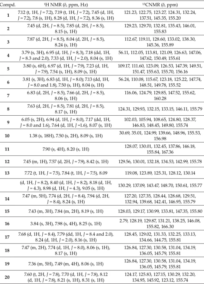
All compounds were characterized by hydrogen (¹H) and carbon (¹³C) NMR. The main chemical shit s for each compound are shown in Table I. A signal at d 7.27 to 9.05 ppm
Table I. Spectral data of guanylhydrazones
Compd. ¹H NMR (δ, ppm, Hz) ¹³CNMR (δ, ppm)
1 7.12 (t, 1H, J = 7.2), 7.19 (t, 1H, J = 7.2), 7.45 (d, 1H,
J = 7.2), 7.8 (s, 1H), 8.28 (d, 1H, J = 7.2), 8.36 (s, 1H) 121.23, 122.75, 123.27, 124.31, 132.24, 137.51, 145.35, 155.20
2 7.45 (d, 2H, J = 8.15 (s, 1H)8.5), 7.85 (d, 2H, J = 8.5), 129.23, 129.70, 132.81, 135.43, 146.01, 155.83
3 7.87 (d, 2H, J = 8.24 (s, 1H)8.5), 8.04 (d, 2H, J = 8.5), 112.67, 119.11, 128.60, 133.02, 138.30, 145.36, 155.89
4 3.79 (s, 3H), 6.95 (d, 1H, J = 8,3), 7.18 (dd, 1H,
J = 8.3 and 2.0), 7.33 (d, 1H, J = 2.0), 8.04 (s, 1H)
56.11, 112.05, 113.81, 121.09, 126.63, 147.06, 147.62, 150.49, 155.61
5 3.80 (s, 6H), 6.97 (d, 1H, J = 7.9), 7.23 (d, 1H,
J = 7.9), 7.54 (s, 1H), 8.09 (s, 1H) 109.17, 111.60, 123.09, 126.53, 147.39, 149.51, 151.47, 155.63, 155.70, 156.16
6 3.81 (s, 3H), 6.83 (d, 1H, J = 8.0); 7.13 (dd, 1H,
J = 8.0 and 1.8), 7.50 (s, 1H), 8.04 (s, 1H) 56.24, 110.08, 115.67, 123.18, 125.22, 147.74, 148.51, 149.78, 155.52
7 6.83 (d, 2H, J = 8.5), 7.66 (d, 2H, J = 8.5),
8.06 (s, 1H) 116.06, 124.79, 129.85, 147.52, 155.62, 160.28
8 7.63 (d, 2H, J = 8.17 (s, 1H)8.5), 7.81 (d, 2H, J = 8.5), 124.31, 129.93, 132.15, 133.15, 146.11, 155.79
9 6.05 (s, 2H), 6.94 (d, 1H, J = 8.0), 7.17 (dd, 1H,
J = 8.0 and 1.6), 7.64 (d, 1H, J =1.6), 8.07 (s, 1H)
102.03, 105.94, 108.65, 124.80, 128.37, 146.83, 148.45, 149.80, 155.74
10 1.38 (s, 18H), 7.50 (s, 2H), 8.09 (s, 1H) 30.69, 35.01, 124.99, 139.66, 148.96, 155.53, 156.98
11 7.90 (s, 4H), 8.20 (s, 1H) 128.07, 130.01, 132.45, 137.86, 146.18, 155.84, 167.36
12 7.45 (m, 1H), 7.57 (d, 2H, J = 7.9), 8.42 (s, 1H) 129.56, 130.01, 132.18, 134.53, 142.99, 155.78
13 7.72 (t, 1H, J = 7.5), 7.84 (t, 1H, J = 7.5), 8.09 119.08, 123.89, 125.31, 128.12, 130.14 (d, 1H, J = 8.2), 8.40 (d, 1H, J = 8.2), 8.18 (d, 1H,
J = 4.3), 8.98 (d, 1H, J = 4.3), 9.05 (s, 1H) 130.29, 137.09, 143.47, 148.70, 150.61, 155.77
14 7.47 (m, 5H), 7.74 (d, 2H, J = 8.4), 7.94 (d, 2H,
J = 8.4), 8.24 (s, 1H)
127.20, 127.35, 128.44, 128.68, 129.51, 132.94, 139.68, 142.41, 146.95, 155.79
15 7.43 (m, 3H), 7.84 (m, 2H), 8.19 (s, 1H) 128.03, 129.17, 130.99, 133.81, 147.35, 155.80
16 3.84 (s, 3H), 7.98 (s, 4H), 8.25 (s, 1H) 2.79, 128.19, 129.87, 131.21, 138.25, 146.08, 155.82, 166.30
17 7.68 (d, 1H, J = 8.4), 7.79 (dd, 1H, J = 8.4 and 2.0),
8.24 (d, 1H, J = 2.0), 8.16 (s, 1H) 128.45, 129.02, 131.33, 132.25, 133.13, 134.66, 144.75, 155.81
18 7.47 (m, 2H), 7.74 (d, 1H, 8.17 (s, 1H)J = 8.0), 8.06 (s, 1H), 126.84, 127.30, 130.58, 131.04, 134.19, 136.05, 145.79, 155.81
19 7.36 (m, 5H), 7.49 (m, 4H), 8.06 (s, 1H) 126.84, 127.30, 130.58, 131.04, 134.19, 136.05, 145.79, 155.81
20 7.60 (t, 2H, (d, 1H, J = 7.8), 7.70 (d, 1H, J = 7.8), 8.12
J = 7.8), 8.21 (s, 1H), 8.31 (s, 1H)
H from imine, since this position is highly deshielded due to the induced anisotropy gene-rated by the bound aromatic ring and the imine double bond. In 13C NMR spectra, the signal related to the imine carbon is usually seen around d 145 ppm, whereas the signal
ranging from d 155 to 175 ppm is associated with the highly deshielded quaternary carbon
of guanidine moiety.
The cell lines were exposed to derivatives guanylhydrazones (0–5 mmol L–1) and
pos-itive control: doxorubicin (5 μmol L–1) for 72 hours. Experiments were performed in tripli-cate.
Phenylaminoguanidine 15 exhibits a very low cytotoxic activity, only 20 % inhibition
of tumor cell lines at 5 mmol L–1. Introduction of an electron-donor group such as hydroxyl
or methoxyl 4, 5, 6 and 7 at ortho- and/or para-position did not increase the cytotoxic
activ-ity. However, bridging between two groups in 9 induced a slight increase in activity to-wards the MDA-MB-435 cell line (Table II).
Replacement of the electron-donor group by electron-withdrawing polar groups such as nitrile 3 or formyl 11 showed no signifi cant improvement in potency relative to the
par-ent phenyl group. Introduction of halogen atoms signifi cantly enhanced the activity, as
seen in compounds 2, 8 and 18.
Compound 6 has shown higher selectivity against MDA-MB-435 than the other two
cell lines. This facts, may be related to hydroxyl at position 3 in the phenyl ring, showing superior activity compared to thecompound 4.
A signifi cant increase in cytotoxic activity was observed, when chlorine was
intro-duced in para- position, since compound 2 was found more potent than 18 (meta-position). However, disubstituted 3,4-dichloro analogue 17 led to bet er activity, with 100 %
inhibi-tion of tumor cell lines at 5 mmol L–1. Interestingly, guanabenz (trade name, Wintensy®) 12, a drug developed as an alpha-2 adrenergic agonist was signifi cantly less active than
com-pound 19.
Introduction of a second phenyl ring at para-14 or ortho-19 position led to a set of more
potent compounds. This eff ect highlights the importance of the lipophilic character, which probably plays an important role in cytotoxic activity. In this sense, compound 10 was also
highly active, since the presence of the bulky tert-butyl groups increased lipophilic
char-acter to the molecule, resulting in the best compound of the series.
Finally, the replacement of the phenyl ring by a heteroaromatic ring as in 1 and 13 led
to activity decrease compared to compound 15.
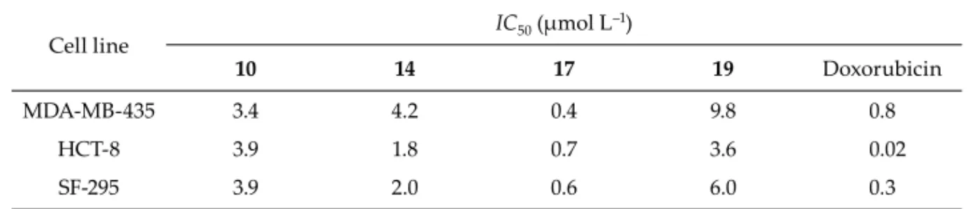
The most powerful compounds recognized in cytotoxic assays against human cancer cell lines at 5 mmol L–1 (Table II) were tested to obtain IC50values, which were compared to
those of doxorubicin (control), as shown in Table III. Each assay was carried out in
Table II. Cytotoxicity of guanylhydrazone derivatives against human cancer cell lines
Compd.
Inhibition (%)a
Ar Yield
(%) HCT-8 MDA-MB-435 SF-295
1 – – 24.0 71
2 72.9 80.2 72.1 71
3 25.4 38.8 28.6 89
4 3.5 0.2 25.3 93
5 – 17.3 19.5 98
6 – 32.4 2.5 92
7 – – 11.5 95
8 67.3 81.6 58.0 83
9 – 53.2 19.6 81
10 97.2 92.6 97.8 85
cate and the means and respective confi dence intervals were obtained by nonlinear
regres-sion analyses.
In order to investigate the eff ect of guanylhydrazone derivatives on non-tumor cells, compounds 10, 14, 17 and 19 were evaluated on macrophages J774 at er 24 hours of
incuba-12 45.2 15.3 51.9 89
13 42.1 29.8 52.0 91
14 100 100 100 90
15 28.3 26.5 24.7 89
16 43.1 48.6 37.8 92
17 100 100 100 92
18 52.1 46.0 52.0 91
19 96.9 95.2 93.8 92
20 52.7 33.7 45.5 88
Doxorubicin 97.3 96.9 87.6 – –
tion and data obtained are displayed in Fig. 2. Results suggest that 10 shows low toxicity
to non-tumor cells, because it is only able to decrease cell viability at a high concentration of 1 mmol L–1. The other three compounds, 14, 17 and 19,showed higher cytotoxicity for non-tumor cells since they reduced cell viability at all tested concentrations (10, 100 and 1000 mmol L–1). This confi rms that compound 10 acts selectively on tumor cell lines and
bears the most promising profi le regarding cytotoxic activity.
Fig. 2. J774 cell viability at er 24 h in the presence of selected guanylhydrazone compounds (mean ± SD, n = 2).
Table III. IC50 values of guanylhydrazone derivatives in three human cancer cell lines
Cell line IC50 (μmol L
–1)
10 14 17 19 Doxorubicin
MDA-MB-435 3.4 4.2 0.4 9.8 0.8
HCT-8 3.9 1.8 0.7 3.6 0.02
SF-295 3.9 2.0 0.6 6.0 0.3
CONCLUSIONS
The results reported in this study indicate that lipophilic guanylhydrazones 10, 14, 17
and 19 showed the highest cytotoxic activity against diff erent human cancer cell lines.
Compound 10 showed higher selectivity towards tumor cell lines and therefore it may be
considered promising for the development of novel cytotoxic agents based on this scaff old.
Acknowledgments. –This work was supported by the Coordenação de Aperfeiçoamento de Pes-soal de Nível Superior (CAPES), Conselho Nacional de Desenvolvimento Científi co Tecnológico
(CNPq), Fundação de Amparo à Pesquisa do Estado de Alagoas (FAPEAL), and Financiadora de Es-tudos e Pesquisa (FINEP).
REFERENCES
1. F. P. Schmidtchen and M. Berger, Artifi cial organic host molecules for anions, Chem. Rev. 97 (1997)
1609–1646; DOI: 10.1021/cr9603845.
2. U. E. W. Lange, D. Baucke, W. Hornberger, H. Mack, W. Seitz and H. W. Hök en, D-Phe-Pro-Arg type thrombin inhibitors: unexpected selectivity by modifi cation of the P1 moiety, Bioorg. Med. Chem. Let . 13 (2003) 2029–2033; DOI: 10.1016/S0960-894X(03)00347-0.
3. J. L. Jiménez Blanco, P. Bootello, J. M. Benito, C. O. Mellet and J. M. García Fernandez, Urea-, thio-urea-, and guanidine-linked glycooligomers as phosphate binders in water, J. Org. Chem. 71 (2006)
5136–5143; DOI: 10.1021/jo060360q.
4. S. Ekelund, P. Nygren and R. Larsson, Guanidino-containing drugs in cancer chemotherapy: biochemical and clinical pharmacology, Biochem. Pharmacol. 61 (2001) 1183–1193; DOI: 10.1016/
S0006-2952(01)00570-6.
5. A. Andreani, M. Granaiola, A. Leoni, A. Locatelli, R. Morigi, M. Rambaldi, G. Lenaz, R. Fato, C. Bergamini and G. Farruggia, Potential antitumor agents. 37. Synthesis and antitumor activity of guanylhydrazones from imidazo[2,1-b]thiazoles and from the new heterocyclic system thiazo-lo[2’,3’:2,3]imidazo[4,5-c]quinoline, J. Med. Chem. 48 (2005) 3085–3089; DOI: 10.1021/jm040888s.
6. A. L. LaFrate, J. R. Gunther, K. E. Carlson and J. A. Katzenellenbogen, Synthesis and biological evaluation of guanylhydrazone coactivator binding inhibitors for the estrogen receptor, Bioorg. Med. Chem. 16 (2008) 10075–10084; DOI: 10.1016/j.bmc.2008.10.007.
7. P. Ulrich and A. Cerami, Trypanocidal 1,3-arylene diketone bis(guanylhydrazone)s. Structure-activity relationships among substituted and heterocyclic analogues, J. Med. Chem. 27 (1984) 35–
40; DOI: 10.1021/jm00367a007.