Research Article
Preclinical Evidences for an Antimanic Effect of Carvedilol
Greicy Coelho de Souza,
1Julia Ariana de S. Gomes,
1Ana Isabelle de Góis Queiroz,
1Maíra Morais de Araújo,
1Lígia Menezes Cavalcante,
1Michel de Jesus Souza Machado,
1Aline Santos Monte,
1David Freitas de Lucena,
1João Quevedo,
2,3André Ferrer Carvalho,
4and Danielle Macêdo
11Neuropharmacology Laboratory, Department of Physiology and Pharmacology, Faculty of Medicine, Federal University of Cear´a,
Rua Coronel Nunes de Melo 1127, 60431-270 Fortaleza, CE, Brazil
2Laborat´orio de Neurociˆencias, Programa de P´os-Graduac¸˜ao em Ciˆencias da Sa´ude, Unidade Acadˆemica de Ciˆencias da Sa´ude,
Universidade do Extremo Sul Catarinense, 88806-000 Crici´uma, SC, Brazil
3Center for Experimental Models in Psychiatry, Department of Psychiatry and Behavioral Sciences,
he University of Texas Medical School at Houston, Houston, TX 77030, USA
4Psychiatry Research Group, Faculty of Medicine, Federal University of Cear´a, 60430-160 Fortaleza, CE, Brazil
Correspondence should be addressed to Danielle Macˆedo; daniellesilmacedo@gmail.com Received 29 August 2014; Revised 11 November 2014; Accepted 17 November 2014 Academic Editor: Rodrigo Machado-Vieira
Copyright © 2015 Greicy Coelho de Souza et al. his is an open access article distributed under the Creative Commons Attribution License, which permits unrestricted use, distribution, and reproduction in any medium, provided the original work is properly cited. Oxidative imbalance, alterations in brain-derived neurotrophic factor (BDNF), and mitochondrial dysfunction are implicated in bipolar disorder (BD) pathophysiology and comorbidities, for example, cardiovascular conditions. Carvedilol (CVD), a nonselective beta-blocker widely used for the treatment of hypertension, presents antioxidant and mitochondrial stabilizing properties. hus, we hypothesized that CVD would prevent and/or reverse mania-like behavioral and neurochemical alterations induced by lisdexamfetamine dimesylate (LDX). To do this, male Wistar rats were submitted to two diferent protocols, namely, prevention and reversal. In the prevention treatment the rats received daily oral administration (mg/kg) of CVD (2.5, 5 or 7.5), saline, valproate (VAL200), or the combination of CVD5 + VAL100 for 7 days. From the 8th to 14th day LDX was added. In the reversal protocol LDX was administered for 7 days with the drugs being added from the 8th to 14th day of treatment. Two hours ater the last administration the behavioral (open ield and social interaction) and neurochemical (reduced glutathione, lipid peroxidation, and BDNF) determinations were performed. he results showed that CVD prevented and reversed the behavioral and neurochemical alterations induced by LDX. he administration of CVD5 + VAL100 potentiated the efect of VAL200 alone. Taken together these results demonstrate a possible antimanic efect of CVD in this preclinical model.
1. Introduction
Bipolar disorder (BD) is one of the most serious mental illnesses characterized by depressive and manic episodes with spontaneous cycling. he pathophysiology of this mental dis-order remains unclear; however, evidences point towards the involvement of genetics, signal transmission deregulation, deleterious inlammatory proile, dysregulation in oxidative stress, and neurotrophins [1,2].
he disorder presents a chronic course associated with functional decline, elevated mortality, and signiicant disease burden [3, 4]. Importantly, BD patients present an excess
burden of cardiovascular risk and higher rate of hypertension compared to the general population [4]. Additionally, this risk is increased by modern treatments for BD, such as antipsychotics [5].
he number of afective episodes in BD patients is directly related to social and cognitive deicits as well as risk of suicide among others [6]. Still in relation to afective episodes, the number of manic episodes was related to impairment of verbal memory implicating, thus, frontal structures [3]. Besides frontal structures, such as prefrontal cortex, striatum and hippocampus are putative brain areas related to mania as observed in clinical [7] and preclinical studies [8,9].
Based on the importance of manic episodes for BD outcome [10], the preclinical model most widely used to study BD is based on the induction of mania-like episodes by the administration of amphetamine-related compounds, such as d-amphetamine (d-AMPH) [11, 12]. Due to the restrictions on d-AMPH acquisition and use, recently our research group proposed an animal model of mania in rats based on the administration of lisdexamfetamine dimesylate (LDX) [9], a prodrug metabolically converted to d-AMPH [13]. In our previous study [9], LDX caused behavioral and neurochemical alterations similar to those of d-AMPH [11,14] with the alterations prevented and reversed by the mood stabilizer lithium.
Indeed, the administration of amphetamine-related com-pounds to rodents resembles some alterations related to mania [1] such as increase in dopamine (DA) neurotrans-mission with consequent hyperlocomotion, oxidative imbal-ance, and mitochondrial dysfunction, and decrease in brain derived neurotrophic factor (BDNF) in putative brain areas related to BD pathophysiology, namely, prefrontal cortex (PFC), hippocampus (HC), and striatum (ST) [9,14,15].
hus, based on the prooxidant alterations and mitochon-drial dysfunctions observed in BD patients [1,5] and in the animal models of mania [9, 15] as well as on the cardio-vascular risk presented by BD patients, we decided to study the efects of carvedilol (CVD,{ 1-(carbazolyl-(4)-oxy)-3-(2-methoxyphenoxyethyl amino)-propanol-(2)}) against mania induced by LDX.
Carvedilol is prescribed for the treatment of congestive heart failure, mild to moderate hypertension, and myocardial infarction. he drug competitively blocks �1, �2, and � 1-adrenergic receptors while displaying vasodilating proper-ties. A distinctive characteristic of CVD in comparison to other�-adrenergic receptor antagonists is its potent antiox-idant properties [16]. his antioxidant activity of CVD is attributed to its ability to chelate free iron [17]. he drug also presents mitochondria protective [18] and antiapoptotic/anti-inlammatory properties [19].
herefore, herein we aimed to determine the efects of CVD alone and associated with the mood stabilizer drug, valproate, in the prevention and/or reversal of behavioral (hyperlocomotion and social interaction) and neurochemical (reduced glutathione (GSH) and lipid peroxidation) alter-ations in the PFC and ST, as well as hippocampal BDNF levels of animals submitted to the model of mania induced by LDX [9].
2. Materials and Methods
2.1. Drugs. Carvedilol (CVD; Coreg, Roche, Brazil), lisdex-amfetamine dimesylate (LDX; Vyvanse, Shire, USA), and sodium valproate (VAL; Life Pharmaceutical Company) were used. he drugs were made up freshly within 1-2 h of dosing. All other chemicals used were of analytical grade.
2.2. Animals. he experiments were performed in adult male Wistar rats (weighting: 180–250 g) obtained from the Animal House of Federal University of Cear´a. he animals were housed 6 per cage in standard polycarbonate rat cages (42 ×
20.5 × 20cm) and standard environmental conditions (22 ±
1∘C; humidity 60 ± 5%; 12 h light/dark cycle with lights
on at 7:00 am) with access to food (LAB Rat II, FRI-Ribe) and waterad libitum. All experimental procedures were conducted between 8:00 and 14:00 h and were carried out in accordance with the NIH Guide for the Care and Use of Laboratory Animals [20] and the Brazilian College of Animal Experimentation (COBEA). he raters were blind to the experimental groups. his research protocol was approved by the local ethical committee of Federal University of Cear´a.
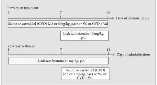
2.3. Study Design. he rats were randomly divided into experimental groups (8–10 animals/group) distributed in two protocols, namely, prevention and reversal treatments
(Figure 1), as described below. he use of LDX 10 mg/kg, p.o.,
to induce a mania-like behavior was based on a previous study from our research group [9]. he doses of CVD used here were calculated based on previous studies showing the neuroprotective efects of this drug [21] and human doses used for hypertension treatment as described elsewhere [22].
2.3.1. Prevention Treatment. In the prevention model, we simulated the maintenance phase of BD treatment, as pre-viously proposed [23]. Briely, diferent groups of animals were treated with CVD (2.5, 5, or 7.5 mg/kg, p.o.) once a day, VAL (200 mg/kg i.p.) twice a day, the association of VAL 100 mg/kg, i.p. (twice a day) + CVD 5 mg/kg, with a 15 min interval between drugs in the irst daily administration of VAL, or saline for 14 days. he reason for choosing the association CVD 5 mg/kg + VAL 100 mg/kg was based, in the case of CVD, on the irst behavioral results obtained where it seemed for us that this was the best dose to conduct this protocol; in the case of VAL the dose was reduced by half to determine a possible potentiation of its efect by CVD. It is important to mention that the dose of VAL usually used in models of mania is 200 mg/kg twice a day [11]. Between the 8th and 14th day, the experimental groups additionally received one oral dose of LDX daily. he time interval between the administration of the drugs and LDX was 30 min.
Locomotor activity using the open ield test (as described
in Section 2.4) and social interaction (as described in
Section 2.5) were measured on the 14th day of drug
adminis-tration, 2 hours ater the last drug administration. Following behavioral determinations, the rats were sacriiced by decap-itation and the prefrontal cortex (PFC), hippocampus (HC), and striatum (ST) were dissected, rapidly frozen, and stored at−70∘C until assayed.
Days of administration
1 7 14
Saline or carvedilol (CVD) (2.5or5mg/kg, p.o.) or Val or CVD +Val
Lisdexamfetamine10mg/kg, p.o.
Days of administration
1 7 14
Lisdexamfetamine10mg/kg, p.o.
Saline or carvedilol (CVD) (2.5or5mg/kg, p.o.) or Val or
CVD +Val Prevention treatment
Reversal treatment
Figure 1: Schematic representation of the experimental design.
between treatments. Locomotor activity and social interac-tion were measured on the 14th day of treatment, 2 hours ater the last drug administration. Ater behavioral determinations the rats were sacriiced and the brain areas dissected for the neurochemical determinations.
In the present study our primary outcome was to deter-mine the behavioral changes induced by CVD alone and associated with VAL in the model of mania induced by LDX. he secondary outcome was to determine the neurochemical alterations underlying these alterations.
2.4. Open Field Test. he locomotor activity was assessed using the open ield test [24]. his test was performed in a
50×50cm open ield surrounded by 50 cm high walls made of acrylic. he open ield loor was divided into four equal parts by black lines. he apparatus was placed in a red light room. he animals were gently placed on the center of the ield and allowed to freely explore the arena for 5 min. Crossings of the black lines (used to determine horizontal activity) and rearing behavior (used to determine vertical activity) were counted, during the 5 min period, by experienced raters who were blinded to treatment.
2.5. Social Interaction Test. he testing apparatus consisted of a60 × 40cm Plexiglas box divided into three chambers. Rats were able to move between chambers through a small opening (6 × 6cm) in the dividers. Iron cages in each of the two side chambers contained, in one side, the probe rat, whereas in the other side, the cage was empty. Test animals were placed in the center chamber. Rats were allowed 5 min of exploration time in the box, ater which an unfamiliar, same-sex probe rat from the same experimental group was placed in one of two restraining cages [25]. he time spent in each of the three chambers was measured, and social preference was deined as follows: (% time spent in the social chamber)−(% time spent in the opposite chamber).
2.6. Neurochemical Determinations
2.6.1. Tissue Preparation. Brain tissue samples were homog-enized (10 times (w/v) with ice-cold 0.1 M phosphate bufer (pH 7.4). he homogenates were centrifuged at 10,000 rpm for 15 minutes, and aliquots of supernatants were separated and used for determination of oxidative stress parameters.
For enzyme immunoassay determinations (ELISA) 20 times (w/v) homogenates prepared in cold phosphate-bufered saline (PBS, pH 7.4) were used. A protease inhibitor cocktail (Sigma-Aldrich, St. Louis, USA) was added to the bufer and the homogenate was centrifuged at 14,000 rpm for 30 min.
2.6.2. Determination of Reduced Glutathione (GSH) Lev-els. Reduced glutathione levels were evaluated to estimate endogenous defenses against oxidative stress. he method was based on Ellman’s reagent (DTNB) reaction with free thiol groups [26]. he brain areas were diluted in EDTA 0.02 M bufer (10% w/v) and added to a 50% trichloroacetic acid solution. Ater centrifugation (3,000 rpm/15 min), the supernatant of the homogenate was collected and mixed with 0.4 M tris-HCl bufer, pH 8.9, and 0.01 M 5,5-dithiobis 2-nitrobenzoic acid (DTNB). he yellow color product was read immediately at 412 nm using a spectrophotometer (Beckman coulter UV/Visible). Results were calculated based on a standard glutathione curve and are expressed as ng of GSH/g wet tissue.
Results are expressed as�mol of malonaldehyde (MDA)/g tissue.
2.6.4. Determination of Hippocampal BDNF Levels. he levels of BDNF (ELISA; Millipore, USA) were determined in each sample by enzyme immunoassays according to the speciic manufacturers’ directions. Results are expressed as pg/g tissue.
2.7. Statistical Analysis. Statistical analyses were performed with GraphPad Prism 6.0 for Windows, GraphPad Sotware (San Diego, CA, USA). he results of the behavioral and neurochemical studies are expressed as means±SEM (stan-dard errors of the mean). Regular two-way ANOVA with “treatment protocol” and “experimental groups” as factors was performed. Tukey’s test was used as post hoc test. Before ANOVA, D’Agostino-Pearson omnibus test was conducted to verify the normal distribution of the data. For all analyses, the signiicance level was set at� = 0.05.
3. Results
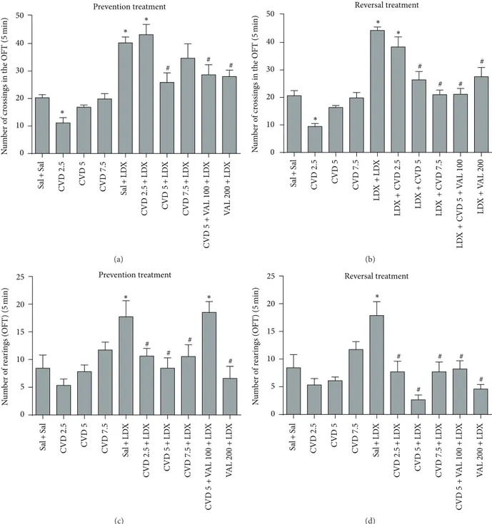
3.1. Carvedilol Alone and Associated with VAL Prevented and Reversed Hyperlocomotion and Alterations in Social Interac-tion Induced by LDX. Locomotor agitation and increased sociability are common features of mania [28]. In the present study two-way ANOVA of the number of crossings revealed a signiicant interaction between “treatment protocol” and “experimental group” [�9,88= 2.287,� = 0.0425] with signif-icant main efect of “treatment protocol” [�1,88 = 4.210,
� = 0.0431] and “experimental group” [�9,88 = 21.02,� <
0.001]. Regarding rearing behavior there was no signiicant interaction between factors [�9,85 = 1.57,� = 0.1671], but signiicant main efects of “treatment protocol” [�1,85= 10.02,
� = 0.0022] and “experimental groups” [�9,85 = 9.77,
� < 0.0001] were observed. Post hoc analysis showed that in both prevention and reversal protocols LDX caused hyper-locomotion (Figures 2(a) and 2(b)) and increased rearing behavior (Figures2(c)and2(d)) as compared to control (Sal + Sal) rats. In the prevention treatment the administration of CVD 5, the combination of CVD5 + VAL100, and VAL200 signiicantly prevented the hyperlocomotion induced by LDX (� < 0.001). Regarding rearing behavior CVD 2.5, CVD5, CVD7.5, and VAL200 prevented the increase in this parameter (� < 0.001). In the reversal treatment CVD5, CVD7.5, CVD5 + VAL100, and VAL200 signiicantly reversed the hyperlocomotion induced by LDX (� < 0.001). he increase in rearing behavior was reversed by CVD 2.5, CVD5, CVD7.5, CVD5 + VAL100, and VAL200 (� < 0.001). Ater the administration of CVD alone we could observe that only CVD 2.5 signiicantly decreased the number of crossings in both treatments.
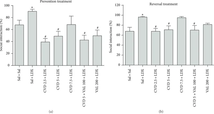
In the evaluation of social interaction two-way ANOVA revealed a signiicant interaction between factors [�6,72 =
2.24,� = 0.0490] with signiicant main efect of “treatment protocol” [�1,72 = 19.48, � < 0.0001] and “experimental group” [�6,72 = 5.28, � = 0.0001]. Post hoc analysis
showed that the administration of LDX in both prevention and reversal treatments signiicantly increased the percent of social interaction as compared to control (Sal + Sal) animals. In the prevention paradigm CVD2.5, CVD5, CVD5 + VAL100, and VAL200 signiicantly prevented the alteration induced by LDX (� < 0.01) (Figure 3(a)). On the other hand in the reversal paradigm CVD2.5, CVD5, and CVD5 + VAL100 signiicantly reversed the alterations induced by LDX (� < 0.05) (Figure 3(b)).
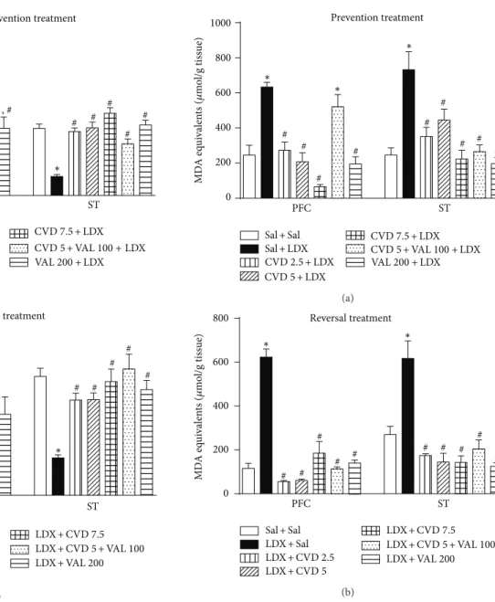
3.2. Carvedilol Alone and Associated with VAL Prevented and Reversed the Alterations in GSH and Lipid Peroxidation Induced by LDX in the Prefrontal Cortex and Striatum of Rats. Two-way ANOVA of GSH levels revealed in the PFC a signiicant interaction between “treatment protocol” and “experimental group” [�6,83 = 5.95,� < 0.0001] with sig-niicant main efect of “treatment protocol” [�1,83 = 13.97,
� = 0.0003] and “experimental group” [�6,83 = 4.99,� =
0.0002]. In the ST a signiicant interaction between factors [�6,98 = 2.68, � = 0.0188] with signiicant main efect of “treatment protocol” [�1,98 = 13.73, � = 0.0003] and “experimental group” [�6,98 = 8.34, � < 0.0001] was also observed. In relation to MDA levels in the PFC a signiicant interaction between “treatment protocol” and “experimental group” [�6,70 = 3.58, � = 0.0038] with signiicant main efect of “treatment protocol” [�1,70 = 19.21,� < 0.0001] and “experimental group” [�6,70 = 18.67, � < 0.0001] was observed. In the ST there was no signiicant interaction between factors [�6,92 = 1.26,� = 0.2841], but signiicant main efects of “treatment protocol” [�1,92 = 10.69, � =
0.0015] and “experimental group” [�6,92= 18.64,� < 0.0001] were observed.
Post hoc test showed that the administration of LDX in both prevention and reversal treatments signiicantly decreased the levels of GSH (Figure 4) as well as increased MDA levels (Figure 5) in the PFC and ST when compared to control (Sal + Sal) rats, as expected for an animal model of mania.
Regarding GSH levels, in the prevention treatment, the administration of CVD 2.5, CVD 5, CVD5 + VAL100, and VAL200 not only signiicantly prevented the decrement in GSH levels induced by LDX (� < 0.001) in the PFC
(Figure 4(a)) but also signiicantly increased GSH levels in
these experimental groups in relation to control animals (� <
0.001). In the ST all treatments prevented the decrease in GSH induced by LDX (� < 0.001). In the reversal treatment only VAL200 prevented the decrease in GSH levels induced by LDX in the PFC. In the ST CVD2.5, CVD5, CVD5 + VAL100, and VAL200 reversed the GSH decrement induced by LDX (� < 0.001) (Figure 4(b)).
he evaluation of lipid peroxidation revealed that in the PFC and ST of the animals subjected to the prevention
(Figure 5(a)) and reversal (Figure 5(b)) treatments both doses
of CVD and VAL200 and the association of CVD + VAL100 prevented and reversed the alterations induced by LDX (� <
0 10 20 30 40 50 Prevention treatment ∗ ∗ ∗ # # # Sal + Sal CVD 2.5 CVD 5 CVD 7. 5 Sal + LD X CVD 2.5 + LD X CVD 5+ LD X CVD 7. 5 + LD X CVD 5+ V AL 100 + LD X V AL 200 + LD X N u m b er o f cr ossin
gs in t
h e O FT ( 5 min) (a) 0 10 20 30 40 50 Reversal treatment ∗ ∗ ∗ # # # # Sal + Sal CVD 2.5 CVD 5 CVD 7. 5 LD X + LD X CVD 2.5 + LD X CVD 5 + LD X CVD 7. 5 + LD X CVD 5+ V AL 100 + LD X V AL 200 + LD X N u m b er o f cr ossin
gs in t
h e O FT ( 5 min) (b) Prevention treatment 0 5 10 15 20 25 ∗ ∗ # # # # Sal + Sal CVD 2.5 CVD 5 CVD 7. 5 Sal + LD X CVD 2.5 + LD X CVD 5+ LD X CVD 7. 5 + LD X CVD 5+ V AL 100 + LD X V AL 200 + LD X N um b er o f r ea rin gs (O FT) ( 5 min) (c) Reversal treatment 0 5 10 15 20 25 ∗ # # # # # Sal + Sal CVD 2.5 CVD 5 CVD 7. 5 Sal + LD X CVD 2.5 + LD X CVD 5+ LD X CVD 7. 5 + LD X CVD 5+ V AL 100 + LD X V AL 200 + LD X N um b er o f r ea rin gs (O FT) ( 5 min) (d)
Figure 2: Number of crossings and rearings in the open ield test in animals submitted to the prevention ((a), (c)) and reversal ((b), (d)) treatments. Data were analyzed by one-way ANOVA followed by Student-Newman-Keuls post hoc test. Values represent mean±SEM (6–8 animals/group).∗� < 0.05versus Sal + Sal;#� < 0.05versus Sal + LDX or LDX + LDX. CVD: carvedilol; LDX: lisdexamfetamine; OFT: open
ield test; Sal: saline; VAL: valproate.
CVD5 + VAL100 in which the alterations in MDA levels were not prevented by this treatment.
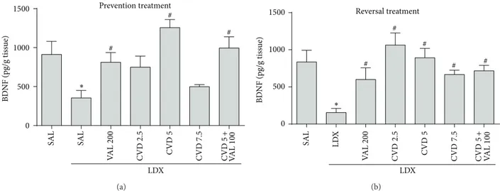
3.3. Carvedilol Alone and Associated with VAL Prevents and Reverses the Alterations in the Hippocampal Levels of BDNF Induced by LDX. Two-way ANOVA of hippocampal BDNF levels revealed no signiicant interaction between factors [�6,62 = 1.93,� = 0.0905], but main efect of “experimental
group” [�6,62 = 7.48, � < 0.0001] was observed. BDNF is being considered a potential candidate as a biomarker for bipolar disorder [1]. In this context, as observed in
Figure 6, post hoc analysis showed that the administration
0 20 40 60 80 100
Prevention treatment
S
o
cial in
terac
tio
n
(%)
∗
# #
# #
Sal
+
Sal
Sal
+
LD
X
CVD
2.5
+
LD
X
CVD
5+
LD
X
CVD
7.
5
+
LD
X
CVD
5+
V
AL 100
+
LD
X
V
AL 200
+
LD
X
(a)
S
o
cial in
terac
tio
n
(%)
0 20 40 60 80 100
120 Reversal treatment
∗
# #
#
Sal
+
Sal
Sal
+
LD
X
CVD
2.5
+
LD
X
CVD
5+
LD
X
CVD
7.
5
+
LD
X
CVD
5+
V
AL 100
+
LD
X
V
AL 200
+
LD
X
(b)
Figure 3: Percent of social interaction in animals submitted to the prevention (a) and reversal (b) treatments. Data were analyzed by one-way ANOVA followed by Student-Newman-Keuls post hoc test. Values are expressed as mean±SEM (6–8 animals/group).∗� < 0.05versus Sal +
Sal;#� < 0.05versus Sal + LDX or LDX + LDX. CVD: carvedilol; LDX: lisdexamfetamine; Sal: saline; VAL: valproate.
levels induced by LDX (� < 0.05). In the reversal treatment both doses of CVD as well as CVD5 + VAL100 and VAL200 reversed the decrease in these neurotrophin levels induced by LDX (� < 0.01).
4. Discussion
he results of the present work demonstrated, to the best of our knowledge, for the irst time that CVD is efective in the prevention and reversal of LDX-induced mania-like behavioral and neurochemical alterations. Additionally, CVD potentiated the efects of the mood-stabilizing VAL.
Manic states include complex and multifaceted symptoms such as overactivity, hypersexuality, irritability, and reduced need for sleep [29]. To date, the evaluation of these symp-toms in preclinical models of mania is almost restricted to the observation of hyperlocomotion; nevertheless this was recently criticized [12]. hus, in our study we decided to evaluate the social interaction of the animals since this feature is increased in mania [28].
Based on our behavioral results we observed that the administration of CVD 5 mg/kg, VAL 200 mg/kg, and the combination of VAL 100 mg/kg + CVD 5 mg/kg prevented and reversed hyperlocomotion and the increased sociability induced by LDX, with the exception of VAL alone that was not able to reverse the LDX-induced alteration in social interaction. Regarding the mood-stabilizing drugs, when we irst proposed this LDX-induced model of mania [9] lithium was the only drug used to assess the predictive validity of the model. Based on a previous preclinical study that demonstrated a potentiation of anticonvulsant efects by
the administration of VAL 100 mg/kg and CVD 5 mg/kg, we decided to use, in the present study, VAL as mood-stabilizing drug [30].
he behavioral alterations observed in BD patients are linked to central mechanisms, such as the following [7,31]: (i) alterations in monoamine levels, for example, the dopamine dysregulation syndrome; (ii) mitochondrial dysregulation; (iii) alterations in calcium homeostasis; (iv) neuroinlam-mation; (v) oxidative imbalance; and (vi) dysregulation in neurotrophin’s levels. hese mechanisms are also involved in the neuroprogression of this mental disorder [32], emerging, mainly the last ive, as important targets for BD treatment [1,33].
Focusing on the aforementioned targets for BD treat-ment, CVD is a drug that presents antioxidant and mitochon-drial stabilizing properties as well as regulating intracellular calcium that may be considered as an important option for the treatment of BD. Additionally, in the last ten years the neuroprotective properties of this drug began to be studied although with incipient indings to date [34,35].
Prevention treatment
0 1000 2000 3000 4000
GS
H (n
g/g tissue)
PFC ST
∗ ∗
# #
#
# #
Sal +Sal Sal +LDX
VAL 200 +LDX CVD2.5 +LDX
CVD5 +LDX
CVD7.5 +LDX CVD5 +VAL 100 +LDX
∗, # ∗, #
∗, #
∗, #
(a)
GS
H (n
g/g tissue)
Reversal treatment
0 500 1000 1500 2000
PFC ST
∗
∗
# # #
# #
Sal +Sal
LDX +Sal
LDX +VAL 200 LDX +CVD2.5
LDX +CVD5
LDX +CVD7.5 LDX +CVD5 +VAL 100
(b)
Figure 4: Levels of reduced glutathione (GSH) in the prefrontal cortex (PFC) and striatum (ST) of mice submitted to the prevention (a) and reversal (b) treatments. Data were analyzed by one-way ANOVA followed by Student-Newman-Keuls post hoc test. Values are expressed as mean ±SEM (6–8 animals/group).∗� < 0.05 versus Sal + Sal;#� < 0.05versus Sal + LDX or LDX + LDX. CVD:
carvedilol; LDX: lisdexamfetamine; Sal: saline; VAL: valproate.
to reduced expression of BDNF. he mechanisms linking oxidative stress to decreased BDNF levels involve several factors such as the following: (i) decrease of DNA-binding activities of activator protein-1 and cAMP response element-binding protein (CREB), BDNF transcription factor, which is associated with reduction of BDNF gene expression [39], and (ii) energy imbalance which causes a dysfunction in the N-methyl-D-aspartate (NMDA) channel resulting in decreases in BDNF gene expression [40].
Regarding BDNF, the serum levels of this neurotrophin in BD patients are decreased in depressive and manic episodes,
Prevention treatment
0 200 400 600 800 1000
PFC ST
MD
A eq
ui
valen
ts (
𝜇
mo
l/g tissue)
∗
∗
∗
# #
# #
# #
# #
#
Sal +Sal Sal +LDX
VAL 200 +LDX CVD2.5 +LDX
CVD5 +LDX
CVD7.5 +LDX CVD5 +VAL 100 +LDX
(a)
MD
A eq
ui
valen
ts (
𝜇
mo
l/g tissue)
0 200 400 600 800
PFC ST
∗ ∗
# # # #
#
# #
#
# #
Sal +Sal
LDX +Sal
LDX +VAL 200 LDX +CVD2.5
LDX +CVD5
LDX +CVD7.5 LDX +CVD5 +VAL 100
Reversal treatment
(b)
Figure 5: Levels of malondialdehyde (MDA), a lipid peroxidation marker, in the prefrontal cortex (PFC) and striatum (ST) of mice submitted to the prevention (a) and reversal (b) treatments. Data were analyzed by one-way ANOVA followed by Student-Newman-Keuls post hoc test. Values are expressed as mean± SEM (6–8 animals/group).∗� < 0.05versus Sal + Sal;#� < 0.05versus Sal + LDX or LDX + LDX. CVD: carvedilol; LDX: lisdexamfetamine; Sal: saline; VAL: valproate.
returning to normal levels in euthymia [41] with a similar pattern of alteration in animal models of mania [9, 14]. Together, these mechanisms have been extensively implicated in the pathophysiology of schizophrenia and BD [37,42].
Prevention treatment
SAL SAL
V
AL 200
CVD
2.5
CVD
5
CVD
7.5
CVD
5+
V
AL 100
0 500 1000 1500
LDX
BD
NF (pg/g tis
sue)
∗
#
#
#
(a)
SAL LD
X
V
AL 200
CVD
2.5
CVD
5
CVD
7.
5
CVD
5+
V
AL 100
LDX 0
500 1000 1500
BD
NF (pg/g tis
sue)
Reversal treatment
∗
#
# #
# #
(b)
Figure 6: Levels of brain derived neurotrophic factor (BDNF) in the hippocampus of rats submitted to the prevention (a) and reversal (b) treatments. Data were analyzed by one-way ANOVA followed by Student-Newman-Keuls post hoc test. Values are expressed as mean±SEM (6–8 animals/group).∗� < 0.05versus Sal + Sal;#� < 0.05versus Sal + LDX or LDX + LDX. CVD: carvedilol; LDX: lisdexamfetamine; Sal:
saline; VAL: valproate.
of the antioxidant N-acetyl cysteine improved functional outcomes [43]. herefore, this adds further evidences for the importance of CVD, a drug that treats cardiovascular conditions besides being antioxidant, in BD treatment.
Of note, the use, in the present study, of the association of half the dose of VAL (100 mg/Kg, twice a day) with CVD 5 maintained the efect of VAL 100 in a similar pattern to the one presented by VAL 200 mg/kg twice a day. One exception was noticed in the social interaction test where in the reversal protocol the association reversed the alteration induced by LDX whereas VAL alone did not. his is an important inding because VAL presents important side efects, such as tremor, weight gain, alopecia, more frequent sedation, and infection [44] that could be alleviated by the use of lower doses. Indeed a previous preclinical study suggested that CVD potentiates the anticonvulsant activity of VAL possibly by a pharmacodynamics interaction [30]. In this previous study the association of CVD 5 + VAL 100 was the best for the increase in seizure threshold induced by pentylenetetrazole, an efect that was accompanied by increase in GSH levels and decrease in lipid peroxidation [30].
he combination CVD 5 + VAL 100 did not prevent LDX-induced rearing alteration. his result does not compromise the antimanic-like efect of this combination observed in this study, since the parameter number of crossings is the most reliable [45, 46]. On the contrary, CVD 7.5 did not prevent LDX-induced alterations in the number of crossings, social interaction, GSH levels in the PFC, and BDNF. One possible explanation in that when compared to humans on an mg/m2 basis, 7.5 mg/kg CVD in rats corresponds approximately to 75 mg/daily CVD in humans. Of note, the total daily doses of 6.25 to 50 mg CVD are the most prescribed for cardiovascular conditions [47]. hus, based on this evidence possibly 7.5 mg/kg CVD in rats was an excessive dose.
Our study has some limitations: irst, animal models in psychiatry are fair representations of real complexity of
the disorders. hat is to say, clinical studies are necessary before we can conclude that CVD could bring tangible beneits to BD patients. Furthermore, our study evaluated a correlation between intervention and outcome and was not equipped to parse possible mechanistic pathways. herefore, we could not establish the real mechanisms that are necessary and adequate to explain the behavioral efects of CVD; for example, mitochondrial protection by CVD was not evaluated here.
In conclusion, CVD was able to prevent and reverse oxidative imbalance and BDNF levels in the model of mania induced by LDX. Overall, the results presented here give the irst preclinical evidences for the future design of clinical trials investigating the use of CVD in mania.
Conflict of Interests
he authors declare that there is no conlict of interests regarding the publication of this paper.
Acknowledgments
by Department of Psychiatry and Behavioral Sciences, the University of Texas Medical School at Houston.
References
[1] B. N. Frey, A. C. Andreazza, J. Houenou et al., “Biomarkers in bipolar disorder: a positional paper from the International Soci-ety for Bipolar Disorders Biomarkers Task Force,”Australian and New Zealand Journal of Psychiatry, vol. 47, no. 4, pp. 321– 332, 2013.
[2] R. F. Bachmann, R. J. Schloesser, T. D. Gould, and H. K. Manji, “Mood stabilizers target cellular plasticity and resilience cas-cades: implications for the development of novel therapeutics,” Molecular Neurobiology, vol. 32, no. 2, pp. 173–202, 2005. [3] A. Mart´ınez-Ar´an, E. Vieta, M. Reinares et al., “Cognitive
func-tion across manic or hypomanic, depressed, and euthymic states in bipolar disorder,”he American Journal of Psychiatry, vol. 161, no. 2, pp. 262–270, 2004.
[4] A. M. Kilbourne, J. R. Cornelius, X. Han et al., “Burden of general medical conditions among individuals with bipolar disorder,”Bipolar Disorders, vol. 6, no. 5, pp. 368–373, 2004. [5] M. Weiner, L. Warren, and J. G. Fiedorowicz, “Cardiovascular
morbidity and mortality in bipolar disorder,”Annals of Clinical Psychiatry, vol. 23, no. 1, pp. 40–47, 2011.
[6] M. Berk, “Neuroprogression: pathways to progressive brain changes in bipolar disorder,”International Journal of Neuropsy-chopharmacology, vol. 12, no. 4, pp. 441–445, 2009.
[7] H. K. Manji, J. A. Quiroz, J. L. Payne et al., “he underlying neu-robiology of bipolar disorder,”World Psychiatry, vol. 2, pp. 136– 146, 2003.
[8] D. S. Macˆedo, C. D. Medeiros, R. C. Cordeiro et al., “Efects of alpha-lipoic acid in an animal model of mania induced by d-amphetamine,”Bipolar Disorders, vol. 14, no. 7, pp. 707–718, 2012.
[9] D. S. Macˆedo, D. F. de Lucena, A. I. G. Queiroz et al., “Efects of lithium on oxidative stress and behavioral alterations induced by lisdexamfetamine dimesylate: relevance as an animal model of mania,”Progress in Neuro-Psychopharmacology and Biological Psychiatry, vol. 43, pp. 230–237, 2013.
[10] R. H. Perlis, M. J. Ostacher, J. K. Patel et al., “Predictors of recurrence in bipolar disorder: primary outcomes from the Sys-tematic Treatment Enhancement Program for Bipolar Disorder (STEP-BD),”American Journal of Psychiatry, vol. 163, no. 2, pp. 217–224, 2006.
[11] B. N. Frey, S. S. Valvassori, G. Z. R´eus et al., “Efects of lithium and valproate on amphetamine-induced oxidative stress gen-eration in an animal model of mania,”Journal of Psychiatry & Neuroscience, vol. 31, no. 5, pp. 326–332, 2006.
[12] J. W. Young, B. L. Henry, and M. A. Geyer, “Predictive animal models of mania: hits, misses and future directions,”British Journal of Pharmacology, vol. 164, no. 4, pp. 1263–1284, 2011. [13] M. Pennick, “Absorption of lisdexamfetamine dimesylate and
its enzymatic conversion to d-amphetamine,”Neuropsychiatric Disease and Treatment, vol. 6, pp. 317–327, 2010.
[14] B. N. Frey, A. C. Andreazza, K. M. M. Ceres´er et al., “Efects of mood stabilizers on hippocampus BDNF levels in an animal model of mania,”Life Sciences, vol. 79, no. 3, pp. 281–286, 2006. [15] S. S. Valvassori, G. T. Rezin, C. L. Ferreira et al., “Efects of mood stabilizers on mitochondrial respiratory chain activity in brain of rats treated with d-amphetamine,” Journal of Psychiatric Research, vol. 44, no. 14, pp. 903–909, 2010.
[16] T.-L. Yue, H.-Y. Cheng, P. G. Lysko et al., “Carvedilol, a new vasodilator and beta adrenoceptor antagonist, is an antioxidant and free radical scavenger,”Journal of Pharmacology and Exper-imental herapeutics, vol. 263, no. 1, pp. 92–98, 1992.
[17] N. Noguchi, K. Nishino, and E. Niki, “Antioxidant action of the antihypertensive drug, carvedilol, against lipid peroxidation,” Biochemical Pharmacology, vol. 59, no. 9, pp. 1069–1076, 2000. [18] A. Kumar, S. Dogra, and A. Prakash, “Efect of carvedilol on
behavioral, mitochondrial dysfunction, and oxidative damage against D-galactose induced senescence in mice,” Naunyn-Schmiedeberg’s Archives of Pharmacology, vol. 380, no. 5, pp. 431– 441, 2009.
[19] R. F. D. A. J´unior, T. O. Souza, C. A. X. de Medeiros et al., “Carv-edilol decrease IL-1� and TNF-�, inhibits MMP-2, MMP-9, COX-2, and RANKL expression, and up-regulates OPG in a rat model of periodontitis,”PLoS ONE, vol. 8, no. 7, Article ID e66391, 2013.
[20] NIH,Guide for the Care and Use of Laboratory Animals, Institute of Laboratory Animal Research, National Research Council, National Academies Press, 1996.
[21] S. I. Savitz, J. A. Erhardt, J. V. Anthony et al., “he novel� -blocker, carvedilol, provides neuroprotection in transient focal stroke,”Journal of Cerebral Blood Flow & Metabolism, vol. 20, no. 8, pp. 1197–1204, 2000.
[22] S. Reagan-Shaw, M. Nihal, and N. Ahmad, “Dose translation from animal to human studies revisited,”he FASEB Journal, vol. 22, no. 3, pp. 659–661, 2008.
[23] B. N. Frey, M. R. Martins, F. C. Petronilho, F. Dal-Pizzol, J. Quevedo, and F. Kapczinski, “Increased oxidative stress ater repeated amphetamine exposure: possible relevance as a model of mania,”Bipolar Disorders, vol. 8, no. 3, pp. 275–280, 2006. [24] J. Archer, “Tests for emotionality in rats and mice: a review,”
Animal Behaviour, vol. 21, no. 2, pp. 205–235, 1973.
[25] K. Radyushkin, K. Hammerschmidt, S. Boretius et al., “Neuroli-gin-3-deicient mice: model of a monogenic heritable form of autism with an olfactory deicit,”Genes, Brain and Behavior, vol. 8, no. 4, pp. 416–425, 2009.
[26] G. L. Ellman, “Tissue sulhydryl groups,”Archives of Biochem-istry and Biophysics, vol. 82, no. 1, pp. 70–77, 1959.
[27] H. Ohkawa, N. Ohishi, and K. Yagi, “Assay for lipid peroxides in animal tissues by thiobarbituric acid reaction,”Analytical Bio-chemistry, vol. 95, no. 2, pp. 351–358, 1979.
[28] L. G. Sylvia, L. B. Alloy, J. A. Hafner, M. C. Gauger, K. Verdon, and L. Y. Abramson, “Life events and social rhythms in bipolar spectrum disorders: a prospective study,”Behavior herapy, vol. 40, no. 2, pp. 131–141, 2009.
[29] R. A. Kowatch, E. A. Youngstrom, A. Danielyan, and R. L. Fin-dling, “Review and meta-analysis of the phenomenology and clinical characteristics of mania in children and adolescents,” Bipolar Disorders, vol. 7, no. 6, pp. 483–496, 2005.
[30] R. Goel and A. Goel, “Interactions between carvedilol and sodium valproate along with neurobehavioural co-morbidities in various epilepsy models,”Drug Invention Today, vol. 5, no. 2, pp. 87–91, 2013.
[31] A. R. Newberg, L. A. Catapano, C. A. Zarate, and H. K. Manji, “Neurobiology of bipolar disorder,”Expert Review of Neurother-apeutics, vol. 8, no. 1, pp. 93–110, 2008.
[33] C. A. Zarate Jr. and H. K. Manji, “Bipolar disorder: candidate drug targets,”Mount Sinai Journal of Medicine, vol. 75, no. 3, pp. 226–247, 2008.
[34] K. Yamagata, S. Ichinose, and M. Tagami, “Amlodipine and carvedilol prevent cytotoxicity in cortical neurons isolated from stroke-prone spontaneously hypertensive rats,” Hypertension Research, vol. 27, no. 4, pp. 271–282, 2004.
[35] Y. Ouyang, Z. Chen, M. Tan et al., “Carvedilol, a third-gener-ation �-blocker prevents oxidative stress-induced neuronal death and activates Nrf2/ARE pathway in HT22 cells,” Biochem-ical and BiophysBiochem-ical Research Communications, vol. 441, no. 4, pp. 917–922, 2013.
[36] J. N. Keller and M. P. Mattson, “Roles of lipid peroxidation in modulation of cellular signaling pathways, cell dysfunction, and death in the nervous system,”Reviews in the Neurosciences, vol. 9, no. 2, pp. 105–116, 1998.
[37] O. M. Dean, M. van den Buuse, A. I. Bush et al., “Role for glutathione in the pathophysiology of bipolar disorder and schizophrenia? Animal models and relevance to clinical prac-tice,”Current Medicinal Chemistry, vol. 16, no. 23, pp. 2965– 2976, 2009.
[38] A. C. Andreazza, C. Cassini, A. R. Rosa et al., “Serum S100B and antioxidant enzymes in bipolar patients,”Journal of Psychiatric Research, vol. 41, no. 6, pp. 523–529, 2007.
[39] E. Iwata, M. Asanuma, S. Nishibayashi, Y. Kondo, and N. Ogawa, “Diferent efects of oxidative stress on activation of transcription factors in primary cultured rat neuronal and glial cells,”Molecular Brain Research, vol. 50, no. 1-2, pp. 213–220, 1997.
[40] M. Roceri, W. Hendriks, G. Racagni, B. A. Ellenbroek, and M. A. Riva, “Early maternal deprivation reduces the expression of BDNF and NMDA receptor subunits in rat hippocampus,” Molecular Psychiatry, vol. 7, no. 6, pp. 609–616, 2002.
[41] I. Grande, G. R. Fries, M. Kunz, and F. Kapczinski, “he role of BDNF as a mediator of neuroplasticity in bipolar disorder,” Psychiatry Investigation, vol. 7, no. 4, pp. 243–250, 2010. [42] R. Machado-Vieira, A. C. Andreazza, C. I. Viale et al., “Oxidative
stress parameters in unmedicated and treated bipolar subjects during initial manic episode: a possible role for lithium antioxi-dant efects,”Neuroscience Letters, vol. 421, no. 1, pp. 33–36, 2007. [43] P. V. Magalh˜aes, O. M. Dean, A. I. Bush et al., “Systemic illness moderates the impact of N-acetyl cysteine in bipolar disorder,”Progress in Neuro-Psychopharmacology and Biological Psychiatry, vol. 37, no. 1, pp. 132–135, 2012.
[44] K. A. Macritchie, J. R. Geddes, J. Scott, D. R. Haslam, and G. M. Goodwin, “Valproic acid, valproate and divalproex in the maintenance treatment of bipolar disorder,” he Cochrane Database of Systematic Reviews, vol. 10, Article ID CD003196, 2001.
[45] R. E. Riegel, S. S. Valvassori, G. Elias et al., “Animal model of mania induced by ouabain: evidence of oxidative stress in submitochondrial particles of the rat brain,” Neurochemistry International, vol. 55, no. 7, pp. 491–495, 2009.
[46] R. Lien, S. Flaisher-Grinberg, C. Cleary, M. Hejny, and H. Einat, “Behavioral efects of Bcl-2 deiciency: implications for afective disorders,”Pharmacological Reports, vol. 60, no. 4, pp. 490–498, 2008.
Submit your manuscripts at
http://www.hindawi.com
Neurology
Research International Hindawi Publishing Corporation
http://www.hindawi.com Volume 2014
Alzheimer’s Disease
Hindawi Publishing Corporation
http://www.hindawi.com Volume 2014
Scientifica
Hindawi Publishing Corporation
http://www.hindawi.com Volume 2014
Hindawi Publishing Corporation
http://www.hindawi.com Volume 2014
BioMed
Research International
Hindawi Publishing Corporation
http://www.hindawi.com Volume 2014
Research and Treatment
The Scientiic
World Journal
Hindawi Publishing Corporationhttp://www.hindawi.com Volume 2014
Hindawi Publishing Corporation
http://www.hindawi.com Volume 2014
Neural Plasticity
Hindawi Publishing Corporation
http://www.hindawi.com Volume 2014
Parkinson’s
Disease
Hindawi Publishing Corporationhttp://www.hindawi.com Volume 2014 Research and Treatment
Autism
Sleep Disorders
Hindawi Publishing Corporationhttp://www.hindawi.com Volume 2014
Hindawi Publishing Corporation
http://www.hindawi.com Volume 2014
Neuroscience
Journal
Epilepsy Research and Treatment
Hindawi Publishing Corporation
http://www.hindawi.com Volume 2014
Hindawi Publishing Corporation
http://www.hindawi.com Volume 2014
Psychiatry
Journal
Hindawi Publishing Corporation
http://www.hindawi.com Volume 2014
Computational and Mathematical Methods in Medicine
and Treatment
Hindawi Publishing Corporation
http://www.hindawi.com Volume 2014
Hindawi Publishing Corporation
http://www.hindawi.com Volume 2014
Brain Science
International Journal ofResearch and Treatment Hindawi Publishing Corporation
http://www.hindawi.com Volume 2014
Neurodegenerative
Diseases
Hindawi Publishing Corporation
http://www.hindawi.com Volume 2014
Journal of
Cardiovascular Psychiatry and Neurology
Hindawi Publishing Corporation