ABSTRACT: The aim of this study was to evaluate the inluence of using the surgical operating microscope (SOM) for detection of the mesiolingual (ML) canal oriice in extracted irst maxillary permanent molars. One hundred and eight human irst maxillary permanent molars were randomly selected and mounted onto a dental chair mannequin. Conventional access cavity was prepared and an attempt was made to locate the mesiolingual canal oriice using only a sharp explorer, a mirror and a #10 K-ile. A mesiolingual canal oriice was either located or not located. If not located, the teeth were then evaluated by using a surgical operating microscope (SOM). The mesio-buccal roots of all teeth where the ML canal oriice had not been located were sectioned in an axial plane and the sections were explored with an adjunctive use of the SOM at a 25 X magniication. ML canal oriices were detected in 58 teeth using only a sharp explorer, a mirror and #10 K-ile. In the remaining 50 teeth, 37 ML canal oriices were located by using the SOM and 3 ML canal oriices were located after root sectioning. In 10 teeth, the ML canal oriices were not found. The results of this study showed a high incidence of a ML canal in the mesiobuccal roots of the irst maxillary molars (90.7%) and demonstrated that the adjunctive use of the SOM increased the ability of the dental clinician to locate the ML canal oriice.
DESCRIPTORS: Incidence; Dental pulp cavity; Microscopy.
RESUMO: O objetivo deste estudo foi avaliar a inluência do uso do microscópio cirúrgico na localização do canal mesiopalatino (MP) em primeiros molares superiores humanos permanentes extraídos. Cento e oito primeiros molares superiores permanentes foram selecionados aleatoriamente e montados em um manequim dental. Uma cavidade de acesso convencional foi realizada e uma tentativa de se localizar o canal mesiopalatino foi feita, uti-lizando-se somente uma sonda exploradora aiada, um odontoscópio e uma lima tipo K tamanho 10. Quando não localizado o referido canal, os dentes foram então avaliados com auxílio de um microscópio cirúrgico (MC). As raízes mésio-vestibulares de todos os dentes nos quais o canal MP não foi localizado foram então seccionadas transversalmente e exploradas com auxílio do MC, com um aumento de 25 X. Os canais MP foram detectados em 58 dentes em que se usaram somente uma sonda exploradora aiada, um odontoscópio e uma lima tipo K tamanho 10. Nos 50 dentes restantes, 37 canais MP foram localizados com o auxílio do MC e 3 canais MP foram localizados após a secção das raízes. Em 10 dentes os canais MP não foram localizados. Os resultados deste estudo mostra-ram uma alta incidência do canal MP na raiz mésio-vestibular do primeiro molar superior permanente (90.7%) e demonstraram que o uso adjunto do MC aumentou a capacidade do operador em localizar o canal MP.
DESCRITORES: Incidência; Cavidade da polpa dentária; Microscopia.
INTRODUCTION
The goals of successful endodontics are the total obliteration of the canal space and the per-fect sealing of the apical foramen with an inert filling material. For this, the location and nego-tiation, with subsequent cleaning and shaping,
of the root canal system are necessary. Pécora et al.11 (1992) affirms that one of the main reasons
for the failure of root canal therapy is the lack of sufficient knowledge concerning the anatomy of teeth, both internal and external. The first
* PhD, Department of Endodontics; **Specialists in Endodontics; ****MSc, Department of Endodontics – Rio de Janeiro State University.
*** PhD, Department of Endodontics, University of Fortaleza.
The influence of the Surgical Operating Microscope in locating
the mesiolingual canal orifice: a laboratory analysis
Influência do Microscópio Cirúrgico na localização do canal
mesiopalatino: uma análise laboratorial
Tauby Coutinho Filho* Renata Sá La Cerda**
lary molar is the most bulky teeth in the mouth, and has many anatomical variations. Usually both the distobuccal root and the palatal root present only one canal. The mesiobuccal root presents more anatomical variations, such as the number and disposition of the canals. Bjorndal, Skidmore2
(1983)affirmed that the difficulty in locating the mesiolingual canal (ML) during the first maxillary permanent molar endodontic treatment may have implications for the long-term prognosis.
Clinically, the presence or absence of the mesiolingual canal is limited by the conditions in which locating of the orifice is carried out. The ability to locate the mesiolingual canal depends on the skill of the operator, the complexity of the anatomy and the use of high power illumination and magnification techniques, such as that per-formed with the surgical operating microscope. A literature review has demonstrated wide variation in the prevalence of the ML canal mostly in in vi-tro researches. Hess6 (1925), in a classical study,
reported finding 4 canals in 54% of first maxil-lary molars. Weine et al.16 (1969) evaluated first
maxillary molars and located 4 canals in 62% of the teeth. Pineda, Kuttler12 (1972) evaluated the
number of canals in first and second molars and found 4 canals in 51.5% of the teeth. Fogel et al.4
(1994) evaluated the use of 2.5 X magnification telescopes with fiberoptic headlamps for locating the mesiolingual canals in first maxillary molars in vivo. They found that 71.2% of the mesiobuccal roots had two canals. Stropko15 (1999) found 73%
to 93% of mesiolingual canals in a recent clinical study. Baldassari-Cruz et al.1 (2002) evaluated the
influence of the dental operating microscope in locating the mesiolingual orifice. This study dem-onstrated that the adjunctive use of the dental operating microscope increased the ability of the clinician to locate a mesiolingual canal.
The purpose of this study was to evaluate whether the adjunctive use of the surgical oper-ating microscope would increase detection of the mesiolingual canal orifice in the mesiobuccal root of first maxillary permanent molars.
MATERIALS & METHODS
For this study, 108 human first maxillary left and right molars were selected randomly from the tooth bank of the Department of Endodontics, Rio de Janeiro State University.
The teeth were stored in 10% neutral formalin. The sex and race of the patients from whom these
teeth were obtained were unknown. The teeth were mounted onto a dental chair mannequin (Columbia Dentoform, Long Island, NY, USA). No isolation of the teeth by rubber dam was done. Without using magnification or headlamps, a conventional access cavity was prepared using a #6 high-speed hand-piece spherical bur (Dentsply-Maillefer, Ballaigues, Switzerland), a sharp endodontic explorer, a mirror, a #10 K-file (Dentsply-Maillefer, Ballaigues, Switzer-land) and water irrigation. After locating the mesio-buccal, distobuccal and palatal canals, an attempt was made to locate the mesiolingual canal orifice us-ing only a sharp explorer, a mirror and a #10 K-file. If the mesiolingual canal orifice was not located, a #700l low-speed hand-piece bur (Dentsply-Maillefer, Ballaigues, Switzerland) was used 2 or 3 mm into the orifice of the mesiobuccal canal where a trench was prepared in a lingual and slightly mesial direc-tion through the mesial dentinal shelf1. The root was
again explored by using only a sharp endodontic explorer, a mirror and a #10 K-file in an attempt to locate a mesiolingual canal orifice.
A mesiolingual canal orifice was either located or not located. If not located the teeth were then evaluated by using a surgical operating microscope (Dental F. Vasconcelos, M900 –25 X, São Paulo, Brazil) at a magnification of 25 X. Again, an ML canal orifice was either located or not located. The mesiobuccal roots of all teeth where the ML canal orifice was not located were sectioned in an axial plane 6 mm below the cemento-enamel junction. The sections were explored using a sharp endodon-tic explorer, a mirror and a #10 K-file (Figure 1F) with the adjunctive use of the surgical operating microscope at a magnification of 25 X to determine the actual presence or absence of the orifice of the ML canal. In this methodology, each tooth served as its own control.
RESULTS
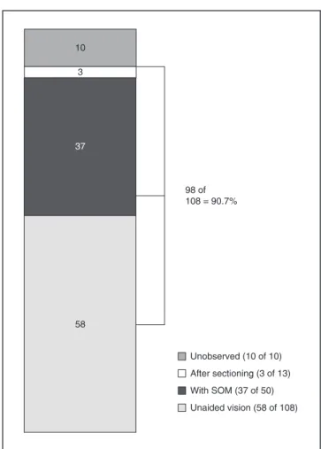
the remaining 13 teeth (23%). These 3 canals were located neither with the traditional methods nor with the SOM evaluation. A total of 98 ML canal orifices were identified out of 108 experimental teeth (90.7%). (Graph 1).
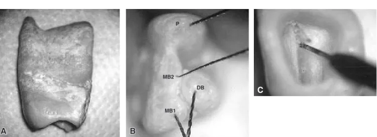
Figure 1A shows one of the first maxillary mo-lars used in this experiment and the presence of 4 distinct foramina (Figure 1B). Figure 1C shows an example of the difficulty in locating the ML canal orifice in the first maxillary molar, and Figure 1D summarizes the results of this work.
DISCUSSION
Successful endodontic treatment demands an adequate cleaning, shaping and filling of the root canal system. For this, the endodontist must have comprehensive knowledge about root canal morphology. Many types of root curvatures and other anatomical variations may be present in
teeth subjected to endodontic treatment. If a root canal system is not located, this may reduce the chance of treatment success. In that perspective, the incidence of the ML canal in the mesiobuccal root of the first maxillary molar is always a matter of interest to the endodontic community. Baldas-sari-Cruz et al.1 (2002) related that the ML canal
in mesiobuccal roots of maxillary first molars can be extremely difficult to locate clinically.
There is a significant difference in the inci-dence of the ML canal of the mesiobuccal root of first maxillary molar when evaluated in vitro and in vivo. Seidberg et al.14 (1973) reported a clinical
incidence of 35%, varying to 69% in vitro. Pomer-anz, Fishelberg13 (1974) related findings closer to
those of Seidberg et al.14 (1973), an incidence of
approximately 69% of the ML canal in the mesio-buccal roots of first maxillary molars in vitro, and only 31% after in vivo evaluation. Hartwell, Bellizzi5
(1982) observed a divergence between the clinical incidence of the ML canal in first maxillary molars and the in vitro incidence. These findings show that locating of the mesiolingual canal is a difficult step in the first maxillary molar root canal treatment. Kulild, Peters7 (1990) found the incidence of a
sec-ond canal in the mesiobuccal roots of the first and second maxillary molars to be approximately 95%. The attention required for locating the ML canal is greater in young patients between 20 and 40 years of age, in accordance with Pineda, Kuttler12 (1972)
and Neaverth et al.8 (1987).
The results of the present study demonstrate that 53.7% of the ML canal orifices were detected by using a sharp endodontic explorer, a mirror and a #10 K-file. With the adjunctive use of the SOM, the incidence increased from 53.7% to 87.96%. This result showed the efficacy of this clinical procedure. Carr3 (1992) affirms that the operating microscope
has greatly improved the ability of the endodontist to visualize and treat periapical pathology in endo-dontic surgery. It has also enhanced the practice of nonsurgical endodontics. The higher magnification and illumination can be useful for access cavity preparation, instrumentation and obturation. It can improve the clinician’s view of the complexity of the root canal anatomy and aid in the location of additional canals, fins or ribbons. Thus, the use of the SOM to detect the ML canal orifice of first and second maxillary molars may enhance the success of endodontic procedures.
In a recent study, Baldassari-Cruz et al.1
(2002), using a very similar methodology to that of this study, observed a prevalence of 90%. However,
10
58
37
3
98 of 108 = 90.7%
Unobserved (10 of 10)
After sectioning (3 of 13)
With SOM (37 of 50)
Unaided vision (58 of 108)
MB2
DB
MB1
another group of studies demonstrated a reduced incidence of the ML canal, around 50%5,6,9,10,16.
We believe that these different values can be ac-counted for by the different methodology adopted by those researches, especially regarding the dif-ficulty in obtaining appropriate standardization of the variables of anatomical researches.
Conservative or small access cavity prepara-tions are not recommended because some missed canals can lead to root canal therapy failure. Weller, Hartwell17 (1989) have stated that there
is an increased probability of finding the mesio-lingual canal if the initial access is changed from a classical triangular shape to a more rhomboi-dal shape. Modification of the access cavity (to a rhomboidal shape) to include a trench preparation from the mesiobuccal canal to a mesiopalatal di-rection, where the ML canal orifice may be typi-cally found, increases the frequency of ML canal orifice detection. Once a rhomboidal access shape
has been established and all major canals have been located, a careful examination of the pulpal floor should be conducted. Baldassari-Cruz et al.1
(2002)related that different access cavity shapes increase the frequency of locating the ML canal in the mesiobuccal root of the first maxillary molar (Figure 1E). The surgical operating microscope is very useful in performing this task. Combined with the knowledge about root canal system morphol-ogy and accessibility, enhanced vision to the area allows the operator to achieve maximum results. This is confirmed by the high prevalence of the ML canal orifice found in this study.
The negotiation as well as the cleaning and shaping of the ML in the mesiobuccal roots of first maxillary permanent molars was not part of this study. We believe that a great number of these canals are impossible to be treated by methods used in endodontics nowadays. This represents an interesting theme for future researches.
FIGURE 1A THROUGH C - One of the irst maxillary molars used in this experiment (A). By inspecting the roots fo-ramina, we can see the presence of four canals (B). Surgical microscope with the aid of a long neck bur to ind the
ML canal, after the unaided vision technique had been used and failed (C).
FIGURE 1D THROUGH F - Aspect of the inal access cavity (D). Locating of a fourth canal in an unusual anatomical area, next to the palatal canal (E). Sectioned root showing the mesiobuccal and the mesiolingual canals, which are distinct canals (F).
A B
C
D E F
CONCLUSION
Our study showed a high incidence of the ML canal in the mesiobuccal roots of first maxillary
molars (92%) and demonstrated that the adjunc-tive use of the SOM increases the ability to detect an ML canal orifice.
REFERENCES
1. Baldassari-Cruz LA, Lilly JP, Rivera EM. The influence of dental operating microscope in locating the mesiolingual canal orifice. Oral Surg Oral Med Oral Pathol Oral Radiol Endod 2002;93(2):190-4.
2. Bjorndal AE, Skidmore AE. Anatomy and morphology of human teeth. Research summary pamphlet. Iowa City: University of Iowa Press; 1983.
3. Carr GB. Microscopes in endodontics. J Calif Dent Assoc 1992;20(11):55-61.
4. Fogel HM, Peikoff MD, Christie WH. Canal configuration in the mesiobuccal root of the maxillary first molar: a clinical study. J Endod 1994;20(3):135-7.
5. Hartwell G, Bellizzi R. Clinical investigation of in vivo en-dodontically treated mandibular and maxillary molars. J Endod 1982;8(12):555-7.
6. Hess W. The anatomy of the root canals of the teeth of the Permanent dentition. New York: Williams Wood; 1925. 7. Kulild JC, Peters DD. Incidence and configuration of
ca-nal systems in the mesiobuccal root of maxillary first and second molars. J Endod 1990;16(7):311-7.
8. Neaverth EJ, Kotler LM, Katenbach RF. Clinical investi-gation (in vivo) of endodontically treated maxillary first molars. J Endod 1987;13(10):506-12.
9. Nosonowitz DM, Brenner MR. The major canals of the me-siobuccal root of the maxillary first and second molars. N Y J Dent 1973;43(1):12-5.
10. Okumura T. Anatomy of the root canals. J Am Dent Assoc 1927;14(4):632-6.
11. Pécora JD, Woelfel JB, Sousa Neto MD, Issa EP. Mor-phologic study of the maxillary molars. Part II: Internal Anatomy. Braz Dent J 1992;3(1):53-7.
12. Pineda F, Kuttler Y. Mesiodistal and buccolingual roentgenographic investigation of 7,275 root canals. Oral Surg Oral Med Oral Pathol 1972;33(1):101-10.
13. Pomeranz H, Fishelberg G. The secondary mesiobuccal canal of maxillary molars. J Am Dent Assoc 1974;88(1):119-24. 14. Seidberg BH, Altman M, Guttuso J, Suson M.
Fre-quency of two mesiobuccal root canals in maxillary per-manent first molars. J Am Dent Assoc 1973;87(4):852-6. 15. Stropko JJ. Canal morphology of maxillary molars:
clinical observations of canal configurations. J Endod 1999;25(6):446-50.
16. Weine FS, Healey HJ, Gerstein H, Evanson L. Canal configuration in the mesiobuccal root of the maxillary first molar and its endodontic significance. Oral Surg Oral Med Oral Pathol 1969;28(3):419-25.
17. Weller RN, Hartwell GR. The impact of improved access and searching techniques on detection of the mesiolingual canal in maxillary molars. J Endod 1989;15(2):82-3.